
Article
Methylene blue in gastrointestinal endoscopy and chromoendoscopy
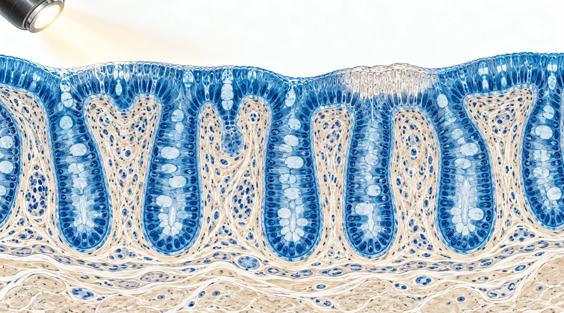
Two blue liquids share one molecule and almost nothing else. In an endoscopy suite, a physician sprays dilute methylene blue onto the lining of the colon to make flat lesions easier to see. At home, someone squeezes blue drops from a retail bottle into water as a wellness routine. Confusing the two is a conceptual error, not just a dosing error: one is a diagnostic visualization procedure performed with clinical preparations, the other a consumer product. This article explains the endoscopy side, where the dye is a tool for seeing, not a treatment for what is seen. For the broader map of established and experimental medical applications, see the overview of methylene blue in medicine.
Surface staining: a vital dye, not a highlighter
Dye-assisted visualization during endoscopy is called chromoendoscopy. Methylene blue performs what pathologists call vital or absorptive staining: living absorptive epithelial cells take the dye into their cytoplasm, so healthy mucosa stains relatively evenly while dysplastic, inflamed, or malignant areas take up less dye or stain patchily. The contrast reveals mucosal architecture that white light alone can miss, particularly flat lesions.
That mechanism differs from indigo carmine, the other classic chromoendoscopy dye. Indigo carmine is barely absorbed; it pools in pits and grooves and exaggerates surface topography, which is contrast staining rather than vital staining. Reviews often pool both dyes under dye-based chromoendoscopy, so a reported benefit of chromoendoscopy in general is not automatically a benefit of methylene blue in particular.
The practical protocol is fussy. In pancolonic methylene blue chromoendoscopy, the endoscopist sprays roughly 0.1% dye segment by segment through a catheter, waits about a minute for uptake, and suctions away the excess before inspecting. Concentrations up to about 0.5% appear in targeted or magnification protocols, but 0.1% is the conventional whole-colon regimen, and the whole exercise demands excellent bowel cleansing plus extra handling time. Those drawbacks motivated a formulation that stains the colon before the procedure starts.
The tablet that stains from the inside
Lumeblue, also called methylene blue MMX, is an oral prolonged-release tablet formulation authorized in the EU as a diagnostic agent for enhancing visualization of colorectal lesions during screening or surveillance colonoscopy, with marketing authorization from August 2020 described in the EMA medicine overview. The dose is eight 25 mg tablets, 200 mg in total, swallowed intact during or after PEG-based bowel preparation on the evening before colonoscopy, in a staggered 3-3-2 sequence.
The MMX matrix is the point of the design. Its coating survives stomach acid and breaks down around pH 7 near the terminal ileum, then releases dye slowly so the colonic mucosa gets prolonged, fairly uniform exposure. By the time the colonoscopy begins, the colon is already stained. That makes Lumeblue a regulated diagnostic medicine with a fixed dose and a defined indication, which is a different concept from both the endoscopist's sprayed solution and any retail wellness product. The three preparations answer different questions and are not interchangeable; retail solutions are discussed separately in the supplements and labelling guide.
| Preparation | How it reaches the mucosa | Purpose | Setting |
|---|---|---|---|
| Sprayed methylene blue (~0.1%) | Endoscopist sprays dye during the procedure | Vital staining for lesion detection | Colonoscopy, operator dependent |
| Lumeblue / MB-MMX (200 mg oral) | Patient swallows prolonged-release tablets with bowel prep | Pre-stained mucosa for lesion detection | Authorized diagnostic medicine |
| Methylene-blue-tinted injectate | Endoscopist injects under the lesion | Visual contrast for the resection plane | EMR/ESD procedures |
What detection studies found in the colon
The strongest methylene-blue-specific colorectal evidence is the phase III MB-MMX trial reported by Repici and colleagues (PubMed 30742834): 1,205 screening and surveillance patients randomized to 200 mg MB-MMX or placebo. Histologically confirmed adenoma or carcinoma detection was 56.29% versus 47.81%, an absolute difference of about 8.5 percentage points, with the clearest gains in nonpolypoid and diminutive lesions and no significant advantage for larger or polypoid ones.
Earlier dye-spray evidence comes largely from inflammatory bowel disease surveillance rather than average-risk screening. Kiesslich and colleagues randomized 165 patients with long-standing ulcerative colitis to conventional colonoscopy or 0.1% methylene blue chromoendoscopy and found 32 versus 10 intraepithelial neoplasias (PubMed 12671882). In IBD colitis, targeted dye-spray biopsies similarly outperformed random sampling in the trial by Marion and colleagues (PubMed 18844620). These are colitis-surveillance results and belong in that context, alongside the dedicated discussion of methylene blue and inflammatory bowel disease.
Broader meta-analyses favor dye-based chromoendoscopy for detection while mostly testing indigo carmine: the 2016 Cochrane review found higher odds of finding at least one neoplastic lesion (PubMed 27056645), and the 2022 Antonelli meta-analysis of ten randomized trials reported a relative increase in adenoma detection of about 20% (PubMed 35588768). Useful for the concept, but not methylene-blue-specific proof.
Detection is not treatment
Every result above concerns finding lesions, not treating cancer. Lumeblue is authorized as a diagnostic agent, and its registration trial measured detection rates, not tumor response, recurrence, or survival. EMA's own phrasing is conditional: better detection may lead to lower cancer risk if the detected lesions are removed. An 8.5-point detection difference cannot be restated as an 8.5% reduction in cancer or mortality. Readers evaluating treatment claims should keep this boundary sharp, as the laboratory-versus-patient evidence guide for methylene blue and cancer explains.
The upper-gastrointestinal story is weaker
In Barrett's esophagus, methylene blue stains intestinal metaplasia blue, which suggested targeting biopsies at stained areas instead of taking blind four-quadrant samples. Early single-center results looked promising, but larger and better-controlled studies did not confirm a dysplasia-detection advantage, and a 2009 meta-analysis of nine studies found no statistically significant incremental yield for intestinal metaplasia, dysplasia, or high-grade dysplasia and early cancer over systematic biopsy (PubMed 19215918).
Guidelines moved on. Current ACG, ESGE, and ASGE Barrett's surveillance guidance recommends chromoendoscopy in the sense of acetic acid or electronic virtual chromoendoscopy, always alongside the structured Seattle biopsy protocol, not methylene blue spray as a substitute for random biopsies. Saying guidelines recommend chromoendoscopy therefore does not mean they recommend methylene blue chromoendoscopy.
The blue cushion under the lesion
A blue layer seen during endoscopic mucosal resection or submucosal dissection is a third concept again. There, the lift comes from saline or another fluid injected beneath the lesion to separate it from the muscle wall; a few drops of blue dye are added so the operator can see the cushion, the dissection plane, and the deep margin. The color marks where the injectate went. It says nothing about metaplasia or dysplasia, and its purpose is mechanical safety during resection, a topic covered with other operative uses in the surgery and diagnostic procedures guide.
A genuine safety footnote: light plus dye
Methylene blue is a photosensitizer, and two studies gave endoscopists pause. Olliver and colleagues showed that 0.5% methylene blue with white-light endoscopy increased DNA damage in Barrett's mucosa on comet assays, while dye without light did not (PubMed 12907012). Davies and colleagues reproduced the pattern in the colon: 0.1% methylene blue plus white light damaged colonocyte DNA in vitro and in vivo, while indigo carmine did not (PubMed 17172595).
The honest reading is narrow: demonstrated acute DNA damage, not demonstrated cancer. No study showed methylene blue chromoendoscopy causing cancer in patients. But combined with the weak Barrett's benefit, an avoidable genotoxic signal made the dye unattractive there, which is part of why guidelines favor other techniques. General handling and adverse-effect context is collected in the safety and side-effects reference.
After the procedure: blue is expected
Methylene blue is a strong dye, and transient blue or green urine and stool afterward are expected effects of the dye rather than complications; urine can look green because blue dye mixes with yellow urinary pigments. Dye on skin leaves temporary blue staining. One caution survives the reassurance: new generalized blue skin accompanied by breathing difficulty or altered consciousness is a different situation from visible surface staining and needs urgent assessment, not cosmetic patience. And a final conceptual caution: never infer which dye was used from color alone. Blue appearance does not identify methylene blue, since other agents tint tissue blue too.
What to take away
Think in three separate boxes: surface staining that exploits epithelial uptake, oral diagnostic formulations that pre-stain the colon under a marketing authorization, and tinted submucosal fluid that marks a resection plane. Ask which box a claim belongs to, whether its evidence measured detection or treatment, and which preparation and concentration were actually tested. Studies behind these distinctions are catalogued in the methylene blue research library.