
Article
Methylene blue in surgery and diagnostic procedures: mapping, leak tests, and dye tracing
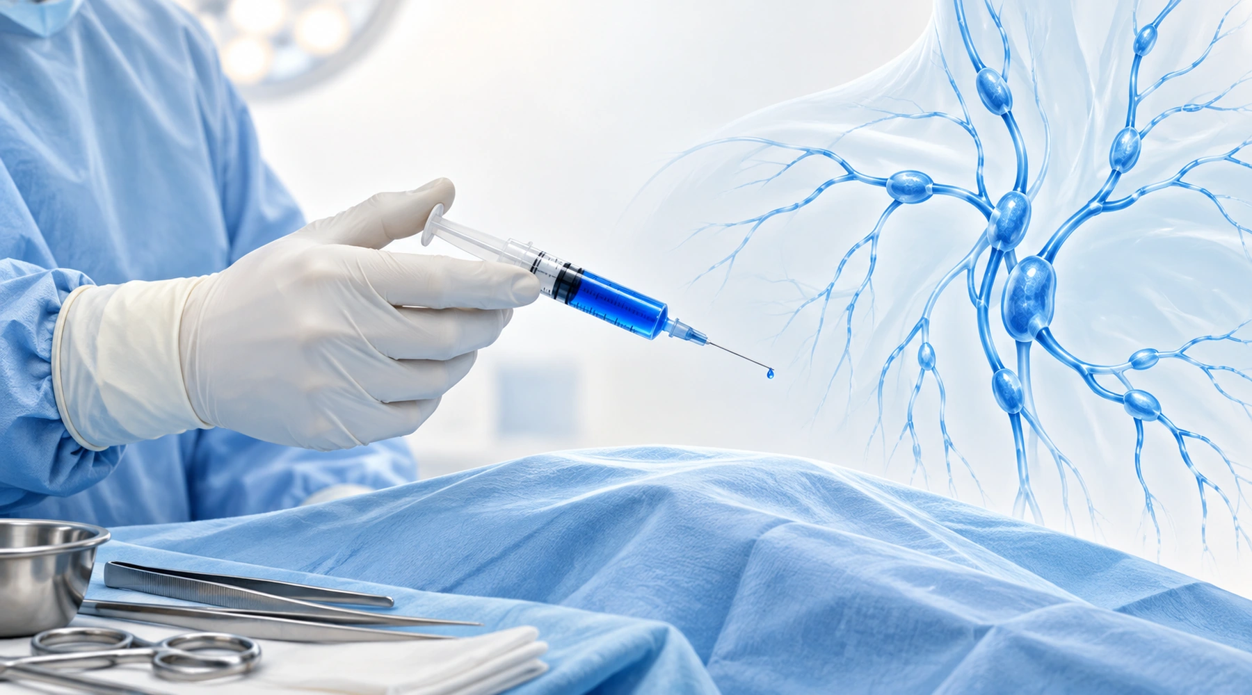
A patient preparing for breast surgery reads that a "blue dye" will map her lymph nodes. A relative having a fistula investigated hears about "fistulography" and assumes dye will be involved. Someone with a tracheostomy is offered a "blue dye test" for swallowing safety. All three hear the word blue and assume it is the same procedure with the same dye. It is not.
Methylene blue in the operating room is best understood as one idea applied in unrelated places: a dye that makes the invisible visible. It follows lymphatic drainage, stains a target gland, colors leaking test fluid, marks a ruptured airway cuff, or demonstrates a hidden connection between two body cavities. The evidence, the amount used, and the risk vary enormously between these applications. And some procedures called "blue dye tests" use other dyes entirely, while radiologic fistulography uses no methylene blue at all. This article maps each use separately. For the broader question of which medical uses are established and which remain investigational, see authorized and investigational medical uses.
Procedure map: visualization, not treatment
| Procedure | What the dye does | Typical exposure | Key limit |
|---|---|---|---|
| Sentinel lymph node biopsy | Stains draining lymphatics and nodes | ~1 mL of 1% solution locally | Lower identification rate alone than combined with radiotracer |
| Parathyroid localization (historical IV) | Stains abnormal glands systemically | 3–7.5 mg/kg IV | Selective adjunct; serotonergic interaction risk |
| Parathyroid localization (local) | Marks a known target in scarred necks | ~0.2 mL of ~0.2% solution | Small observational series only |
| Saline-load test | Colors fluid escaping a joint wound | Dilute, non-standardized | No proven gain in test sensitivity |
| Laser airway cuff | Tints cuff saline so puncture shows blue | Sealed inside cuff | Does not make a tube laser-resistant |
| Swallow blue dye test | Colors food to reveal aspiration | Non-standardized | False negatives; "blue dye" may be another dye |
| Fistula dye tracing | Shows a tract by direct staining | Site-specific, dilute | Different test from radiopaque fistulography |
None of these is treatment with methylene blue. The labeled therapeutic intravenous indication in the United States is acquired methemoglobinemia, generally 1 mg/kg IV, as stated in the DailyMed prescribing information. Surgical tracing applications are mostly off-label or device-specific uses of the same coloring property.
Sentinel lymph node biopsy: following the drainage
After local injection near a tumor, methylene blue enters lymphatic vessels and stains the draining channels and the first (sentinel) nodes blue, so the surgeon can identify them by sight. In breast cancer, blue dye remains an established tracer, traditionally combined with a technetium-99m radiocolloid, and current NICE guidance recommends the dual isotope plus blue-dye technique. Note that "blue dye" in guidelines does not always mean methylene blue specifically; patent blue V and isosulfan blue are also used.
Representative breast protocols use 1% methylene blue (10 mg/mL), often around 1 mL, with placement varying between subareolar, periareolar, intradermal, or peritumoral injection. Mapping with methylene blue alone works, but pooled evidence favors adding a radiotracer: a meta-analysis of methylene blue alone reported a 91% identification rate with a 13% false-negative rate, while combined techniques detect more reliably. In melanoma, mapping relies principally on preoperative technetium lymphoscintigraphy with an intraoperative gamma probe, and blue dye is an adjunct; the ASCO/SSO technical guidance describes 1–2 mL of vital blue dye injected intradermally with about 5–10 minutes allowed for lymphatic transit, listing methylene blue and isosulfan blue as options.
Newer fluorescence mapping with indocyanine green (ICG) visualizes lymphatics in real time without ionizing radiation but requires near-infrared equipment, and guideline adoption remains heterogeneous. The practical reading: methylene blue is the cheap, visually intuitive tracer, and its weakness is that nodes are only visible once surgically exposed.
Methylene blue has specific downsides here. It can cause prolonged skin and tissue staining, inflammatory change, fat necrosis, and occasionally skin necrosis, and postoperative breast imaging can show irregular dye-associated lesions that mimic recurrence. Surgeons should inform radiology and pathology whenever dye has been injected into or near a specimen. Blue dyes can also cause immediate hypersensitivity; a meta-analysis of over 60,000 sentinel-node procedures found an overall blue-dye anaphylaxis rate of about 0.061%, with methylene blue associated with lower rates than isosulfan blue. Broader safety data are collected in side effects and safety.
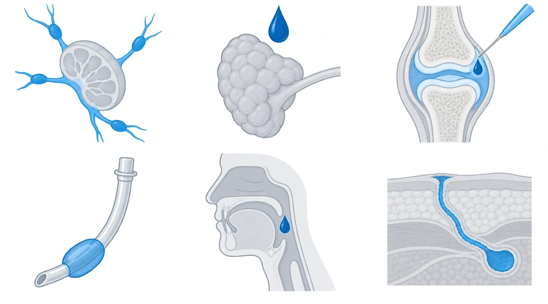
Parathyroid localization: an older systemic use, now selective
Intravenous methylene blue to find abnormal parathyroid glands is an older technique that survives only as a selective adjunct. Historically, fairly large intravenous doses were given before neck exploration so abnormal tissue turned blue; published protocols range roughly from 3 to 7.5 mg/kg IV. A systematic review found 39 observational studies and no randomized trials: staining rates were reportedly high, but no incremental value alongside modern localization had been established.
Current practice relies instead on preoperative ultrasound and other imaging, operative anatomy, and intraoperative parathyroid hormone monitoring. The AAES guideline lists methylene blue among several possible visualization adjuncts rather than as a primary localization method.
This is also the use where the drug-interaction warning matters most. Methylene blue is a potent reversible monoamine-oxidase inhibitor, and the systematic review reported neurotoxicity in 25 patients, all taking serotonergic medication. A separate literature review of methylene blue-associated serotonin syndrome found 50 cases, all involving serotonergic antidepressants, most connected to parathyroid surgery or vasoplegia treatment. Historical parathyroid doses can substantially exceed the current 1 mg/kg therapeutic dose, so this interaction belongs to systemic exposure, not to a drop of dye on tissue.
A distinct, much smaller-exposure technique has been described for reoperative or scarred necks: ultrasound-guided injection directly into a known target, with small series using about 0.2 mL of a 1:5 dilution of 1% solution (approximately 0.2%) just before surgery. Evidence is limited to small observational series.
Saline-load test: the dye does not make the test work
In a saline-load test for traumatic arthrotomy, saline is injected into a joint and the wound is watched for leaking fluid. Methylene blue is sometimes added simply to make escaping fluid easier to see. The critical finding is that the dye has not been shown to improve the test: in a randomized prospective knee study of 58 patients, false-negative rates were 69% with methylene blue versus 66% with plain saline, with similar volumes required for a positive test. A 2025 systematic review found 8 of 23 saline-load studies had used the dye, with performance driven by joint, wound location, injected volume, and technique rather than by dye addition. There is no validated standard concentration for this purpose. Methylene blue here is an optional coloring agent in historical or experimental protocols, not the component that makes the test sensitive.
Laser airway tubes: dye sealed inside the cuff
This use works differently from every other on this page: the dye is normally placed inside the endotracheal tube cuff, not into the patient. For airway laser surgery, anesthesia fire-safety guidance recommends an appropriate laser-resistant tube and, where feasible, filling the cuff with saline rather than air. Tinting that saline with methylene blue means a laser puncture produces an immediately obvious blue leak. No universal concentration is specified; the goal is visible tinting, as described in the ASA operating-room fire practice advisory.
Two cautions matter. Some historical laser tubes incorporated dye crystals into the cuff or pilot system, and a published case describes undissolved crystals obstructing a pilot tube so the cuff could not deflate, which is why device-specific instructions matter. And methylene blue itself does not make an ordinary tube laser-resistant; fire prevention also depends on tube construction, cuff protection, and minimizing oxidizer concentration.
Swallow tests: "blue dye" may not be methylene blue
In a typical bedside test for a patient with a tracheostomy, food or liquid is colored and tracheal secretions are checked for blue discoloration; a positive result supports aspiration. The terminology is easy to misread: published "blue dye test" and Modified Evans Blue Dye Test protocols have used Evans blue, methylene blue, and food dyes, so "blue dye test" does not automatically mean methylene blue.
The main problem is false negatives. Sensitivity across heterogeneous protocols ranges from roughly 38% to 95%, and guidance treats the test mainly as an adjunctive rule-in signal when positive: a negative test does not exclude aspiration. Definitive assessment, when needed, uses instrumental studies such as fiberoptic endoscopic evaluation of swallowing or videofluoroscopy. There is no validated optimal dye quantity.
Do not conflate this with the older practice of continuously tinting enteral feeds with FD&C Blue No. 1. That is a different dye and a different practice, associated with systemic absorption and serious toxicity in critically ill patients and subsequently discouraged.
Fistula tracing is not fistulography
Local methylene blue tracing and radiologic fistulography are different diagnostic approaches that are often conflated. In local tracing, dye is placed into a suspected fistula opening or organ cavity and the clinician watches for blue emerging elsewhere or follows stained tissue at surgery: injection through an external opening to locate an anal fistula tract, coloring bladder irrigation to reveal leakage into the vagina in suspected vesicovaginal fistula, and similar site-specific applications. Protocols are site-specific and concentrations are not standardized.
A true fistulogram or sinogram is an imaging procedure: radiopaque contrast is injected into the tract and fluoroscopy or CT maps its anatomy. It is not a methylene blue test. Neither approach necessarily substitutes for MRI, CT, endoscopy, or examination under anesthesia in a complex fistula.
One route-specific trap deserves emphasis: rectally administered methylene blue can be absorbed and later excreted into urine. A volunteer study demonstrated this after a 50-mg enema and concluded that intrarectal dye should not be used to confirm a suspected colovesical fistula, because blue urine can occur without any fistula. State the anatomical site and the imaging method rather than treating every fistulography question as the same test.
Cross-cutting limits worth remembering
Systemic exposure carries pharmacologic considerations that local tracing does not. Enough methylene blue in the bloodstream absorbs light used by monitoring devices, so pulse oximetry may underestimate oxygen saturation. Systemic administration can produce severe hemolysis in people with G6PD deficiency, and current injectable labeling lists G6PD deficiency as a contraindication. Again, the risk from minute local injections or dye sealed inside an airway cuff is not equivalent to several mg/kg given intravenously, but "local" does not automatically mean zero systemic absorption: route, quantity, and tissue integrity matter.
A final point concerns dye identity. Surgical visualization depends on knowing exactly which dye is in the syringe: methylene blue, isosulfan blue, patent blue V, Evans blue, and food dyes behave differently and carry different risks, yet all appear in procedures casually called "blue dye" tests. The same discipline applies outside the hospital. Products sold as methylene blue vary widely in grade and documentation, and textile-grade material is not a substitute for material manufactured and tested for its stated purpose. When purity matters, look for pharmaceutical manufacturing, full USP testing with a lot-specific certificate of analysis, and independent verification rather than a heavy-metals screen alone. The guide to reading a certificate of analysis explains which fields to check. Deeper sourcing is indexed in the research library.