
Article
How methylene blue works: electron transfer, and where the electrons land
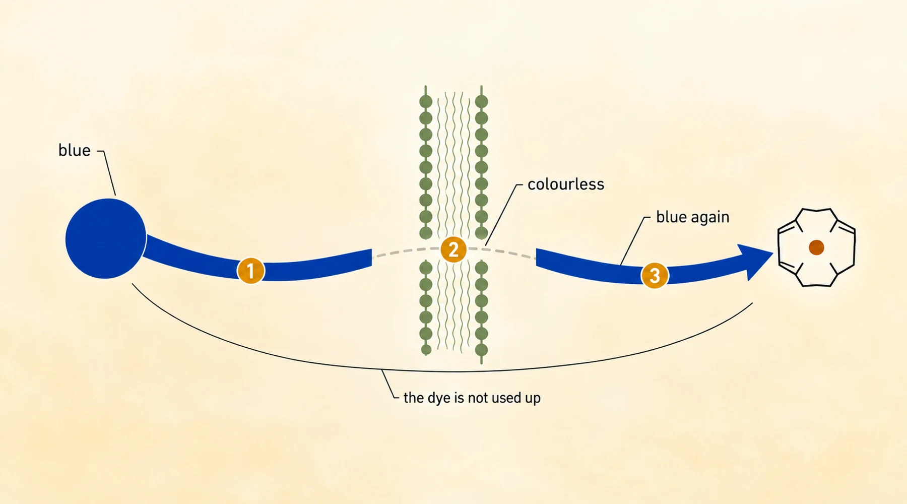
A stoppered flask on a bench is the shortest honest introduction to how methylene blue works. Water, glucose, a pinch of sodium hydroxide, and enough dye to turn the liquid a deep, unmistakable blue. Wait a few minutes and the blue drains away until the flask looks like a flask of water. Shake it, and the colour snaps back. Then it fades again, and it will keep doing that for as long as there is glucose willing to give up electrons and oxygen willing to take them.
Nothing about the dye has changed except the number of electrons it is carrying. Oxidised, it is blue. Reduced, it is not. Almost everything else in the therapeutic and research literature on this molecule follows from that one reversible step, because a molecule that can take electrons from one partner and hand them to another is a relay, and a relay does nothing on its own. What it does depends entirely on what is standing at each end of it.
Two forms, and why the pair matters
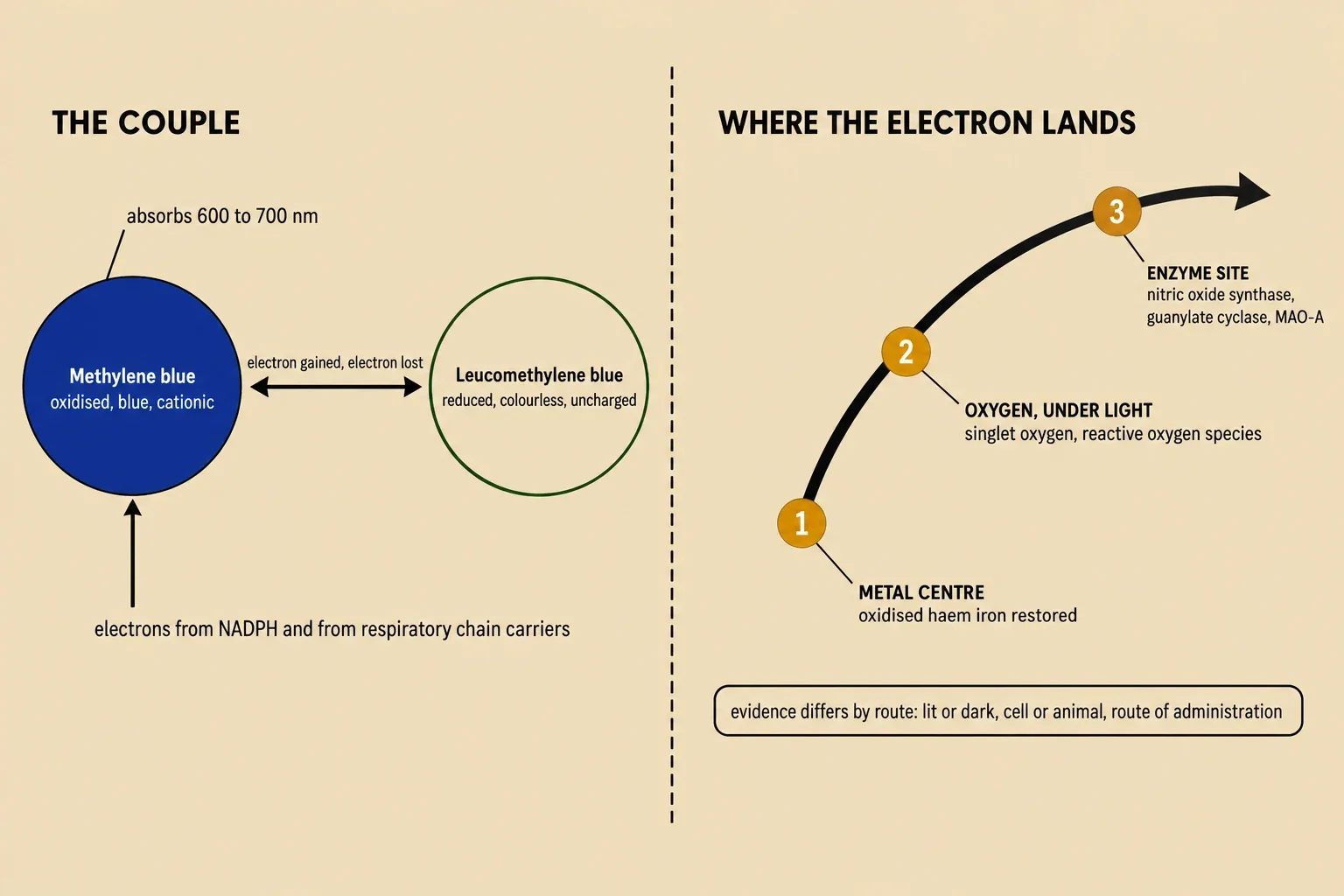
The oxidised form is a planar, positively charged ring system. That flat, charged shape is why it stacks against nucleic acids, why it binds to sialylated structures on cell membranes, and why it absorbs visible light strongly between roughly 600 and 700 nanometres, which is the part of the spectrum the human eye reads as blue.
The reduced form is a different proposition. It carries no charge, it dissolves in fat rather than water, and it is colourless. Its ionisation is very low at the pH inside the body, on the order of a few per cent. So the reduced molecule crosses a cell membrane easily by diffusion, and once inside, where the environment is more oxidising, it can be re-oxidised and effectively trapped. The oxidised ring system is flat; the reduced one can twist. Those structural consequences, and the colour change itself, belong to the story of the two forms in their own right, and they are covered in more detail in the comparison of methylene blue and leucomethylene blue.
Accepting electrons
The oxidised dye will accept electrons from a wide range of donors. Glucose in the demonstration flask is one. Inside a cell, reduced pyridine nucleotides and components of the mitochondrial electron transport chain are others. When methylene blue is reduced in a tissue that contains oxygen, the cycle it sets up consumes both the reducing agent and oxygen, and produces reactive oxygen species, chiefly hydrogen peroxide. The dye comes back out of the cycle unchanged, ready to run it again.
That last point explains a contradiction that otherwise looks careless. Methylene blue is described in the literature as both a pro-oxidant and an antioxidant, and both descriptions are defensible. It can suppress superoxide production by acting as an artificial electron acceptor, diverting the flow of electrons away from the enzyme sites where oxygen would otherwise be converted into superoxide radicals. In liver cell cultures it has been shown to induce phase-2 antioxidant defence enzymes, including thioredoxin reductase 1 and NADPH quinone oxidoreductase. Which of the two faces you see depends on which end of the relay is short of electrons at that moment. A question like "is methylene blue an antioxidant" is not well formed until you say in which system, at which concentration, with what else present.
Handing electrons on
Once the dye holds a spare electron, three quite different things can happen next, and they are usually described under one heading. They should not be.
It can pass the electron to a metal centre. The clearest case is haemoglobin that has been oxidised from the ferrous to the ferric state. In that state it cannot carry oxygen. In red cells, an enzyme reduces the dye, and the reduced dye then hands electrons to the oxidised haem iron non-enzymatically, restoring working haemoglobin and returning the dye to its oxidised form. The dye is not consumed by this; it cycles. The clinical picture that rests on this reaction is a hospital subject, and the explanation of methylene blue in methemoglobinemia covers how it is used and why it is supervised so closely.
An honest note on the reading: the two reviews this article is built on do not name the same reducing cofactor for the enzyme step, one saying NADH and the other NADPH. The disagreement is real and unresolved in the sources available here, so the safe description is the role each part plays rather than a single cofactor name.
It can pass energy, or an electron, to oxygen, if there is light. This route only exists under illumination. The excited dye reaches a triplet state, and from there two mechanisms are possible. In the first, the excited dye interacts directly with a neighbouring molecule, taking a hydrogen or transferring an electron, which generates radicals; reacting with water this way produces hydroxyl radicals. In the second, the excitation energy is handed to oxygen itself, producing singlet oxygen, a highly electrophilic species that decays within microseconds and is generally accepted as the major route by which illuminated methylene blue damages microbial cells. Reactions with cell envelope lipids and peptides, and with guanine bases in DNA, are among the documented outcomes.
The practical consequence of this route is that illumination changes the numbers by orders of magnitude. In the data collected by Wainwright and Crossley, methylene blue against Pseudomonas aeruginosa has a minimum bactericidal concentration around 500 micromolar in the dark, and around 25 micromolar under a light dose of 12 joules per square centimetre. Against one strain of Staphylococcus aureus, dark and illuminated values are both 5 micromolar. In other words, whether the molecule counts as an antimicrobial at all is not a property of the molecule alone; it is a property of the molecule plus the light.
It can act at an enzyme site directly, without the relay. Methylene blue oxidises the ferrous iron bound to nitric oxide synthase, which disables the enzyme. It blocks soluble guanylate cyclase, and between 1 and 10 micromolar is the routine laboratory concentration used for that purpose. It inhibits monoamine oxidase type A with an IC50 between 27 and 180 nanomolar, which is the mechanism behind the well-documented interaction with serotonin reuptake inhibitors and the reason methylene blue's drug interactions are a category of their own.
These three routes are chemically distinct. Collapsing them into a single claim that methylene blue works by redox cycling is the main reason so much writing about it reads as simultaneously confident and vague.
Four distinctions that decide whether a mechanism statement means anything
Which form. The reduced dye is not interchangeable with the oxidised dye. Acetylcholinesterase inhibition illustrates the trap: in a long in vitro incubation, methylene blue is progressively reduced to its colourless form, and the inhibition falls away, because the reduced molecule does not inhibit that enzyme. An assay run long enough to change the form will report a weaker effect and invite the wrong explanation.
Which concentration. The laboratory range for soluble guanylate cyclase inhibition is 1 to 10 micromolar. Abolishing glutamate-mediated transmission in rat hippocampal slices took 5 to 50 micromolar. Toxic effects in cell culture are reported above 100 micromolar. These are not the same experiment, and a mechanism confirmed at 50 micromolar is not automatically available to a patient whose plasma concentration is a fraction of that.
Which route. Absolute oral bioavailability is around 72 per cent, and the terminal plasma half-life is 5 to 7 hours. Oral administration produces higher concentrations in the intestine and liver; intravenous administration produces higher concentrations in the brain. Route decides which tissue meets the electrons.
Which model, and whether the lights were on. Much of the mechanistic literature is cell culture and rodent work, and in vitro results on this molecule depend on light exposure, membrane potential and the redox state of the cells, because a microscope lamp is enough to photosensitise it. Between 65 and 85 per cent of an administered dose is reduced to the colourless form in red cells and peripheral tissue. Every one of those variables sits between a clean mechanism in a dish and a claim about a person.
Why the purity question is a chemistry question
Illumination demethylates methylene blue. The first product is Azure B, then Azure A, then thionin, and higher light intensity produces more breakdown than lower. A molecule whose usefulness consists of being able to accept and donate electrons is therefore a molecule whose likely degradation products are close chemical relatives that stay in the same solution. The composition of what is in the container is not a cosmetic detail.
That is the reason conformance to the USP specification is the meaningful test for a buyer. A product that does not meet it is a dye, and a dye carries impurities, including Azure B, heavy metals and residual solvents, that a person swallowing it does not want. The further trap is a seller who tests only for heavy metals and presents the result as full compliance. Blupreme has partnered with a pharmaceutical manufacturer of methylene blue and tests the full specification: identity, purity, organic impurities, residual solvents, elemental impurities, residue on ignition, microbial limits and bacterial endotoxins. The laboratory testing document behind each batch is published, and choosing a supplier is largely a matter of checking whether that document exists and what it covers.
One reversible couple. Several routes out of it. The route decides what the claim can be.