
Article
Methylene blue and mitochondria: electron transport and the evidence
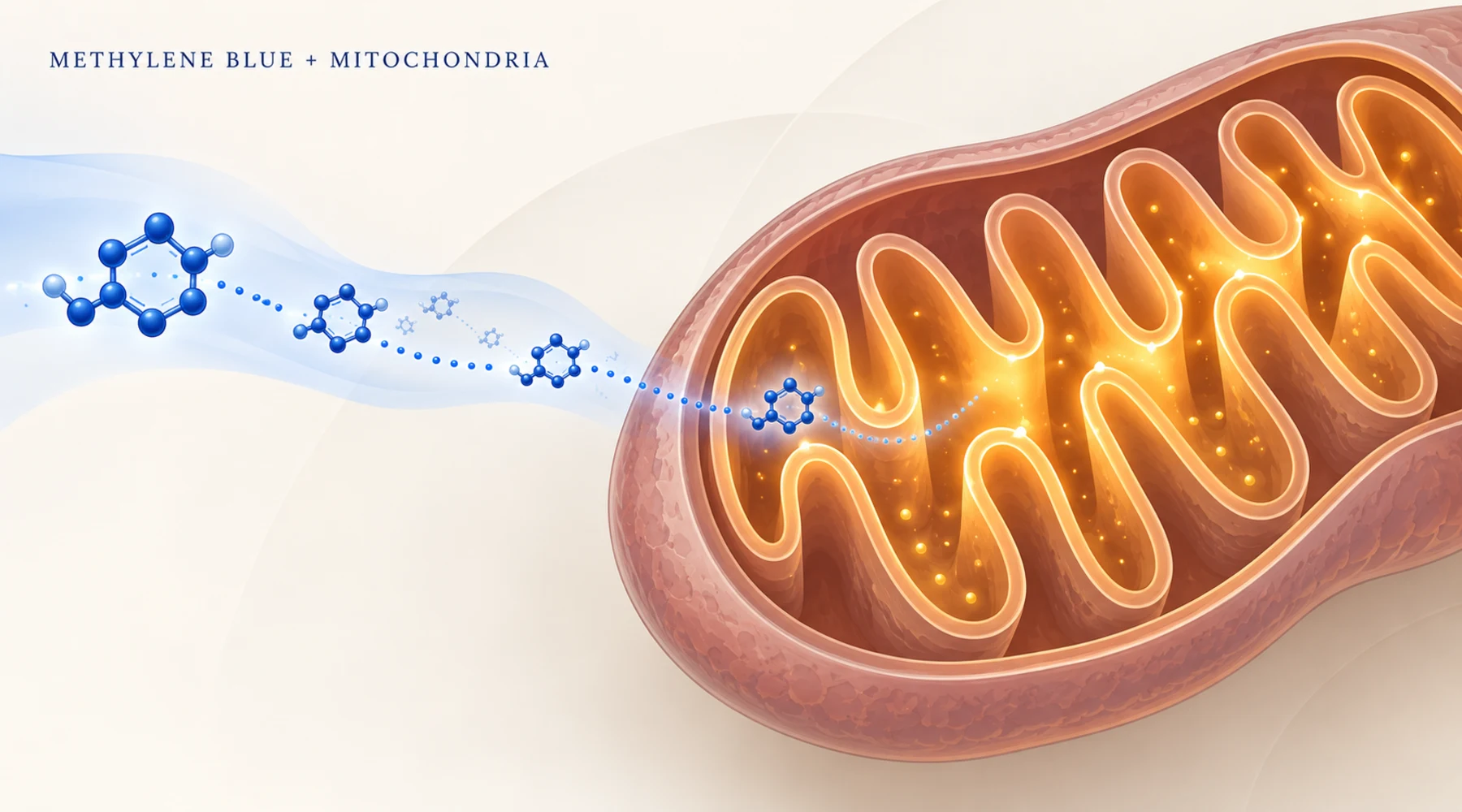
Picture two preparations side by side. Both contain brain mitochondria poisoned with rotenone, a chemical that blocks Complex I of the respiratory chain. Into one, the experimenter pipettes a micromolar dose of methylene blue. Oxygen use partly recovers. The membrane voltage partly returns. A small amount of ATP synthesis restarts. In the neighboring cuvette, a peroxide sensor tells the other half of the story: hydrogen peroxide output has multiplied severalfold, at every dose tested, under every substrate tried.
Both readings are correct. Methylene blue is an electron shuttle that can keep current flowing around a damaged section of the respiratory chain, and the same shuttle charges a toll in peroxide while it does so. Hold both facts at once and the literature on methylene blue mitochondrial function becomes legible. Hold only one and it reads as miracle or menace depending on which paper you opened first.
The shuttle, stated plainly
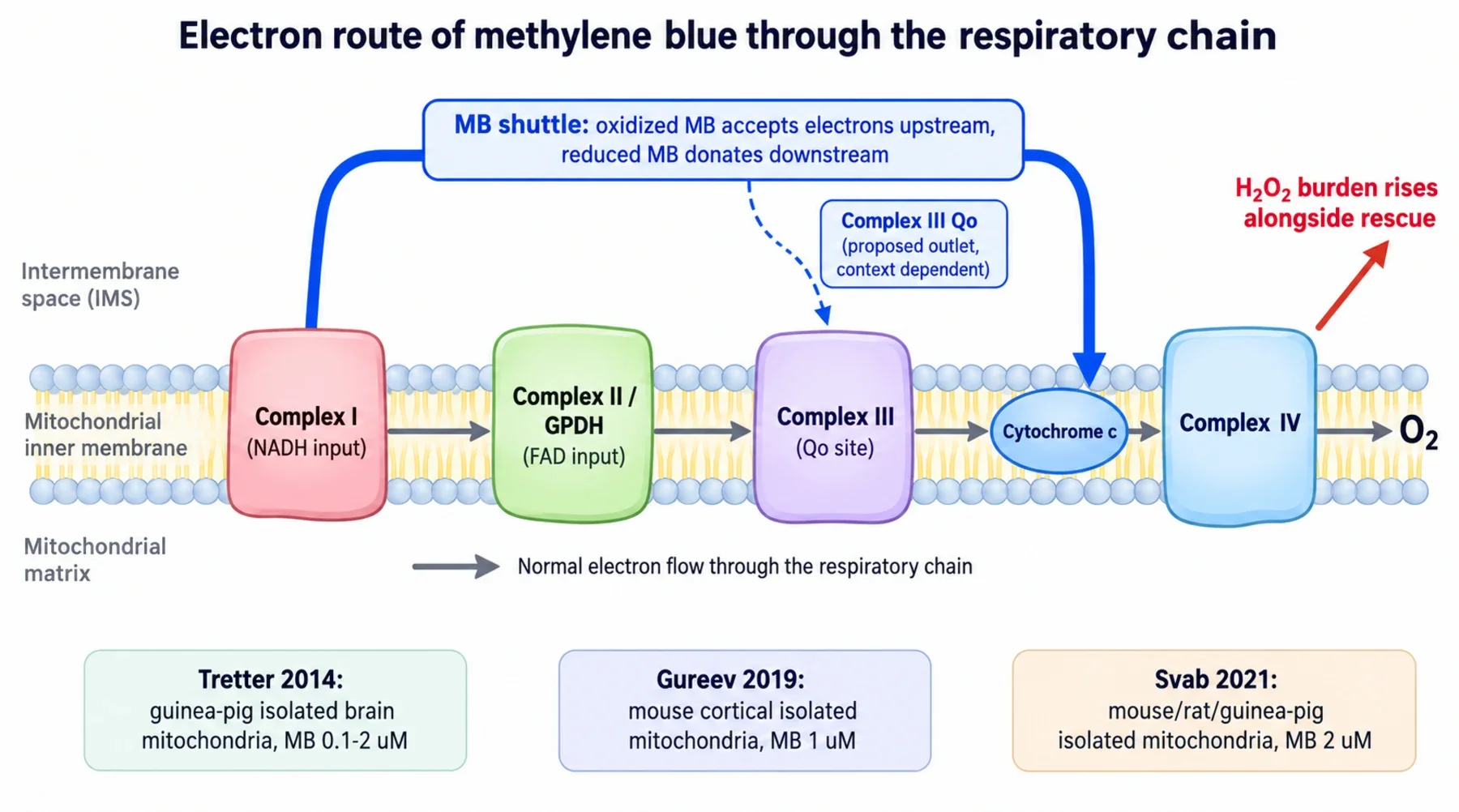
The respiratory chain is a relay line. Electrons enter at Complex I (from NADH) or at Complex II and related flavin enzymes (from succinate and similar donors), pass through Complex III to the mobile carrier cytochrome c, then through Complex IV to oxygen. Each handoff pumps protons across the inner membrane, and the resulting gradient drives ATP synthesis. Block any station and the line upstream goes quiet.
Methylene blue inserts itself as a parallel courier. Its oxidized form picks up electrons from upstream carriers, becoming the colorless reduced form, leucomethylene blue, which then hands those electrons onward downstream and returns to blue. The two forms are described in the account of the redox couple, and the broader relay behavior belongs to the general mechanism explainer. The open question is where the courier re-enters the line: the quinol binding pocket of Complex III known as the Qo site, or cytochrome c directly. Which exit dominates depends on conditions, as the Complex III bypass analysis lays out in detail.
What three rodent studies actually measured
All three studies used isolated brain mitochondria, meaning organelles removed from the cell and suspended in a defined buffer. No intact cells, no dosed animals, no human subjects. Concentrations refer to the bath around the organelles, not to a human oral dose. Keep that frame fixed and the numbers speak clearly.
The first study, in isolated guinea pig brain mitochondria at 100 nanomolar to 2 micromolar, mapped benefit and cost in one preparation. In healthy mitochondria the dye raised resting oxygen use but left ADP stimulated respiration unchanged, so coupled ATP output did not improve. Under Complex I blockade, 2 micromolar restored ATP synthesis from 46 to 93 nanomoles per minute per milligram against a normal rate near 1109. Under Complex III blockade, the same dose restored ATP from about 46 to 63 against a normal rate near 910. Voltage and calcium uptake partly recovered. Cyanide blockade of Complex IV could not be bypassed, which places the useful electron exit upstream of working Complex IV. Every rescue was statistically real and small: single digit percentages of normal ATP output.
The second study, in mouse cortical mitochondria at around 1 micromolar, confirmed the Complex I bypass and drew a boundary at Complex III. Rotenone cut respiration from about 121 to under 2 nanomoles of oxygen per minute per milligram, and the dye lifted it to about 21, roughly one sixth of normal, while membrane voltage recovered almost fully. But after Complex III blockade, neither respiration nor voltage recovered across 1 to 5 micromolar, and the blockade suppressed the dye driven peroxide signal. The authors proposed that reduced dye delivers its electrons at the Qo site of Complex III: with that site occupied, the courier has nowhere to deliver.
The third study, in mouse, rat and guinea pig brain mitochondria at mainly 2 micromolar, tested the disagreement directly and found that a Complex III block can be bridged. With the Qo site inhibitor myxothiazol cutting basal respiration by 88 percent, the dye restored oxygen use to about 62 percent of the unblocked rate. In a direct test, half a micromolar reduced exogenous cytochrome c even while Complex III was blocked, supporting a route from NADH through the dye to cytochrome c and onward to Complex IV. But the rescued respiration behaved abnormally: added ADP lowered oxygen use instead of raising it, and blocking ATP synthesis stimulated respiration. Voltage repolarized only partly. High oxygen use with inverted coupling is not normal energy production.
Why the laboratories disagree, and what settles it
The species explanation fails first. The third group repeated the work in three rodents and obtained the same qualitative bypass in all of them, including the mouse strain where the bypass had been reported absent. What differs instead is concentration (1 versus 2 micromolar), inhibitor doses, buffers, isolation methods, and above all the energetic state compared: the bypass is clearest before ADP addition, exactly where the paradoxical ADP response appears. The honest diagram therefore shows two condition dependent exits, one at the Qo site and one at cytochrome c. The courier delivers where the local chemistry lets it deliver.
The peroxide toll
The cost side is as consistent as the rescue side. In the guinea pig work the dye raised peroxide release under every condition tested, resting, ATP synthesizing and chemically impaired, across all three substrates. During ATP synthesis, output rose from about 148 to over 1500 picomoles per minute per milligram at 1 micromolar, and even 100 nanomolar more than quadrupled the signal. The dye also slowed peroxide disposal roughly 2.6 fold, so the signal reflects faster formation plus slower clearance. The proposed chemistry is direct reduction of oxygen by reduced dye without detectable superoxide, though that remains an interpretation rather than a proven step.
The mouse work complicates the picture in a useful way. Peroxide rose steeply with dose, from a baseline near 76 to nearly 2000 picomoles per minute per milligram at 2 micromolar, yet Complex III blockade suppressed the dye driven component. If peroxide came only from reduced dye reacting with oxygen, blocking the chain should not matter so much. It does, so the chain participates. The antioxidant and pro-oxidant review places this duality in wider context, and the oxygen versus ATP analysis explains why oxygen numbers alone never settle an energy claim.
Rescue is not repair
The phrase that needs retiring is "does methylene blue repair mitochondria." Repair would mean durable correction: rebuilt complexes, coupling that persists after the drug leaves, recovery in intact cells, then improved function in a living organism. None of the three papers shows that. They show acute rerouting in freshly isolated organelles during artificial chemical blockade, with ATP output at small fractions of normal and sometimes frankly strange coupling. Rotenone and antimycin are laboratory tools, not models of aging or human mitochondrial disease, and nothing here translates into more energy for a healthy person, which is a separate evidence question with its own human literature.
Three boundaries keep any biohacking conclusion honest. First, bath concentration is not a dose: 0.1 to 2 micromolar around isolated organelles says nothing about which oral amount produces that free concentration at a human brain mitochondrion, especially since the dye accumulates or releases depending on membrane voltage itself. Second, oxygen is not energy: the dye can raise oxygen use through cycling and leak as well as through productive phosphorylation, so respirometry without ATP and voltage readouts cannot license an energy claim. Third, benefit and burden share one sentence: any statement of rescued voltage or ATP that omits the concomitant peroxide rise misreports the experiment. Related qualifiers include NAD redox balance, dose response shape and the assay guide.
One practical footnote: the shuttle is a property of the exact molecule. Demethylated relatives such as Azure B are redox active too, with different behavior, so a partly degraded solution is a different experiment from intact dye. That is one reason to insist on the full pharmacopeial specification and a published batch document rather than a metals only screen. Light driven chemistry adds a parallel caveat for illuminated uses, covered under red light combinations.
The narrow claim survives all of this and is worth keeping: methylene blue can act as a conditional electron courier in damaged isolated mitochondria, partially preserving flow at a peroxide price. Everything beyond that, energy, longevity, repair, awaits evidence these studies were never built to give.