
Article
Methylene blue and leucomethylene blue: one redox couple, two colors
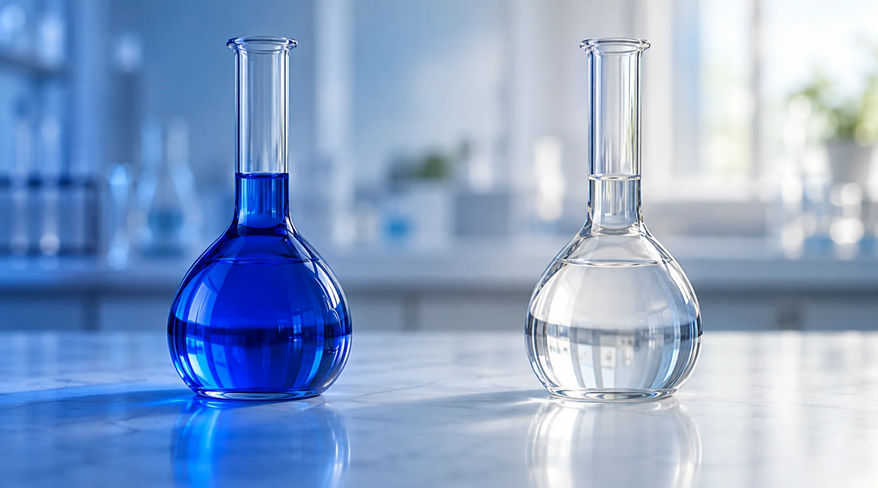
A student adds a few drops of blue dye to a flask, swirls, and watches the color vanish. She shakes the flask and the blue returns. Nothing was added or removed between the two states except electrons and contact with air. That flask contains the whole subject of this article: methylene blue and its reduced partner, leucomethylene blue, interconverting in a reversible redox couple where color is the readout.
The practical point comes first. Color change is not a side effect here. It is the analytical signal. Anyone who formulates with methylene blue, titrates with it, or teaches with it should be able to write the half-reaction from memory, state the conditions that push it left or right, and name the impurities that blur the result.
The couple in one equation
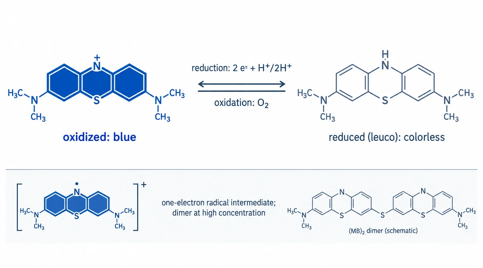
Oxidized methylene blue is the methylthioninium cation: a flat phenothiazinium system with sulfur and nitrogen in the central ring and a dimethylamino group at each end, supplied as the chloride salt. PubChem records it as methylthioninium chloride, C.I. Basic Blue 9. In water the monomer absorbs near 664 nm, which is the blue the eye sees.
Reduced leucomethylene blue (LMB) is the same skeleton with the central ring nitrogen protonated and the chromophore broken. It is uncharged near neutral pH and nearly colorless, with negligible visible absorbance and a strong ultraviolet band near 258 nm (PubChem entry for leucomethylene blue, CAS 613-11-6). The proton count in the half-reaction depends on pH, because the reduced form protonates in acid (approximate pKa values near 4.5 and 5.8). Three regimes cover the practical range:
MB(+) + 2 e(-) + H(+) <=> LMB, blue <=> colorless (near neutral pH, 1 proton)
MB(+) + 2 e(-) + 2 H(+) <=> LMBH(+), blue <=> colorless (moderately acidic, 2 protons)
MB(+) + 2 e(-) + 3 H(+) <=> doubly protonated leuco species (strongly acidic, 3 protons)
All three writings describe the same couple under different protonation states, a point the redox behavior of thiazine dyes literature treats systematically. The pH dependence follows the Nernst equation for Ox + mH(+) + 2e(-) <=> Red, giving a slope of about 29.6, 59.2, or 88.7 mV per pH unit for m = 1, 2, or 3. Measured slopes near 28, 58, and 87 mV per pH unit confirm the assignment, so a reported potential without its pH is meaningless.
What the equation tells a formulator
Three facts fall out of the stoichiometry. First, two electrons per molecule means methylene blue is a two-electron indicator and mediator, not a one-electron shuttle, in its net reaction. Second, protons participate, so the formal potential moves with pH as described above. Third, the formal potential at pH 7 sits near +0.011 V against the standard hydrogen electrode (about +0.53 V at pH 0), which places the couple among mild oxidants: strong enough to take electrons from good donors such as NAD(P)H or ascorbate, weak enough to hand them to dissolved oxygen or ferric centers. That middle position is exactly why the dye cycles instead of getting stuck at one end.
Concentration adds one more wrinkle. Above roughly one hundred micromolar in water, consistent with a reported monomer dimer dissociation constant near 170 to 250 uM at pH 7, the oxidized cation stacks into dimers and higher aggregates with a shifted absorption band near 610 to 615 nm. A spectrum that looks off peak may therefore mean aggregation, not impurity. Dilute first, then judge.
Conditions that drive each direction
The table below is the bench reference. Reduction needs an electron donor; reoxidation needs an electron acceptor, and air usually suffices.
| Direction | Reagent or condition | What happens | Notes |
|---|---|---|---|
| Reduction | Glucose in alkaline solution | Blue fades on standing | The classic blue bottle system; shaking with air restores the color |
| Reduction | Ascorbic acid | Blue fades | Basis of vitamin C clock and titration demonstrations |
| Reduction | Sodium dithionite | Rapid, complete bleaching | Strong reductant for analytical work; excludes air afterward |
| Reduction | NAD(P)H with a suitable catalyst or enzyme | Blue fades | Models the biological reduction route |
| Reduction | Zinc in hydrochloric acid | Bleaching in strongly acid medium | Preparative route to the leuco form |
| Oxidation | Shaking with air or pure oxygen | Colorless returns to blue | Dissolved O2 is the default oxidant; the classroom proof of reversibility |
| Oxidation | Ferric ion, persulfate, or dichromate | Rapid return to blue | Strong oxidants for quantitative reoxidation |
Every entry obeys the same equation. Only the driving force changes. That is the sense in which the couple is reversible: the path is the same, traveled in opposite directions under opposite conditions.
The blue bottle, explained
The demonstration deserves its mechanism stated plainly because it is the couple in miniature. In alkaline glucose solution, base promotes glucose enolization and the resulting enediolate reduces methylene blue to leucomethylene blue, so the flask goes colorless. Shaking dissolves oxygen from the headspace, oxygen reoxidizes the leuco form, and the blue returns. Standing still depletes dissolved oxygen near the surface reaction zone and glucose wins again. The cycle repeats until the glucose is consumed, and each shake is a visible Nernst equation: raise the oxidant activity and the equilibrium shifts toward blue.
The radical in the middle
The net reaction moves two electrons, but elementary steps move one at a time. Between the blue cation and the colorless leuco form sits a semiquinone radical intermediate that mostly disproportionates back to the pair rather than accumulating. Its one-electron potential near minus 0.23 V at pH 7 lies far below the net two-electron midpoint, which is why the radical stays a minor steady-state species. It matters for two reasons. Under illumination the dye reaches excited states that open radical pathways, which is why methylene blue solutions are light sensitive and why photochemistry belongs in any stability protocol. And at high concentration the oxidized form dimerizes, which changes the spectrum and the apparent kinetics without changing the underlying couple. Neither fact breaks reversibility. Both explain observations that otherwise look like irreversibility.
Demethylated relatives shift the scale
Strip methyl groups from the two dimethylamino arms and the couple survives with shifted properties. The absorption maximum shifts monotonically blueward with each demethylation: methylene blue near 664 nm, Azure B near 654, Azure A near 630, Azure C near 615, and thionine near 600 nm. Azure B, missing one methyl group, is the closest relative: a major methylene blue metabolite and a documented impurity in methylene blue material. Azure A and Azure C continue the series with the same redox-active skeleton. Thionine, with both amino groups fully demethylated, sits at the end with a formal potential near +0.06 V at pH 7 against methylene blue at +0.011 V, appreciably easier to reduce. Redox potentials across the series depend on medium and electrode, so compare values only within one set of conditions. This is why impurity profiling is a redox issue, not just a purity checkbox. A few percent of Azure B means a few percent of a different redox couple in the bottle, and azure B as impurity and metabolite documents how that fraction is controlled.
Where biology plugs in
Inside a red blood cell, NADPH reduces methylene blue to leucomethylene blue, and the leuco form passes electrons to ferric heme iron, restoring the ferrous state of hemoglobin. That single electron relay is the chemical basis of methylene blue use in acquired methemoglobinemia. Everything downstream of that relay, energy metabolism, mitochondrial electron flow, and any biological outcome, belongs to mitochondrial electron transport and is deliberately left there.
Buy the couple, not just the color
A redox dye is only as good as its specification. High purity matters because non-USP methylene blue is typically textile grade, carrying Azure B, heavy metals, or residual solvents that shift redox readings and blur endpoints no staining protocol screens for. Most sellers that claim USP conformance test only heavy metals and declare victory. Full conformance covers identity, assay, organic impurities, residual solvents, elemental impurities, residue on ignition, microbial limits, and bacterial endotoxins, and how to choose pharmaceutical-grade methylene blue walks through reading that documentation. Blupreme works with a pharmaceutical manufacturer of methylene blue, tests the full USP specification, and publishes the complete COA. When the color is the measurement, that paperwork is part of the instrument.