
Article
Methylene blue and red light therapy: photochemistry and clinical evidence
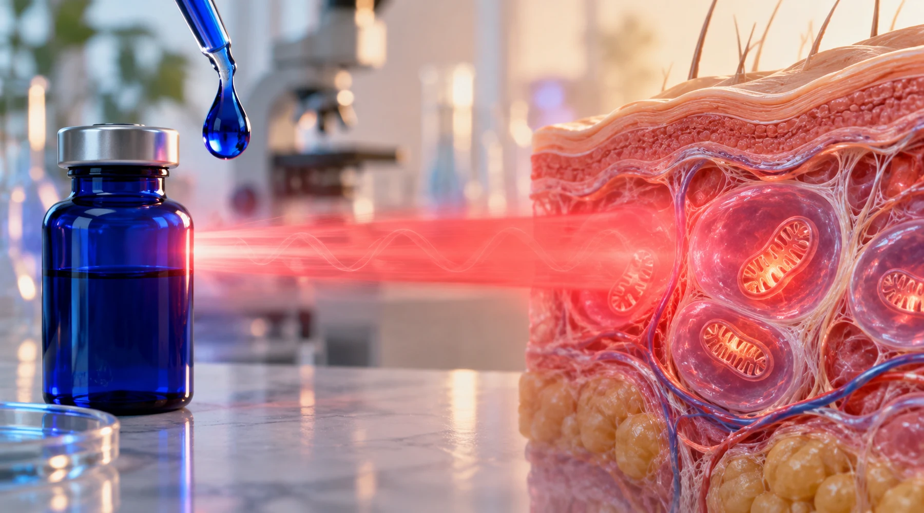
Two readers type methylene blue and red light therapy into a search box and mean opposite things. One imagines a red panel that helps mitochondria work better, with a blue dye added for extra effect. The other imagines a dermatology procedure where light activates a dye to destroy something unwanted. Both use the same words. They describe different concepts, and the confusion matters because light plus dye can either signal to a cell or injure it.
The distinction is the subject of this review. Photobiomodulation, usually called PBM, uses red or near infrared light to modulate endogenous signaling without adding a photosensitizer. Photodynamic therapy, usually called PDT, uses three parts together: light at a wavelength the sensitizer absorbs, a photosensitizer such as methylene blue, and oxygen to create a burst of reactive species that damages the target. Methylene blue red light therapy sits in both camps in online discussion. Photochemistry decides which camp a specific combination belongs in.
Absorption decides whether light and dye interact
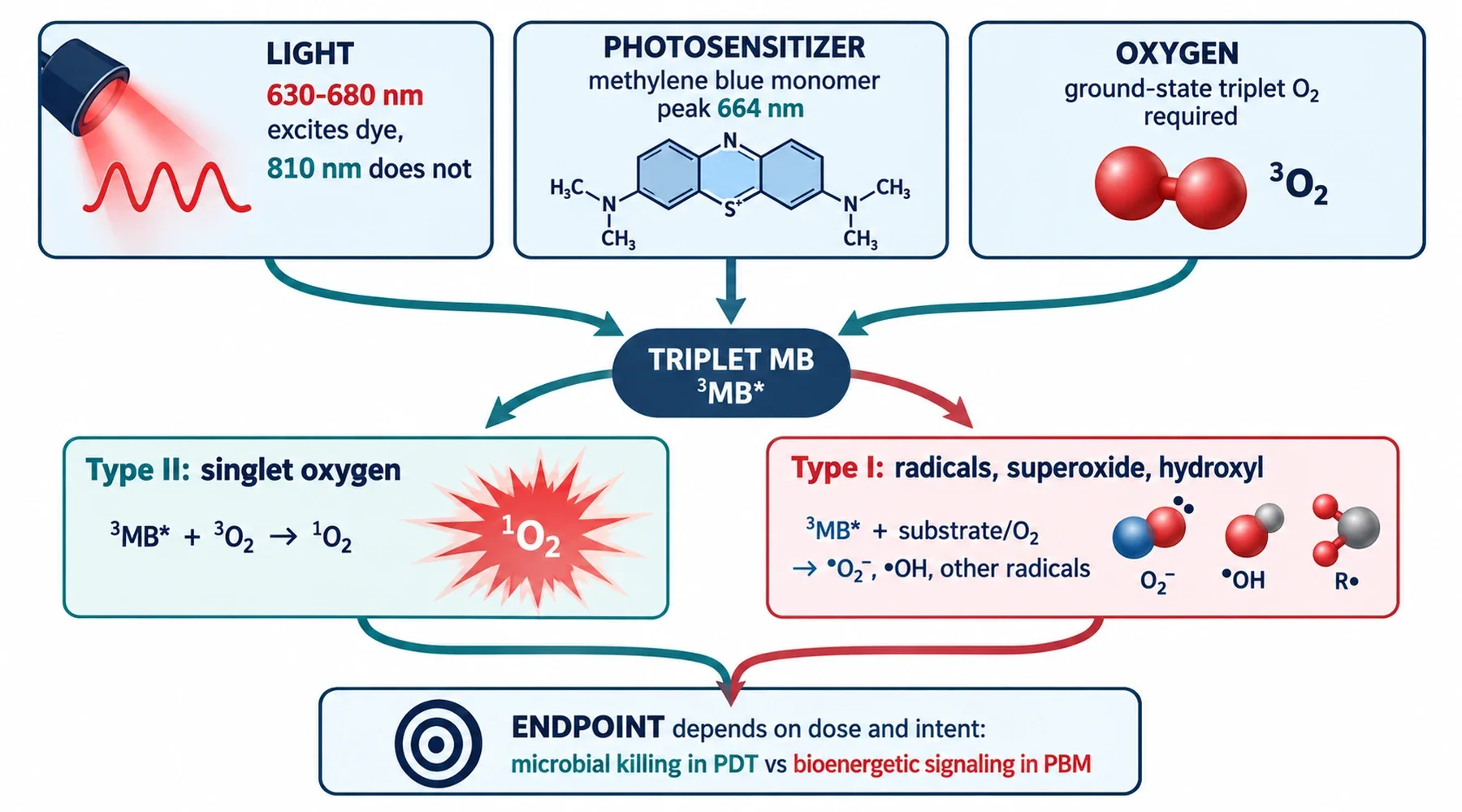
Monomeric methylene blue in water absorbs red light strongly, with a peak near 664 to 665 nm and a shorter wavelength shoulder near 610 nm that grows as the dye aggregates. Molar extinction near the peak is on the order of 65,000 to 85,000 M-1 cm-1, which is why micromolar solutions are intensely blue. This is not trivia. It is the precondition for any photosensitized reaction.
Absorption puts the dye into a short lived singlet state, followed by crossing to a longer lived triplet state, with reported triplet yields near 0.5. In deoxygenated solution that triplet persists for tens of microseconds. In aerated solution it falls to about 2 microseconds because oxygen quenches it. Quenching is where the two photochemical paths diverge.
In Type II chemistry, triplet dye transfers energy to ground state oxygen and produces singlet oxygen, with a commonly cited yield near 0.52. In Type I chemistry, the triplet dye instead transfers an electron or hydrogen to a substrate, which leads to radicals including superoxide and hydroxyl species. Both paths can run at once. Type II requires molecular oxygen by definition, and conventional PDT as a biological effect stays strongly oxygen dependent even when Type I contributes.
The practical consequence is a wavelength rule. Red light from about 630 to 680 nm overlaps the dye band well, with 660 to 665 nm near the monomer maximum, so it can drive photosensitizer chemistry in tissue that contains appreciable dye. Near infrared at 810 nm lies well outside that ground state band, so it is a poor wavelength for direct excitation of methylene blue. An 810 nm exposure plus methylene blue should not be described as photoactivation of the dye. It is better described as two mitochondrial interventions used at the same time. That difference structures everything below, and it is also covered from the chemistry side in the methylene blue chemistry reference.
Two concepts that share mitochondria but differ in mechanism
A widely cited review of mitochondria as a neuroprotection target pairs the two approaches because they converge on respiration while starting from different places. In the standard PBM model, the proposed chromophore is cytochrome c oxidase at Complex IV. The leading hypothesis holds that photons promote dissociation of inhibitory nitric oxide from the enzyme, which increases electron flux and ATP synthesis. That model should stay labeled as a hypothesis. Direct demonstrations remain limited, so confidence here should stay modest.
Methylene blue acts differently in the dark. At low dose it behaves as a catalytic redox cycler and alternative electron carrier. Reduced pyridine nucleotides reduce it to leucomethylene blue, which can hand electrons onward and recycle, effectively supporting flow around compromised segments between Complex I and Complex III while reducing electron leak. Photon absorption by the dye plays no role in that description. It is chemistry, not photochemistry, and the broader relay is explained in how methylene blue works as an electron relay.
PDT adds the third part back. Light excites the sensitizer, the sensitizer engages oxygen and substrates, and the objective is localized oxidative injury. PBM uses red or near infrared light at nonthermal exposure to alter bioenergetics without deliberately adding an exogenous sensitizer to create a cytotoxic burst. The slogan that PDT makes reactive oxygen while PBM does not is false. The real distinction is photosensitized cytotoxic chemistry versus modulatory signaling. Dose alone does not separate them. Human antimicrobial PDT with methylene blue used 6 to 18 J per square cm, which overlaps PBM values. Sensitizer presence, absorbed wavelength, timing, localization, oxygenation, and intent determine the regime. The full photodynamic therapy study collection keeps those parameters visible for each indication.
What the 810 nm combination mouse studies actually show
The two most cited combination papers both use 810 nm light and low dose methylene blue in mice. They are informative exactly because 810 nm is a poor direct activator of the dye. They test combination of distinct mechanisms, not methylene blue PDT.
In a sleep deprivation pretreatment study in mice, 60 male BALB/c mice received daily methylene blue at 0.5 mg per kg for ten days, 810 nm pulsed laser every other day, or both before deprivation. Deprivation impaired T maze, social, and shuttle box performance and reduced hippocampal GAP-43, PSD-95, and synaptophysin. Single treatments largely failed on behavior, while combined pretreatment differed significantly from the deprived group. This is prevention in an imposed deprivation model, not treatment of human impairment.
In a chronic stress study with concurrent treatment, mice underwent chronic mild stress while receiving 810 nm PBM at 8 J per square cm three times weekly, daily methylene blue at 0.5 mg per kg, or both for four weeks. The combination arm applied laser first and injected dye two hours later, specifically to avoid irradiating dye loaded tissue. All three arms reversed the learning deficits. Combination was not generally superior. Additive benefit appeared principally for serum cortisol and brain reactive oxygen species.
Neither paper establishes a human oral dose, a consumer device setting, a drug light interval, or a home schedule. One is pretreatment against a predictable insult. The other treats during stress induction. Extrapolating either to taking dye under a red panel skips every variable that decided the outcome.
Skin evidence is mostly PDT, not enhanced PBM
Human skin data with methylene blue plus light belongs largely on the PDT side. Examples include a 15 patient split face acne trial with topical methylene blue nanoemulgel plus 665 nm diode laser, smaller hidradenitis studies with 630 nm intense pulsed light or 635 nm LED plus methylene blue, a 2025 randomized scar trial comparing microneedling PDT with 0.1 percent versus 1 percent dye, and a 2026 diabetic foot ulcer protocol using 660 nm laser plus 1 percent dye. Wavelengths cluster where the dye absorbs. Intent is local destruction or remodeling. Formulation is topical or intralesional, not systemic.
That framing matters for skincare readers. Low dose dye alone has a separate fibroblast literature, and red light alone has a separate wound literature. But there is no peer reviewed human trial through late 2026 testing systemic oral dye plus red PBM as one combined skin or cognition intervention. Evidence for each part alone is not evidence for the combination.
Why compatible wavelength is not a protocol
The phrase how to use methylene blue with red light therapy deserves a careful answer rather than a recipe. Absorption tells you only that interaction is possible. Outcome also depends on dye concentration, formulation, irradiance and fluence, drug light interval, oxygenation, and depth. At 660 nm, adding dye can convert an otherwise PBM like exposure into photosensitizer driven photochemistry. That is why the 810 nm mouse results do not validate 630 to 680 nm consumer use with dye on board.
Safety reinforces the boundary. Prescription dye carries a boxed warning for serotonin toxicity with serotonergic drugs and some opioids. It is contraindicated in glucose-6-phosphate dehydrogenase deficiency. Phototoxicity is a recognized adverse effect of systemic dye, especially when irradiating tissue near its absorption maximum. Eye safety depends on wavelength, irradiance, and source type. Generic sunglasses are not a substitute for device specified protection. These points are detailed in the safety and interaction guide, and they belong in clinical hands rather than home protocols.
Name the concept before naming the device. No sensitizer plus bioenergetic goal means PBM. Dye at meaningful concentration plus absorbed wavelength plus oxygen plus destructive goal means PDT. Methylene blue plus 810 nm in the published mouse work is a third case: two mitochondrial approaches without direct photoactivation. Keeping those three cases separate prevents the most common error here, which is citing PDT skin clearance as proof that dye enhances PBM, or citing an 810 nm mouse rescue as proof that a 660 nm panel plus dye is a cognition protocol.