
Article
Methylene blue photodynamic therapy: how the treatment is studied
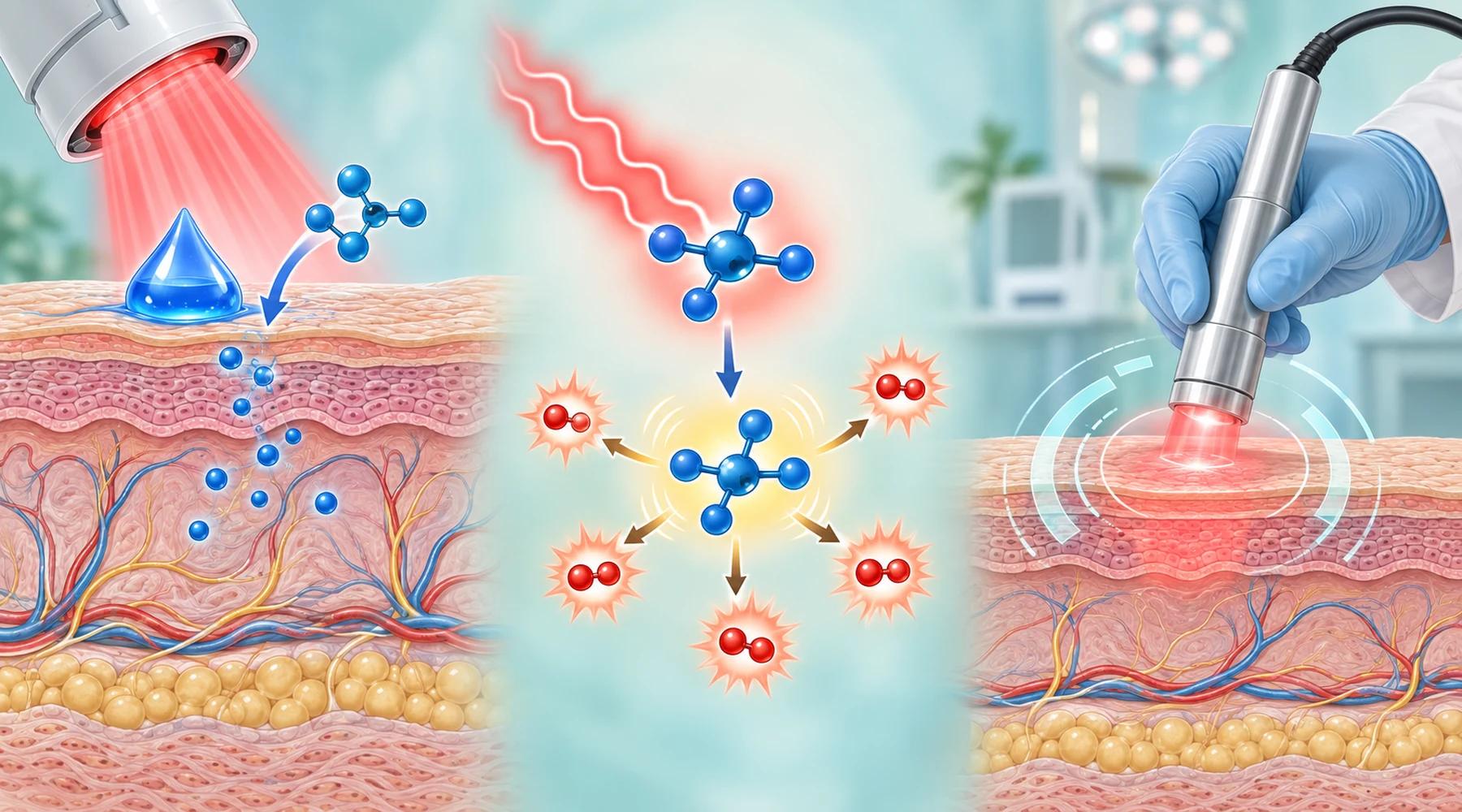
Photodynamic therapy with methylene blue sounds like two steps. Apply blue dye, shine red light. That mental model is wrong, and it is the source of most confusion about this treatment. The studied procedure has three parts that must coincide in the same place at the same time: photosensitizer in its light absorbing form, photons of a suitable wavelength at a measured dose, and molecular oxygen to receive the transferred energy. Remove any one part and the claimed effect has no mechanism.
This article explains how researchers describe that three part system, which parameters they control, and why a published wavelength and energy number cannot be converted into a home procedure. Cancer outcomes belong to a separate review of methylene blue and cancer research, and dental applications are collected in oral and dental photodynamic research. Neither is duplicated here.
The dye is a photosensitizer, not a stain
Methylene blue absorbs red light strongly, with the monomer peak near 664 to 666 nm and very high molar absorptivity. That absorption band is why studies of photodynamic therapy with methylene blue use red sources, typically in the range of about 630 to 680 nm. A lamp outside that band may look bright while delivering almost no usable excitation. The wavelength rules behind that choice are explained in how methylene blue absorbs red light.
Absorption is only the first move. The excited dye crosses to a longer lived triplet state, and from there two photochemical paths are available. In Type I chemistry the excited dye exchanges electrons or hydrogen atoms with nearby substrates and produces radicals, including superoxide chemistry downstream. In Type II chemistry the triplet dye transfers energy directly to ground state oxygen and produces singlet oxygen. Under oxygenated conditions methylene blue acts predominantly as a singlet oxygen sensitizer, with a reported quantum yield near 0.5 in water, while Type I contributions rise or fall with the local environment. The 1993 review of methylene blue photochemistry with biological substrates maps both paths and shows why the balance cannot be read off the dye label.
The same review names the principal nucleic acid target. Guanine has the lowest oxidation potential of the DNA bases, so singlet oxygen oxidizes it preferentially, forming products such as 8-oxo-deoxyguanosine. Intercalation into DNA is not required for that damage. But the review also warns against treating DNA as the whole story. Methylene blue oxidizes proteins, lipids, and membranes, and the lethal target in any given experiment depends on where the dye localizes. Concentration in the bathing solution is therefore a weak proxy for dose at the target. Readers comparing this light driven chemistry with the dark redox roles of the dye should keep the two separate, as set out in how methylene blue works in cells.
A lead compound, not an optimized drug
The second anchor source is the 2005 review of phenothiazinium photosensitizer development, which treats methylene blue as a lead structure with known weaknesses rather than a finished photosensitizer. Small structural changes alter lipophilicity, cellular uptake, intracellular localization, singlet oxygen output, dark toxicity, and resistance to reduction. Methylation of the chromophore, for example, raises lipophilicity and singlet oxygen generation and slows reduction to the inactive leuco form, and methylated analogues showed higher phototoxicity at about 7.2 J per square cm in the underlying work. More lipophilic is still not automatically better, because excessive hydrophobicity hurts solubility, promotes aggregation, and can distort the geometry the photochemistry needs.
The leuco point deserves emphasis because it is invisible. Oxidized methylene blue is blue and absorbs red light. Reduced leuco-methylene blue is nearly colorless and absorbs poorly in the same band, so dye that has been reduced inside cells or in poorly oxygenated tissue is temporarily removed from the photoactive pool until reoxidation. Tissue redox state therefore changes the effective photosensitizer concentration even when the applied concentration is fixed. Formulation history, including early clinical and laboratory uses of the dye family, is covered in the history of methylene blue in medicine.
The study table: what varies and what it means
The table below is the original comparison asset for this article. Each row is one treatment variable that studies must report. Read across to see why changing one setting changes the meaning of all the others.
| Variable | What studies report | Why it changes the outcome |
|---|---|---|
| Photosensitizer formulation | Free dye in water, saline, or buffer; ethanol and Cremophor vehicles in animal work; liposomes, polymer particles, or cyclodextrin carriers in experimental work | Vehicle and carrier change uptake, localization, and penetration through barriers such as biofilm matrix or tumor tissue |
| Applied concentration | Human antimicrobial studies report roughly 0.0003 to 0.06 M with 1 to 5 minute drug light intervals; animal tumor work includes examples near 500 micrograms per mL intratumorally | Higher bulk concentration can increase dimerization near 590 nm and reduce productive monomer photochemistry, so doubling concentration need not double effect |
| Drug light interval | Minutes in topical antimicrobial setups; longer and vehicle dependent in tumor models | Interval decides where the dye sits when light arrives, which decides the dominant biological target |
| Wavelength | Usually about 630 to 680 nm | Must overlap the monomer absorption band; dimer rich solutions shift effective absorption |
| Irradiance | Human antimicrobial reports span about 50 to 750 mW per square cm; animal wound studies commonly at or below 100 mW per square cm | High irradiance can deplete local oxygen faster than perfusion replaces it, which lowers efficiency at the same nominal fluence |
| Fluence | Human antimicrobial reports span about 6 to 18 J per square cm over 8 seconds to 10 minutes; animal wound studies span about 12 to 360 J per square cm | Total energy without irradiance, time, and geometry is an incomplete dose |
| Oxygenation | Rarely controlled directly; inferred from tissue type, fluence rate, and fractionation | Type II chemistry consumes oxygen, so identical light settings can give different effects in well perfused versus hypoxic targets |
| Target and setting | Skin and wound infection models, oral biofilm models, cultured carcinoma and melanoma systems, murine tumors, historical skin tumor reports | Geometry, depth, optics, and tolerated host damage differ so much that antimicrobial numbers never transfer quantitatively to tumor work |
The ranges above are observed study ranges, not a recommended protocol. That distinction is the point of the table. The breadth of the reported light doses is itself evidence that no transferable methylene blue light dose exists. Related laboratory context on light activated biofilm work appears in methylene blue against biofilms, and broader skin and light therapy background sits in skin photosensitivity and light dose basics.
Why identical energy numbers can give different biology
Four confusions recur whenever published parameters are quoted without their context.
First, fluence is not dose. Two exposures at the same wavelength and the same J per square cm differ if one is delivered fast and the other slow, because oxygen depletion, repair during exposure, and dye photobleaching all depend on rate and time. Reporting energy without irradiance and exposure time leaves the experiment underdescribed.
Second, tissue optics rewrite the lamp setting. Absorption, scattering, surface geometry, and backscatter mean the fluence inside tissue can differ materially from the value measured at the lamp aperture, in some circumstances by factors approaching two according to clinical dosimetry literature. A number copied from a paper describes that paper's geometry, not every geometry.
Third, localization decides the target. A cationic dye partitions into membranes, mitochondria, lysosomes, bacterial envelopes, or extracellular matrix depending on formulation, charge, lipophilicity, interval, and tissue. The 1993 photochemistry review documents damage across nucleic acids, proteins, lipids, membranes, organelles, viruses, bacteria, mammalian cells, and tumor systems, which is exactly why the same dye plus light pairing can kill by different routes in different setups.
Fourth, oxygen is a consumable. High fluence rates can outrun resupply by diffusion and perfusion and create transient hypoxia mid exposure. When that happens, adding more light adds little or no additional Type II effect. Fractionation and lower irradiance are studied partly as answers to this constraint, which is also why safety margins and tissue oxygenation belong in clinical safety and supervision standards rather than in a lamp manual.
A clinician led procedure, not a home recipe
The studied treatment is delivered where diagnosis, dosimetry, and supervision coexist. Someone must confirm what the lesion is and how deep it extends, choose a formulation and drug light interval matched to that target, calibrate the source over a defined field with a defined geometry, and monitor skin, eyes, pain, and local tissue response. The underlying phenothiazinium development review frames the whole field as structure activity optimization under controlled conditions, not as dye plus generic red light.
Home imitation fails on every controlled variable at once. Consumer lamps rarely report calibrated spectral irradiance at the treatment distance. Applied dye concentration says nothing about the monomer fraction or the intracellular localization. Tissue oxygenation is unknown and changes during exposure. None of the cited ranges license a home protocol, and this article states no usable recipe by design. Readers weighing general evidence quality should use how to read a methylene blue study before drawing conclusions from any single paper, and readers curious about light only approaches without a photosensitizer should start with red light therapy without a photosensitizer.
What this article does not establish
It does not establish that methylene blue photodynamic therapy cures any skin condition, clears any infection, or treats any tumor. The mechanistic reviews cited here measured photochemistry and compared dye structures; they prescribed no clinical concentration or fluence. The parameter ranges quoted above come from heterogeneous human reports and animal models with different targets, depths, formulations, and endpoints, and they cannot be averaged into guidance. Tumor specific evidence, including the limits of early clinical reports on basal cell carcinoma, Kaposi sarcoma, and melanoma, is assessed separately in the cancer research collection. Questions of purity and sourcing for laboratory grade material are orthogonal to treatment and are handled in how to choose a methylene blue product.