
Article
The history of methylene blue: from dye to medicine and research
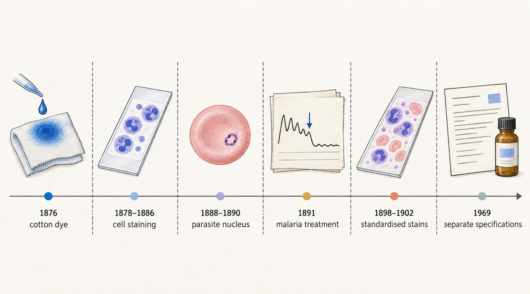
Methylene blue reached a patient before it reached a pharmacy.
The 1891 report by Paul Guttmann and Paul Ehrlich on the dye's use in malaria contains two temperature charts. The second runs from 13 July to 5 August, with the treatment written along the margin day by day. The entries on 18 and 19 July read "1 × 0,05 Methylenblau". The following days list five and seven doses of the same amount, and by 22 July a small quantity of quinine has joined the record. The fever line, which swings between 37 °C and 39.5 °C across the first half of the chart, flattens after the dye appears and stays between about 36 °C and 37.5 °C for the remaining three weeks.
The patient was one of two, described in the paper as a domestic servant and a sailor. The compound they received had been a commercial textile dye for fifteen years, and the two men are among the first people recorded as being treated with a fully synthetic drug aimed at a specific organism.
The dye came first
The first commercially successful synthetic dye was created by William Henry Perkin in 1856. Perkin was eighteen, working in a home laboratory during an Easter holiday, and trying to synthesise quinine. He obtained a black residue instead, dissolved it, and found a colour that fixed brilliantly to silk. He patented it in August 1856 and had a factory supplying a London silk dyeworks by the end of 1857.
In 1875 Adolf von Baeyer synthesised a red dye and named it eosin, from Eos, the Greek word for the dawn. In 1876 Heinrich Caro, then director of the laboratory at Badische Anilin- und Soda-Fabrik, developed methylene blue with Baeyer as an aniline-based dye for cotton. Its chemical structure was not settled until about nine years later.
That origin is still visible in how the substance is named. "Methylene blue" is a trade name from the dye catalogue, not a chemical description. The compound it refers to is methylthioninium chloride, and the distinctions between the name, the salt, and its hydrates matter as soon as anyone weighs the material or reads a specification.
Staining was the bridge
The route from dye works to hospital ran through the microscope, and it started before methylene blue existed. Wilhelm Erb was the first person to apply a synthetic dye to blood, using picric acid in 1865, and saw a reticular pattern inside some red cells.
Paul Ehrlich's doctoral thesis of 1878 compared histological staining with the dyeing of textiles and argued that both were chemical reactions. Working under Robert Koch on the tubercle bacillus, he sorted aniline dyes into acid and basic classes and established that each class coloured different parts of a cell. In 1880 he mixed the two together: a saturated solution of methylene blue poured into acid fuchsin until the supernatant cleared. The result, which he called the neutralen Farbkörper or neutral stain, produced three colours instead of one, revealed granules in the polymorphonuclear cells that had not been visible before, and made the first leukocyte differential count possible. Because those granules took up both components of the mixture, Ehrlich coined the word neutrophil for the cells carrying them.
Two years later Koch used methylene blue to find the tubercle bacillus, and in 1883 Ehrlich improved the method. In 1886 he published a different kind of observation: he injected the dye into live animals and compared how strongly each organ took it up, and concluded that it had a marked affinity for brain and nerve tissue. He attributed that affinity to the dye's oxidation-reduction cycling, which he had observed the previous year. That cycling is also why a methylene blue solution loses its colour when its oxygen is consumed and regains it when air returns. The experiments that first measured the consequences of that cycling belong to a later decade, and the mechanism itself is a separate subject from the history.
The parasite and the purple nucleus
Alphonse Laveran found the malaria parasite in 1880 by examining fresh, unstained blood. Seeing it routinely in a dried smear required a stain, and the stains of the day produced only red and blue.
In 1888 the Polish pathologist Czesław Cheçinski replaced acid fuchsin with eosin, producing a methylene blue and eosin mixture. The case behind his method was a soldier who had died of malaria on 30 September 1887. In the blood films Cheçinski saw blue daisy-like forms, now recognised as Plasmodium malariae, and sickle-shaped forms, now recognised as the gametocytes of Plasmodium falciparum. The result ended two competing explanations of the disease at once: the miasma theory, and the claim by Edwin Klebs and Corrado Tommasi-Crudeli that malaria was caused by a bacterium.
The parasite was visible. Its nucleus was not, which left its nature open. Ernst Malachowski, a physician in Breslau, solved that problem by accident and then on purpose. He noticed differential staining of the plasmodial nucleus in a preparation in 1889 and could not reproduce it. Acidifying the methylene blue did not help. Alkalinising it with borax produced the effect every time. He demonstrated the technique publicly on 15 June 1890 and published it the same year, with a fuller method in August 1891.
Dmitri Romanowsky published his own version in September 1891. His best results came from a neglected, mouldy bottle of methylene blue solution more than two months old. Malachowski's account was deliberate and, in his words, easily reproducible; Romanowsky's was a dead end that worked once. The eponym went to Romanowsky anyway, and the dispute over who produced the purple that made the nucleus visible is a separate story with its own documents.
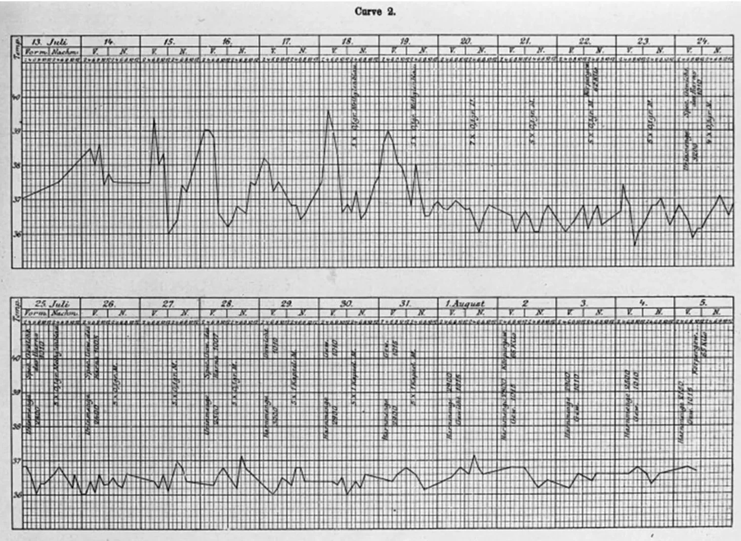
Figure: the second temperature chart from Guttmann and Ehrlich's 1891 report. The upper panel covers 13 to 24 July and the lower panel 25 July to 5 August; the margin entries name the doses given, and the temperature stays near normal for the final three weeks of the record. Reproduced from the report as scanned for Krafts, Hempelmann and Skórska-Stania, Parasitology Research 111:1–6 (2012), PMID 22411634.
Ripening turned out to be demethylation
Nobody involved in the 1890s knew what "ripening" did to methylene blue. The answer came out of industrial chemistry. Heinrich August Bernthsen had synthesised the parent compound phenothiazine in 1883, established in 1885 that methylene blue belonged to that family, and showed in the same period that alkaline conditions strip methyl groups from the dye, producing the compounds now called the azures.
Bernard Nocht extracted the active fraction in 1898 and 1899 and could only call it Rot aus Methylenblau, red from methylene blue. Michaelis identified it as azure I in 1901. Jenner introduced absolute methanol as a solvent for the dye precipitate in 1899, which removed the need to heat-fix blood films; Leishman and Reuter combined both advances; Wright published a simplified method in 1902. Giemsa's contribution, also 1902, was to stop relying on a randomly ripened solution and use measured quantities of known dyes instead. He then declined to publish how he made pure methylene azure and sold the method to Dr. Grübler's laboratory, which marketed the product as azure I and claimed that only its version was authentic Giemsa stain.
Two world wars finished the job by accident. When German dyes became unobtainable, Lillie, Roe and Wilcox worked on the American substitutes and found that the best staining came from three separated components: pure methylene blue, its breakdown product azure B, and eosin. Those three remain the major components of the stain used to diagnose malaria today, under a name that now runs Malachowski, Wright and Giemsa together.
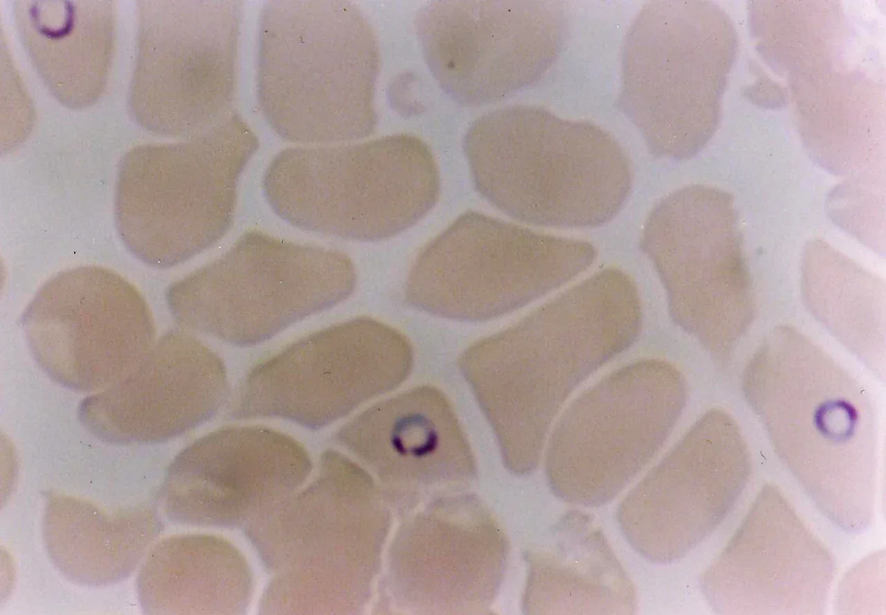
Figure: Plasmodium falciparum ring forms in human red cells, stained with a Romanowsky-type method. The purple structure inside the ring is the chromatin that Malachowski's borax-treated methylene blue made visible for the first time. Image by Ernst Hempelmann, released into the public domain.
One name, several materials
The years that separated those components left a problem that outlasted the chemistry. Ralph Lillie drew the line in 1969 between biological stains, "manufactured to rigid specifications commensurate with their delicate and exacting uses", and non-biological stains for textile colouring, which "require less rigorous and demanding manufacturing standards". Both kinds of product carried the same name, and the difference between them was never a difference of degree. A specification written for cotton and a specification written for a diagnostic reagent are not interchangeable, and a material made to the textile one was not intended to be swallowed.
Azure B is the clearest illustration. It is the monodemethylated form of methylene blue, it has measurable pharmacological activity of its own, and it was present as an uncontrolled constituent of clinical methylene blue preparations for more than a century before anyone separated the two. One group found that human tissue sampled at autopsy after methylene blue administration contained 5 to 14 per cent unchanged methylene blue and 86 to 95 per cent azure B. Whether a given preparation should contain azure B, and at what limit, is a specification question, and the research on azure B as an impurity and a metabolite picks it up from there.
A specification is also where the practical consequence sits. USP conformance is the route to the highest purity the market offers, because it is a complete published test standard rather than a single measurement. It covers identity, assay, organic impurities including the azure dyes, residual solvents, elemental impurities, residue on ignition, microbial limits, and bacterial endotoxins. A certificate that reports a heavy-metal result and calls it USP compliance has reported one line of that list.
Blupreme obtains its methylene blue from a pharmaceutical manufacturer and publishes the full certificate of analysis, testing the whole USP specification rather than heavy metals alone. The guide to reading a certificate of analysis and what the USP designation does and does not establish cover how to check the document against the claim.
What the history licenses, and what it does not
The lineage is real, and it is longer than most product pages imply. Methylene blue treated malaria in 1891, was reported as a sedative in agitated psychosis in 1899, and its side chain runs through quinacrine, chloroquine, promethazine and chlorpromazine. Ehrlich's 1890 paper with Arthur Leppmann described relief of neuralgic and rheumatic pain, and then considered and declined to test the dye in mental illness.
None of that establishes a modern use. The 1891 malaria result was two patients and a temperature chart. Long familiarity with a molecule is a reason to study it, not a result. The history of the compound as a drug and the modern trials in malaria answer different questions from the ones Ehrlich was asking, and the endpoints have to be read on their own terms.
There is one detail in the record that belongs to the present rather than the past. When researchers ran methylene blue malaria trials in Burkina Faso in the 2000s, they found themselves competing for pharmaceutical-grade material, and reported that the price of the raw material had risen by a factor of about a hundred once it carried GMP validation. A dye could be substituted. A drug could not. The distinction Lillie drew in 1969 is the same one that decides what a bottle contains today.