
Article
The experiments that linked methylene blue to cellular respiration
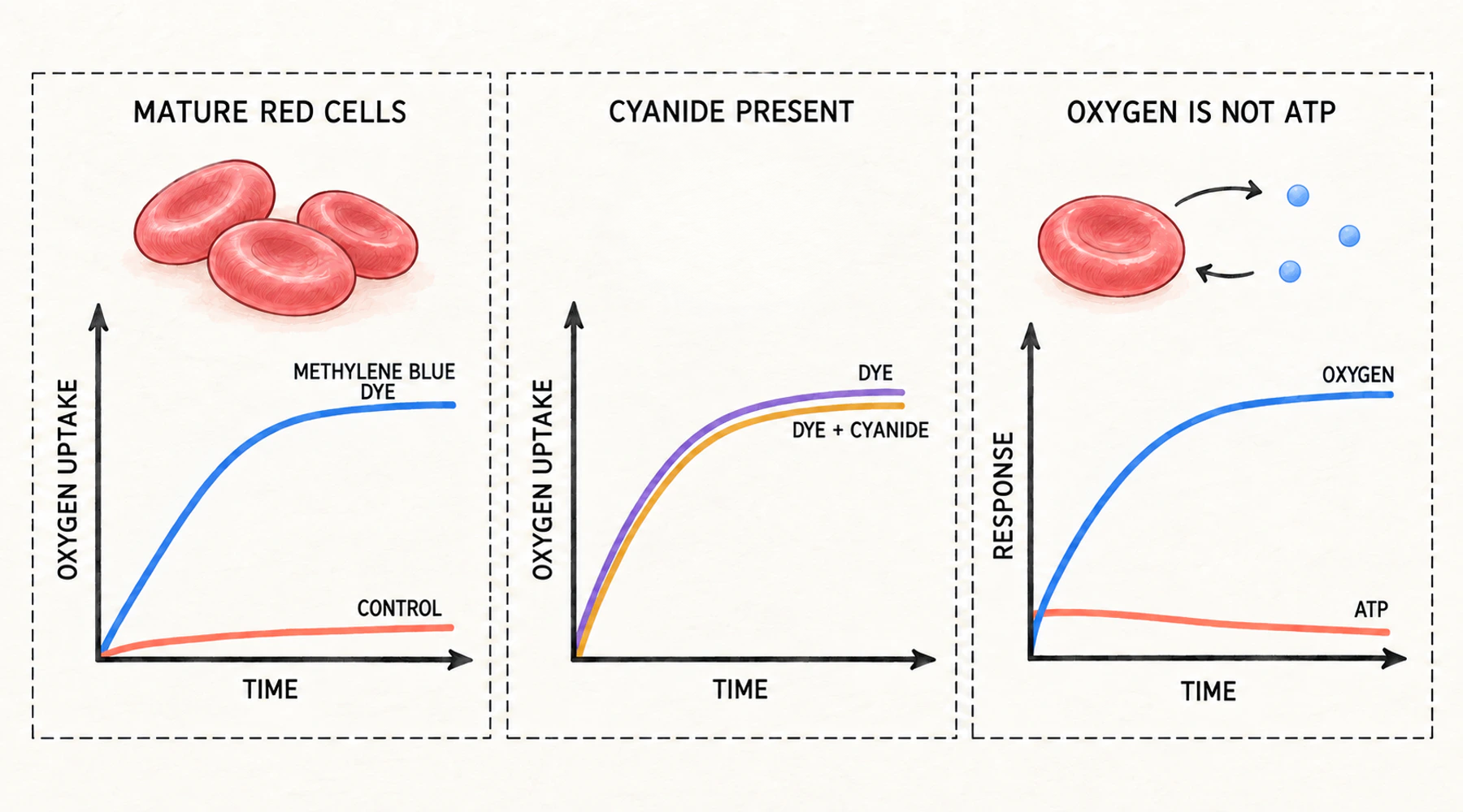
Defibrinated human blood, warmed to 37 °C for three hours, consumes around two per cent of the oxygen it contains. Add a small quantity of methylene blue before the incubation, and the same blood consumes around sixty-five per cent. Nothing about the cells changed. Only the solution did.
That number comes from a 1928 paper by George Harrop and E. S. Guzman Barron at Johns Hopkins, and it is the origin of the widely repeated statement that methylene blue improves cellular respiration. The statement is not false, and it is narrower than it sounds.
What the number in the table actually measures
The endpoint was oxygen uptake, determined by gas analysis. Harrop and Barron filtered freshly drawn blood through cotton wool at 10 °C to strip out the white cells, saturated the sample with oxygen, split it, added dye to one portion, and analysed both after incubation. Leukocyte counts rarely exceeded about a thousand per cubic millimetre. Oxygen consumption was reported as the share of the sample's initial oxygen content that had disappeared.
They also measured carbon dioxide, and there the method fought back. Defibrinated blood released up to two millimoles of carbon dioxide per 100 mL in the first hour of incubation for reasons unrelated to the reaction under study, so respiratory quotients from whole blood were unusable. Only washed cells in Locke's solution gave interpretable figures, and those clustered between 0.73 and 0.80 with and without the dye.
That detail is not trivia. "Respiration" in these papers means gas exchange across a closed vessel. The gap between oxygen consumption and useful energy output is examined separately in what oxygen consumption does and does not tell you about ATP.
The dye added most where the cells respired least
Mature mammalian erythrocytes have almost no measurable oxygen consumption. Nucleated bird erythrocytes do. Harrop and Barron ran both, and the contrast is the backbone of their argument.
In the mammalian samples the dye produced increases of twenty- to fiftyfold. In goose blood, whose cells already respired, it never more than doubled the rate. The higher the starting respiration, the less the dye could add.
The same gradient appears within one species. A human sample from a patient with polycythemia vera, containing immature cells, consumed 11.3 per cent of its oxygen with no dye present, against 2.8 per cent for washed cells from a healthy donor. Dye took it only to 63.4 per cent, a sixfold acceleration where normal cells accelerated nearly thirtyfold. The dye pushed mature cells toward the behaviour of immature ones, which is what the authors said it was doing.
Cyanide should have stopped it
Cyanide was known to stop cellular oxidation. Harrop and Barron added it anyway, alongside the dye.
The beef blood results are representative. Oxygen content fell from 22.61 to 22.03 volumes per cent over three hours with no additions. With methylene blue it fell to 12.84. With methylene blue and one five-thousandth molar potassium cyanide, it fell to 12.74. The dye's effect was essentially untouched.
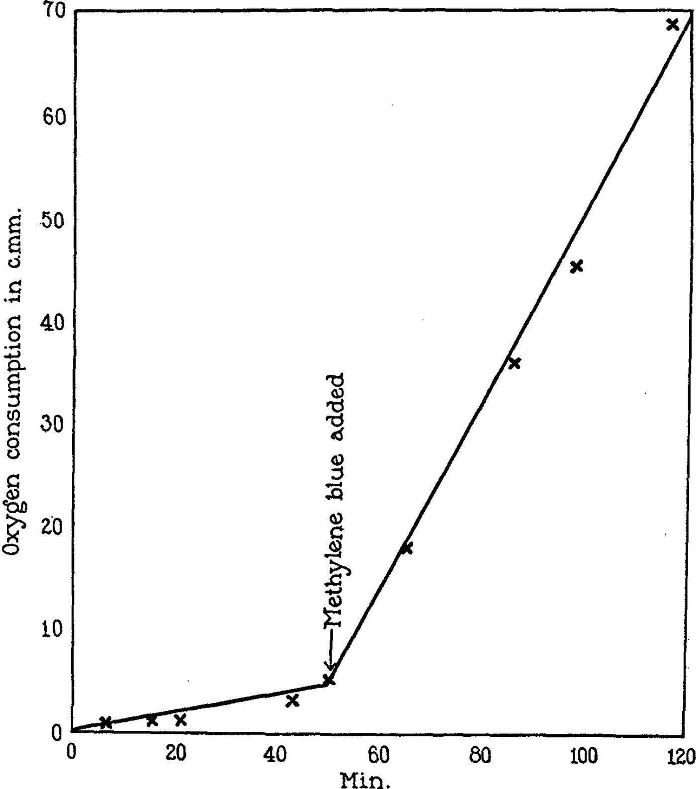
Figure: a continuous microrespiration record of human erythrocytes. The arrow marks the addition of methylene blue. Before it, the trace is nearly flat; afterwards it climbs steadily. Reproduced from Harrop and Barron, Journal of Experimental Medicine 48:207–223 (1928), PMID 19869478.
Their conclusion was that the dye acted as an oxygen carrier, "supplying a substance which has disappeared from adult mammalian non-nucleated erythrocytes." Two years later, Michaelis and Salomon at the Rockefeller Institute put the disagreement in its permanent form. Either the dye's effect was "an accidental property of the dye," or the dye was "a model for some unknown substance in the organisms." That fork has not been closed. What has changed is that the cells can now be described much more precisely.
A liver extract did the same thing
Michaelis and Salomon showed that cold water extracts of organs produce the same stimulation. Rat liver was strongest, followed by kidney, spleen, and testicle. Brain, muscle, and blood serum had little or no effect.
They then tested the extract against carbon monoxide. Respiration induced by either the extract or the dye was unchanged in a mixture of 98 per cent carbon monoxide and 2 per cent oxygen. The same gas mixture inhibited yeast respiration and that of nucleated fowl erythrocytes by roughly 60 to 80 per cent, in line with Warburg's account of a carbon monoxide-sensitive respiration ferment. The unidentified tissue substance was therefore not Warburg's enzyme.
Several candidate identities fell along the way. Reduced glutathione and cysteine produced only the oxygen consumption required by their own oxidation. Harden and Young's coenzyme was ruled out because muscle contains the most of it and showed almost the least effect. Keilin's cytochrome, named a few years earlier, ran into the same contradiction. Their own suggestion, that the active substance came from the cell nucleus, they called a mere suggestion.
The effect had conditions attached
The cells had to be intact. Hemolysis by distilled water or repeated freezing abolished the response to both the dye and the extract, and partial hemolysis reduced it in proportion.
Sugar was required as well. Glucose, fructose, and sucrose supported the dye-induced respiration; mannitol did not. With no sugar present there was no respiration at all, and the dye appeared to damage the cells in the process, so glucose added one or two hours later did not restore it. The authors suggested the dye was oxidising a structural component the cell needed.
Illumination increased the dye's effect considerably, though the effect was clear in the dark. Other reducible dyes behaved differently: toluylene blue matched or exceeded methylene blue, while indigo disulfonate did almost nothing. No clean relationship emerged between a dye's oxidation-reduction potential and the size of its effect, which the authors attributed to membrane permeability and toxicity.
The effect tracked fermentation, not respiration
Barron's 1930 study moved from cell suspensions to tissue slices of normal rat organs and transplanted tumours.
In tissues with no aerobic glycolysis, methylene blue did nothing or slightly suppressed oxygen consumption: kidney by about 14 per cent, pancreas by about 14 per cent, liver by about 5 per cent. In tissues that ferment under oxygen, it increased consumption: spleen by about 8 per cent, brain by about 13 per cent. Tumour increases ran from about 27 per cent in a human breast carcinoma to more than 80 per cent in a Rous chicken sarcoma, roughly in proportion to each tissue's aerobic glycolysis.
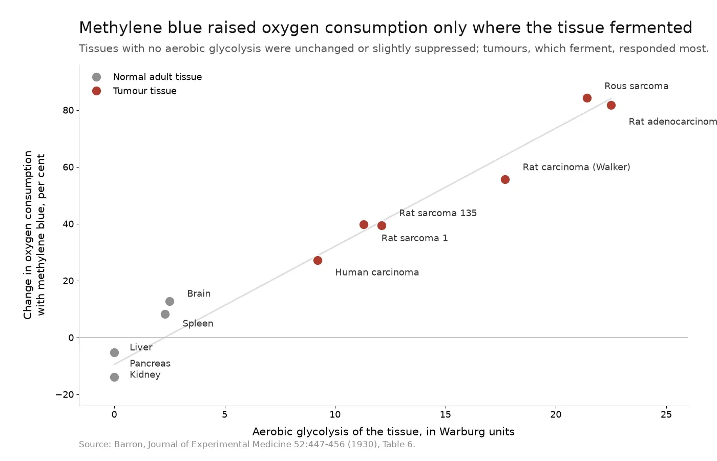
Figure: each point is one tissue type. The horizontal axis is aerobic glycolysis; the vertical axis is the change in oxygen consumption after methylene blue. Data from Barron, Journal of Experimental Medicine 52:447–456 (1930), Table 6, PMID 19869777.
The cyanide experiment then produced the mirror image. When respiration in those previously unresponsive tissues was blocked with cyanide and fermentation came to the surface, the dye increased oxygen consumption again: kidney by about 59 per cent, liver by about 19 per cent, spleen by about 286 per cent, testicle by about 200 per cent.
Barron proposed that the dye could serve as a test for the fermentative power of a cell or tissue, and closed with a speculation: generations of tumour culture in dye-containing medium might one day make a tumour's metabolism permanently like that of normal tissue. That was a research question written in 1930. It is not a treatment claim.
What the experiments could not have been measuring
In 1928 and 1930, "respiration" meant oxygen consumption. Adenosine triphosphate was first reported in 1929, and the demonstration that isolated mitochondria esterify phosphate while transferring electrons came in 1949. None of the early methylene blue papers could have measured energy production, and none of them claimed to.
The cells are the second problem. Mature mammalian erythrocytes contain no mitochondria. During the reticulocyte stage they clear mitochondria and ribosomes by selective autophagy, which is why a mature red cell runs on glycolysis and depends on its pentose phosphate pathway mainly for NADPH rather than for ATP. In the 1928 experiments there was no electron transport chain present to repair. Whatever consumed the oxygen was doing something else.
What that something else was is now reasonably well described. Methylene blue accepts electrons from cellular reducing equivalents and becomes leucomethylene blue, which is reoxidised by molecular oxygen, closing the cycle. In human red cells, NADPH-dependent flavin reductase and related disulfide reductases perform the reduction step. A 1976 study using labelled glucose found that the dye stimulates the hexose monophosphate shunt, that the low-concentration effect depended on oxyhemoglobin, and that a higher concentration, 0.1 mM, generated hydrogen peroxide. A reduced dye meeting oxygen produces peroxide, not phosphate bonds.
In mitochondria the picture is more contested than the popular summaries admit. One line of work holds that the dye takes electrons from the region of complex I and hands them to cytochrome c. Another found that in mouse brain mitochondria it cannot restore respiration after an antimycin block, and placed the electron acceptor at the ubiquinol-binding site of complex III instead. A third found that low concentrations raised resting oxygen consumption while leaving ADP-stimulated respiration unchanged. The most direct statement of the point is that methylene blue-induced oxygen consumption does not depend on oxidative phosphorylation at all: it persisted when the respiratory chain was inhibited, and in preparations without one.
Direct measurements of the gap between the two quantities exist as well. In isolated rat liver mitochondria, increasing dye concentrations lowered respiratory control ratios and caused swelling, which the authors described as uncoupling of oxidative phosphorylation. In perfused rat liver, methylene blue raised oxygen consumption while the ATP-to-ADP ratio fell.
The clinical record carries the same message. Methemoglobinemia is treated with roughly one to two milligrams of methylene blue per kilogram intravenously, by way of an NADPH-dependent reduction pathway, and larger doses cause the condition being treated. The same review notes that the antidotal effect is greatest in intact erythrocytes and diminished when haemolysis is present, which is the 1930 hemolysis finding reappearing seventy-three years later in a hospital context. A compound whose useful and harmful effects sit on opposite sides of a dose threshold is not a compound whose dose should be inferred from an oxygen-consumption measurement.
Why the batch belongs in the discussion
Harrop and Barron noticed something worth a second reading. Methylene blue itself produces a band in the spectroscope between the two characteristic bands of methemoglobin, "which may readily cause confusion on casual inspection." The dye can imitate the substance being looked for.
The same problem has a chemical form. A related phenothiazine present as a minor constituent carries its own redox behaviour, its own permeability, and its own toxicity. One documented case makes the stakes concrete: in a study of methylene blue iodination, samples containing roughly 7 to 8 per cent Azure B produced mainly iodinated Azure B, so the impurity rather than the methylene blue dominated the reaction.
Anyone using methylene blue as a reagent, in a laboratory, at a staining bench, or in an aquarium, is working with an unspecified ingredient unless the batch has been characterised. Related substances, identity, elemental impurities, residual solvents, and residue on ignition are part of the experiment rather than paperwork around it. Blupreme obtains its methylene blue from a pharmaceutical manufacturer and publishes a full certificate of analysis, testing the whole USP specification rather than heavy metals alone; the guide to reading a certificate of analysis and what the USP-grade designation does and does not establish cover how to check the document against the claim.
The number was right
The 1928 and 1930 experiments were careful, and their central observation has survived: a reducible dye can make a cell that barely respires consume oxygen at many times its resting rate, without passing through the step that cyanide blocks.
The mistake appears later, when "respiration" is read with its modern meaning. A gas-exchange measurement tells you that electrons arrived at oxygen. It does not tell you that any of them were used to make ATP, and in a cell with no mitochondria the distance between those two statements is as wide as it gets. The broader map of what methylene blue does in cells puts the same distinction in a wider frame, and the history of the compound from dye to medicine explains how a textile colourant arrived at these experiments in the first place.