
Article
Who developed the purple blood stain? Methylene blue, Malachowski, Romanowsky, and Giemsa
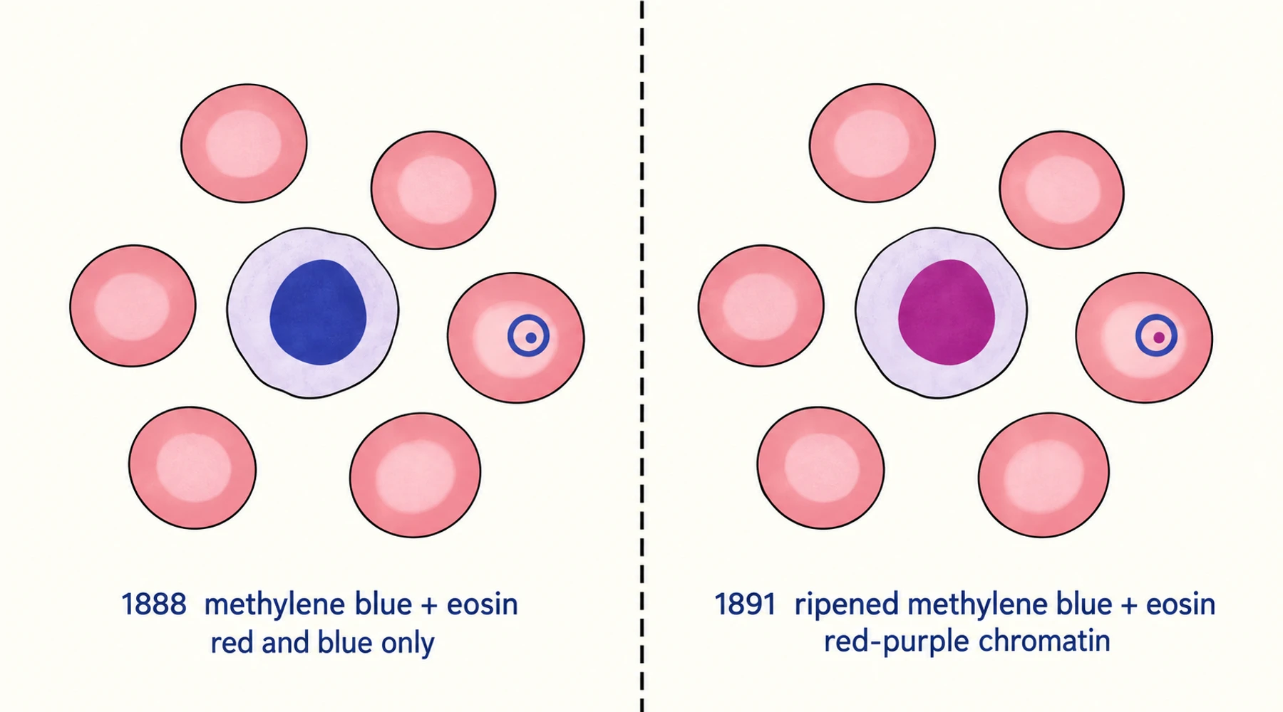
Eosin is red. Methylene blue is blue. Mix the two, apply the mixture to a dried film of blood, and parts of the film come back purple.
Not every part. In a good preparation the nuclei of white blood cells take a red-purple, and inside a malaria parasite the chromatin takes the same colour, while the surrounding red cells stay pink and the cytoplasm stays grey-blue. The purple is what makes the cell types tell themselves apart.
Neither bottle contains purple. The colour appears only when a particular breakdown product of methylene blue meets eosin on the slide, and in the 1880s and 1890s nobody knew that product existed, let alone what it was. The people who chased the colour were working with impure dyes, alone, with no dependable way to find out what was in a bottle after it had sat on a shelf for two months.
That is why the question in the title has more than one defensible answer, and why the answer changed in 2011.
A readout that changed with the mixture
The stain was not invented. It accumulated. Each change to the mixture bought exactly one new piece of visible information, and the sequence of those pieces is the clearest way to read the history.
| Year | What was in the dish | What you could see |
|---|---|---|
| 1865 | Picric acid, the first synthetic dye put on blood, by Wilhelm Erb | A lacy pattern inside some red cells. |
| 1876 | Methylene blue, by Heinrich Caro and Adolf von Baeyer at BASF | Tubercle bacilli, in Robert Koch's hands. Blood cells in one blue tone. |
| 1880 | Methylene blue plus acid fuchsin, Ehrlich's neutral stain | Red cells and eosinophil granules red, nuclei deep blue, lymphocyte cytoplasm pale blue, and dark violet granules that took up both dyes. That is why the cell is called a neutrophil. The first differential white cell count. |
| 1888 | Methylene blue plus eosin. Czesław Chęciński | The parasite itself: blue daisy-shaped forms (Plasmodium malariae) and sickle-shaped forms (P. falciparum gametocytes). Not its nucleus. |
| 1890 | More methylene blue, less alcohol, plus potassium hydroxide. Albert Plehn | A wider and deeper colour range, including rose red cells and rose eosinophil granules. Still red and blue only. |
| 1890 | Eosin plus methylene blue made alkaline with borax. Ernst Malachowski | Red-purple chromatin inside the parasite, nucleus distinguished from cytoplasm. Deliberate, and on his account reproducible. |
| 1891 | Eosin plus methylene blue from a mouldy bottle over two months old. Dmitri Romanowsky | The same purple, once, by a method others could not repeat. |
| 1898 to 1902 | Chloroform-extracted polychrome dye (Nocht); methanol (Jenner); alcoholic redissolution of the precipitate (Leishman, Reuter, Wright); measured dyes with glycerol (Giemsa) | The purple on demand, in a product that behaved the same way in the next laboratory. Jenner's version, without polychromed dye, gave no purple. |
| 1940 to 1943 | Lillie, Roe and Wilcox separated the components and tested them one at a time | Azure B, not the mixture, is the part that produces the purple. |
The pattern in the last column is the important one. Every advance widened the colour range, and every widening made one more structure visible. The purple is not a decoration on the technique; it is the difference between seeing that a parasite is present and seeing what it is made of. How the dye itself travelled out of a BASF dye works and into medicine is a longer history with documents of its own. The full sequence above, with the source behind every entry, is kept as a source-linked contributor timeline.
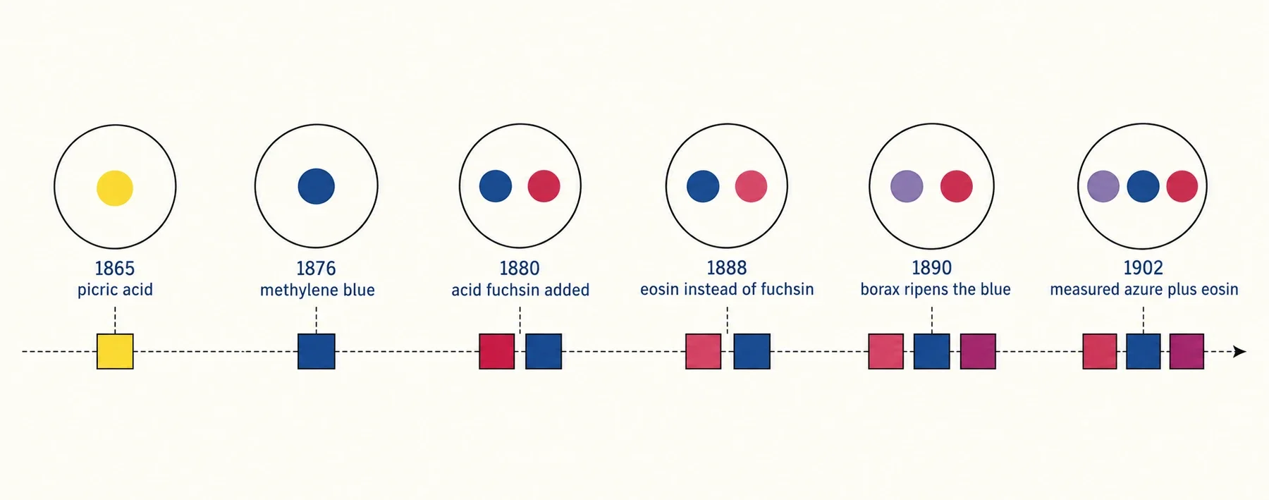
Figure: the same six mixtures drawn as what was in the dish and what came out of it. The circles carry the dyes; the blocks beneath carry the colours those dyes could produce in a blood film. Nothing before 1890 draws a red-purple block, and nothing after it omits one.
The man who used borax on purpose
Ernst Malachowski was a physician in Breslau, now Wrocław, who spent his career in private practice and in a small clinic he ran with a colleague, doing laboratory work in his own time. He was born in Strzelno on 12 March 1857, studied medicine at the University of Breslau, and worked from 1882 to 1887 as an assistant in internal medicine at All Saints Hospital, then the second largest hospital in Germany.
He began in 1888 with the Chęciński and Plehn double stain. In a preparation made in 1889 he saw the chromatin of the plasmodial nucleus stain differently from the cytoplasm, and then found he could not reproduce it. He tried acidifying the methylene blue, which made things worse. He tried alkalinising it with borax, which worked every time. He demonstrated the method publicly on 15 June 1890 and published a fuller description in August 1891, including the borax instruction. He noted that the staining took twenty-four hours and that the results were easily reproducible.
Twenty-four hours is a long time in a diagnostic laboratory, and his paper did not dwell on the purple-red colour of the parasite nucleus. Whatever the reason, nobody took up deliberate alkalinisation of methylene blue until Nocht did it again in 1898. Malachowski never wrote about blood staining again.
The reason so little was known about him until recently is archival rather than mysterious. Almost no trace of his life survived in the medical literature, and what did survive came through one route. His daughter Marie left Germany for London in 1939, served in the British Army, and settled in Canada, and her collection of family papers ended up at the Leo Baeck Institute in New York. Her brother Leo was killed in the First World War. Her sister Eva was deported from Breslau to the Izbica transit camp in April 1942 and never returned. Her mother Hedwig took her own life on 22 April 1942 rather than wait for the same deportation. Malachowski had died in 1934, before any of it.
A family history that ends that way is a family history that stops being cited. The two papers that put Malachowski back into the stain's development, both published in 2011, were assembled from archive documents in Poznań and Wrocław and from Marie's papers in New York.
A mouldy bottle and a lucky Russian
Dmitri Romanowsky studied medicine at the Military Medical Academy in St Petersburg and worked on malaria for most of his career. In 1890 he published a method that was, on paper, nearly identical to Chęciński's: unaltered methylene blue and eosin, with a little more eosin.
In September 1891 he published the method that made his name, and the detail that mattered was buried in the instructions. His best results came from die vorräthige Methylenlösung, a stock solution of methylene blue that had stood for over two months with mould growing on it. The mould and the standing time had partly demethylated the dye. Romanowsky did not know that and could not control it, and other workers who tried his method could not get the purple. His thesis had gone in at St Petersburg in June 1891, which is why the standard account treats 1891 as the year of discovery.
Malachowski's demonstration had been in June 1890, over a year earlier, and his method worked on purpose.
Why the eponym went to the wrong man
The case for Malachowski was assembled by Kristine Krafts, Ernst Hempelmann and Barbara Oleksyn, and it rests on four things. The date of the public demonstration, June 1890 against September 1891. The word deliberate against the word serendipitous. The fact that Malachowski published his method, borax and all, while Romanowsky's depended on an uncontrolled variable. And the observation that the description of the plasmodial nucleus, along with the purple that revealed it, effectively vanished from the literature for seven years, because nobody could reproduce the accidental version.
They are blunt about the conclusion. In the biographical paper they call the attribution "a travesty", and they quote Ralph Lillie, who in 1978 wrote that "perhaps we should refer to this family of stains as Malachowski stains".
The claim is contested, as priority claims usually are, and the shape of the contest is worth explaining rather than resolving. An argument about priority has to say what the discovery was. If the discovery is the purple colour, Malachowski has the earlier documented demonstration. If the discovery is the published method, both men published in 1891 and the eponym went to Romanowsky. If the discovery is the explanation, neither man has a claim, because neither knew that ripening was demethylation and neither could say why the colour appeared at all. That last point is the one popular accounts skip. The active ingredient was an impurity nobody had identified, and the first person to produce the colour deliberately did so from a reproducible empirical recipe, not from an understanding of what the recipe did.
The two 2011 papers are the fullest published statement of the case for Malachowski, and both are free to read at PubMed: the long review at PMID 21235291 and the biographical paper at PMID 21660627. The PubMed record for the review also lists a comment on it, published in the same journal later that year at 86(5):366, which is a fair reminder that the argument was contested in print as soon as it appeared.
What the dispute is not is a disagreement about chemistry. Everyone involved agrees on what the purple is and which components produce it. The disagreement is about who got there first, and about whether an accident that nobody could repeat counts as getting there at all.
What the later names bought
If Malachowski produced the purple first, why does the stain carry the names of Romanowsky, Wright and Giemsa?
Because a stain that works once in the hands of its inventor is not a reagent. The later work turned a temperamental mixture into something a technician in another country could use on a Tuesday.
Nocht got there by reasoning. After failing to get a reliable answer from a simple neutral dye, he concluded that the purple must come from something in the methylene blue that was not methylene blue, and went looking for it. Unna had already noticed that aged methylene blue solutions stained mast cell granules red, and proposed that a new substance formed as the dye ripened. Bernthsen had shown in 1885 that alkaline conditions strip methyl groups from methylene blue, and that these products are what we now call the azures. Nocht extracted the active fraction with chloroform and could call it only Rot aus Methylenblau, red from methylene blue; Michaelis named it azure I in 1901. Jenner solved the logistics. Redissolving the methylene blue and eosin precipitate in absolute methanol gave a single stable solution that fixed the film as it stained and ended heat fixation. Leishman and Reuter combined polychromed dye with an alcoholic solvent and got stability and the purple together. Wright replaced a twelve-hour incubation at 65 °C and ten days of standing with an hour in flowing steam. Giemsa stopped relying on a randomly ripened solution at all, using measured quantities of known dyes with glycerol in the methanol so the product survived tropical heat. He never published how he made pure methylene azure. He sold it to Dr Grübler's laboratory in Leipzig, which marketed it as azure I and claimed that only its version was authentic Giemsa stain.
The identity of that azure is still argued about. Some hold that it was simply polychromed methylene blue, others that it was rich in azure B. The answer came out of a war. When German dyes became unobtainable, a United States Public Health Service team, Ralph Lillie with Maurice Roe and Aimee Wilcox, found that the best staining came from three separated components: unmodified methylene blue, its demethylated breakdown product azure B, and eosin. Azure B replaced the unavailable azure I, and those three components are still the basis of the stain used to diagnose malaria today.
The chemistry of how those three components interact on a slide is a separate question and a different page. The dye roles and chemistry of Romanowsky-type stains covers what azure B and eosin each do and why the pair produces a colour that neither produces alone. What the microscopy and laboratory applications page covers is the practical side: what a given preparation shows, and how to judge whether a particular product is suitable for the method you intend to use.
The part of the history that is still current
One detail in this story did not stop being true in 1902. For roughly fifty years, the most important component of the world's standard malaria stain was an unnamed impurity. It was there by accident, its quantity varied between bottles, its identity was a trade secret for part of that time, and its proportion was argued about until the 1940s. The people using the stain were not careless. They had no way to know what was in the bottle.
The modern version of the same problem is a specification that describes only part of the material. USP conformance is the route to the highest purity the market offers, because it is a complete published standard rather than a single measurement: identity, purity, organic impurities including the azure dyes, residual solvents, elemental impurities, residue on ignition, microbial limits and bacterial endotoxins. Blupreme obtains its methylene blue from a pharmaceutical manufacturer and publishes the full certificate of analysis, so that every line of that list can be checked rather than asserted. A certificate that reports a heavy-metal result and calls it USP compliance is the present-day equivalent of a bottle labelled "ripened methylene blue": possibly a fine reagent, but the label does not tell you what is in it.