
Article
Methylene blue staining: microscopy methods and a real-image atlas
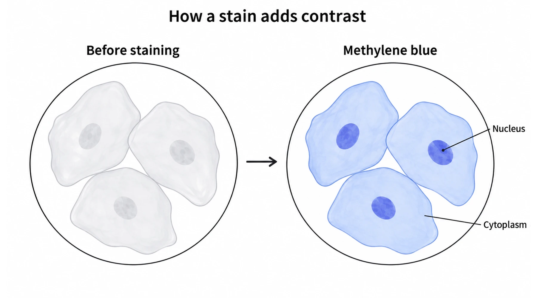
Methylene blue staining makes otherwise faint cellular features easier to distinguish in bright-field microscopy. It can help students locate a cheek-cell nucleus or examine the outline and arrangement of microorganisms. A blue object, however, is an observation. Naming the organism, measuring it, and deciding whether it indicates disease are separate tasks.
The cover illustrates the contrast principle. The atlas below contains real photographs from independently documented specimens, with their original source information.
What does methylene blue reveal?
Methylene blue is a basic dye: its colored ion carries a positive charge. As OpenStax explains in its treatment of specimen staining, basic dyes interact with negatively charged cellular material and can make cells stand out against a lighter background. In suitable preparations, methylene blue emphasizes nucleic-acid-rich regions and other stain-binding structures.
In a simple stain, one dye supplies contrast. Several organisms can consequently look blue without being the same organism. The useful questions are initially descriptive: are the objects round or elongated, isolated or clustered, evenly stained or internally differentiated? See the distinction between simple staining and Gram staining before interpreting color as a classification result.
Contrast and resolution are different. Contrast helps distinguish an object from its surroundings; resolution separates nearby details. A darker image does not guarantee more resolved structures. Nor does every dark granule deserve an organelle label.
Native preparations, stained wet mounts, and smears
An unstained, or native, wet preparation preserves a view without added dye, although removal from the original environment still changes the specimen. A stained wet mount adds reagent while the material remains in liquid under a coverslip. A fixed smear spreads material on a slide and uses a specified fixation method before subsequent processing. OpenStax's preparation overview distinguishes wet mounts from fixation and explains why fixed organisms no longer provide a view of normal movement or metabolism.
These preparations answer different questions:
| Preparation | Useful observation | What the image cannot establish alone |
|---|---|---|
| Native wet mount | Movement, outlines, naturally visible material | Identity from motion or shape alone |
| Stained wet mount | Contrast between stain-binding structures and surroundings | Whether the reagent left normal physiology unchanged |
| Fixed, stained smear | Morphology retained after a defined preparation | How the original living specimen behaved |
| Named differential method | A specified staining reaction with appropriate controls | Species identity or diagnosis without the method's wider evidence |
For a classroom comparison, record an unstained preparation before examining a separately stained preparation made according to the teaching protocol. Keep illumination and image capture settings comparable. If two different cells are photographed, describe them as examples, not as a before-and-after record of one cell.
Microscopy atlas: three real stained specimens
This atlas is an original selection and interpretation of three openly licensed micrographs. The source authors identify the specimens and stains. We have not independently authenticated their specimens. Images are reproduced uncropped as lossless WebP conversions, without added features or color changes.
Plate 1: human cheek cells
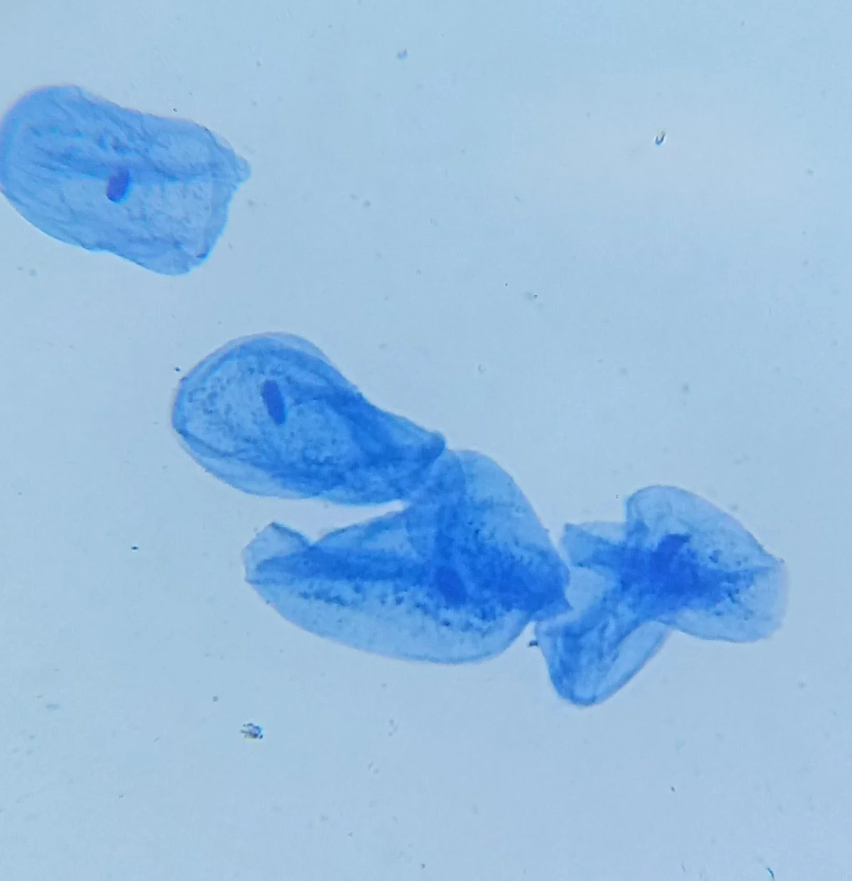
Specimen: human cheek cells. Stain: methylene blue. Magnification: 400×, reported by the photographer. Scale bar: unavailable; the source supplies no calibrated pixel size. Photograph by Fritzmann2002, 2017, from Human Cheek Cells (Methylene Blue Stain), licensed CC BY-SA 4.0. This reproduction retains that license.
.jpg)
Start with an isolated cell: its darker nucleus is easier to distinguish from the surrounding blue cytoplasm. Then inspect the overlapping cells below it. Overlap makes a boundary harder to follow and can create a locally darker region. Use this plate to practice tracing outlines, not counting every dark patch as a nucleus. The cheek-cell observation guide develops the sample-specific exercise.
Plate 2: onion cells
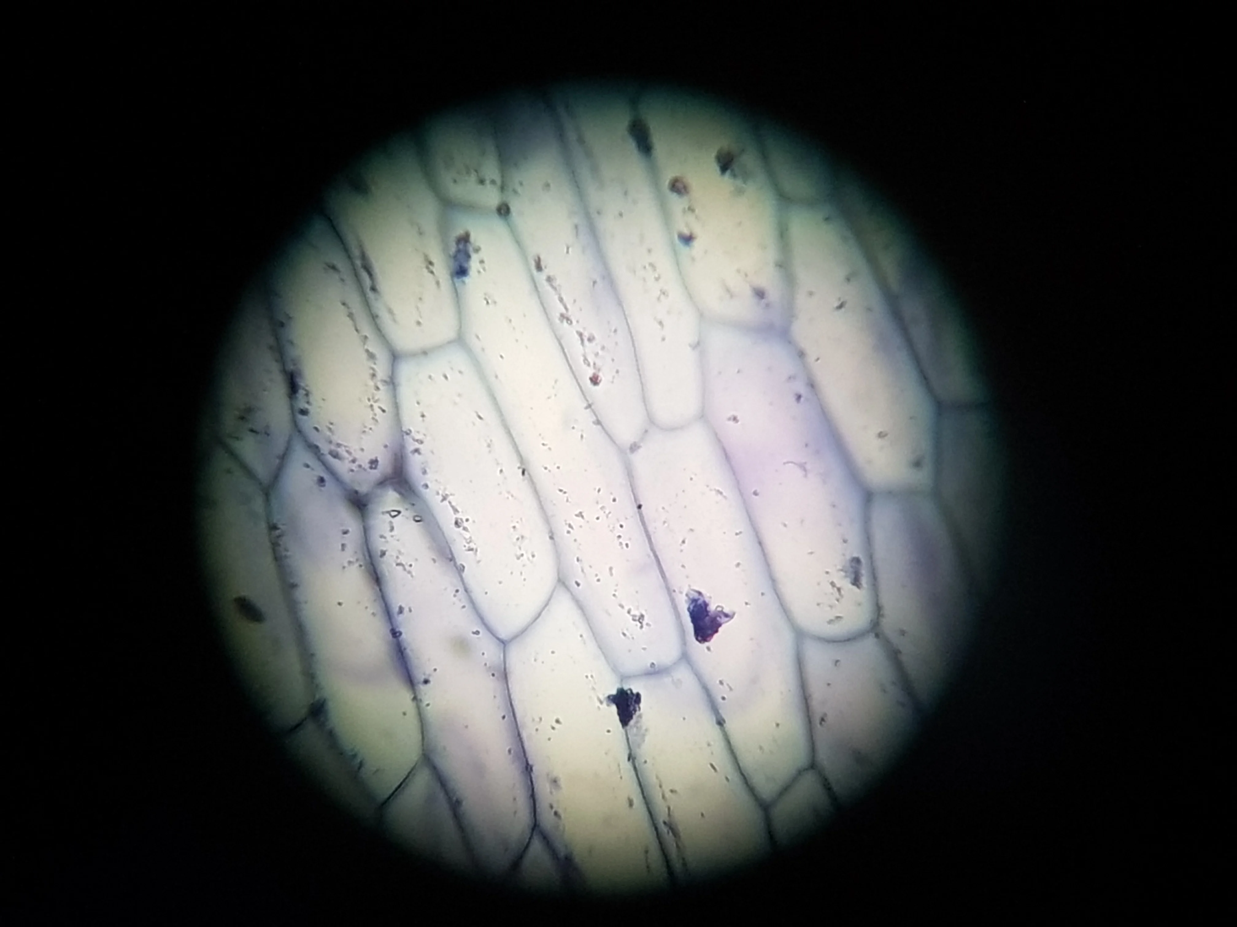
Specimen: onion cells, as identified by the source. Stain: methylene blue. Magnification: “200x” appears in the original filename; it is not independently validated. Scale bar: unavailable; no calibration is supplied. Photograph by Alvy16, 2019, from Onion cell 200x, licensed CC BY 4.0.

The elongated compartments form an organized sheet. Compare their long shared boundaries with the irregular outlines in Plate 1. Also notice the conspicuous dark material and uneven field brightness. The photograph does not establish the identity of every dark deposit. It demonstrates why a useful observation record includes artifacts and uncertainty instead of labeling everything that takes up color.
Plate 3: yeast identified by the source as Candida spp.
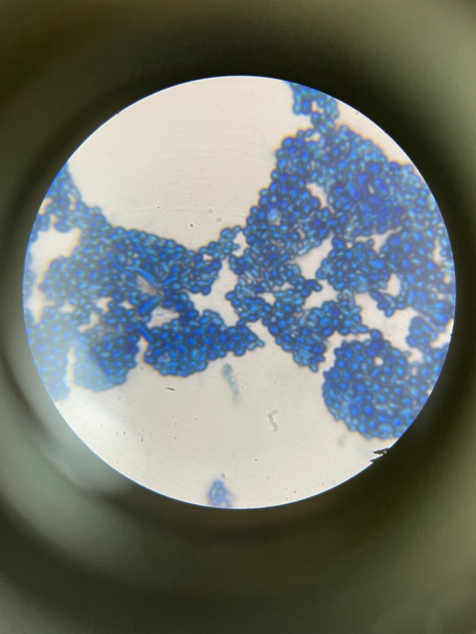
Specimen: Candida spp., according to the photographer. Stain: simple positive staining with methylene blue. Magnification: unavailable. Scale bar: unavailable; no calibrated pixel size is supplied. Photograph by Marta, Wikimedia user Martunia.g, 2021, from Positive staining with methylene blue 2, licensed CC BY 4.0.

Compare the dense blue clusters with the less crowded margins. Individual outlines are easier to follow where objects overlap less. The name Candida comes from the source record, not from a species identification performed on this photograph. This plate also supplies no viability result. Yeast viability and respiration assays require their own conditions, controls, and interpretation.
Reading scale correctly
These plates support visual comparison, not measurements in micrometers or comparisons of absolute cell size between plates. Reported magnification does not recover missing camera calibration. Changing an image's display size changes its apparent enlargement.
For your own images, establish a measurement calibration using a known standard and the relevant optical setup. Nikon's guide to micrometry explains how a stage micrometer converts arbitrary image or reticle divisions into physical lengths. Record the objective, camera configuration, and calibration, and add a scale bar from those data. Do not infer a bar from a textbook's average cell size.
Does the bottle match the method?
A bottle labeled methylene blue is not a complete laboratory specification. Check its concentration and units, solvent, additional ingredients, intended application, batch documentation, and instructions. A reagent's safety data sheet describes hazards; the method instructions establish how to use it for the intended preparation.
A concrete example is Merck's Löffler's methylene blue solution. The manufacturer identifies it as a microscopy reagent and describes its role as a counterstain in acid-fast staining. Its listed flammability also shows why “a blue liquid” is insufficient information for planning handling or heating. Those properties apply to that formulation, not automatically to every aqueous methylene blue solution.
Likewise, eosin methylene blue agar is a formulated culture medium. It is not a slide stain made by adding plain methylene blue to a specimen. For a different imaging objective, compare alternatives to methylene blue for microscopy.
For a beginner microscope kit, start with its documented educational method, known prepared slides, and the specified reagent. If a bottle has lost its label or its composition cannot be established, replace it with the appropriate documented reagent. Use eye protection and the handling instructions supplied for the actual formulation. Human material and unknown environmental samples require the teaching laboratory's sample-handling and disposal procedures; staining is not a substitute for those procedures.
Write the observation before the conclusion
“Blue oval objects in clusters” is a defensible description of an image. “A particular infection” requires evidence the image has not supplied. Keep specimen provenance, preparation, staining reaction, and identification method separate in the notebook. That distinction makes a simple classroom image useful and prevents an attractive micrograph from carrying a claim it cannot support.