
Article
Alternatives to methylene blue for microscopy: choose by task
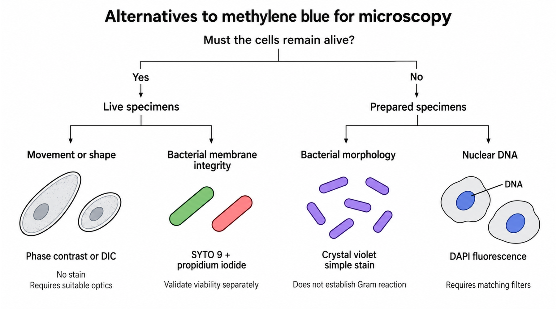
For a simple bacterial smear, crystal violet can replace methylene blue when the aim is to see cell shape. For living cells, the better alternative may be phase contrast with no stain at all. The choice depends on the observation you need to make.
Suppose two people are examining the same bacterial culture. One wants to distinguish rods from round cells. The other wants to know whether a treatment has killed the bacteria. A stain that gives the first person a clear image may give the second person no usable answer.
Before choosing a substitute, complete this sentence: “I need to observe ___ in ___, and the cells must / need not remain alive.” That sentence defines the job the stain must do.
Start with the specimen and the question
First decide whether the cells must remain alive, then identify the feature you need to observe:
- Cells must remain alive: for movement or shape, consider unstained phase contrast or DIC; for nuclear DNA, consider Hoechst 33342 with fluorescence; for membrane integrity, choose a validated assay such as SYTO 9 with propidium iodide.
- Cells need not remain alive: for bacterial morphology, consider a crystal-violet simple stain or nigrosin negative stain; for Gram reaction, use the complete Gram-stain method; for nuclear DNA in fixed cells, consider DAPI fluorescence.
Use this table to compare the limitations of each branch. These are method choices, not instructions to exchange bottles within an existing protocol.
| What you need to observe | Specimen and equipment | Candidate alternative | Main limitation |
|---|---|---|---|
| Bacterial shape and arrangement | Prepared bacterial smear; brightfield | Crystal violet simple stain | Shows morphology; does not establish Gram reaction or viability. |
| Cell outlines against a dark background | Bacteriological preparation; brightfield | Nigrosin negative stain | Preparation and formulation matter; an unstained cell is not necessarily alive. |
| Gram reaction | Prepared bacterial smear; brightfield | Complete Gram-stain method | Requires the full reagent sequence and controls. |
| Movement or changes in living cells | Compatible wet preparation; phase contrast or DIC | No stain | Requires suitable optics; contrast does not identify a molecule. |
| Nuclear DNA | Fixed cells; fluorescence | DAPI | Requires compatible excitation and emission filters. |
| Nuclear DNA in living cells | Live cells; fluorescence | Hoechst 33342 | Cell entry does not establish that the imaging conditions are harmless. |
| Bacterial membrane integrity | Unfixed bacteria; fluorescence | SYTO 9 with propidium iodide | Membrane staining is an indirect measure of viability. |
For a simple smear, replace the method carefully
Crystal violet is a practical candidate when the task is a single-color view of bacterial morphology. The FDA's Bacteriological Analytical Manual lists two crystal-violet formulations suitable for simple staining. Use a documented formulation and its preparation method; the name of the dye alone does not specify a working reagent.
Crystal violet also appears in Gram staining, but that does not make a crystal-violet simple stain a Gram stain. In the ASM Gram-stain protocol, the differential result depends on crystal violet, iodine, decolorization, and counterstaining acting in sequence. Omitting those steps changes what the image can establish. See simple staining versus Gram staining for that distinction.
Nigrosin offers a different way to reveal outlines: it darkens the background while the bacterial cells remain comparatively light. There is a useful detail in Merck's nigrosin instructions that a generic list of substitutes can miss. Its specified working formulation includes formaldehyde. You therefore cannot infer live-cell compatibility from the words “negative stain,” even though the cells themselves appear unstained. Check the complete formulation and specimen preparation.
For living specimens, consider changing the optics
If you want to watch cells move or divide, introducing another dye may complicate the observation. As Nikon explains in its guide to contrast in transparent specimens, phase contrast and differential interference contrast, or DIC, make specimens visible through differences in their optical properties. They can reveal living, unstained cells, provided the microscope has the required components.
This choice preserves an unstained preparation, but it changes the information available. You can follow a boundary or a movement without knowing which molecule produced the contrast. If molecular localization is the question, select an appropriate labeling method instead.
For nuclear DNA, Invitrogen's comparison of DAPI and Hoechst stains distinguishes DAPI's common use with fixed cells from the more cell-permeant Hoechst 33342, which is preferred for live-cell labeling. Both require fluorescence equipment. Neither is a direct replacement for a colored brightfield stain.
Treat dye exposure and illumination as experimental conditions. Compare stained and unstained preparations when the behavior of the cells is your measurement.
A red cell is not automatically a dead cell
SYTO 9 and propidium iodide are often paired to assess bacterial membrane integrity. The BacLight manual associates intact membranes with green staining and damaged membranes with red staining. That interpretation needs validation for the specimen.
A 2019 study by Rosenberg and colleagues illustrates the problem. In 24-hour E. coli biofilms on glass in phosphate-buffered saline, about 96% of cells appeared propidium-iodide-positive in situ. Yet the authors estimated that 82% were cultivable after harvesting. Confocal images showed cells with green interiors beneath red-stained material, implicating extracellular nucleic acids in the misleading signal.
Those percentages describe different measurements on the study's preparations, not a universal correction factor. The useful lesson is to check what generated the signal. In a biofilm, a nucleic-acid dye can detect material outside the cells as well as inside damaged cells. For a viability conclusion, the researchers recommend complementary measurements, such as cultivation or metabolic activity, while acknowledging that each has limitations.
Check what the alternative actually contains
An alternative staining method may still contain methylene blue. Giemsa is an example: Merck describes its product as an azure-eosin-methylene-blue solution, intended for applications including blood and bone-marrow smears. It can be a different method for a different specimen, but it does not satisfy a requirement to exclude methylene blue.
If excluding a compound is part of the experiment, check reagent identity and formulation before comparing images. The guide to methyl blue, methylene blue, and similarly named dyes addresses naming differences.
Verify the substitution before using the result
For a new method, make a small comparison using representative specimens and an appropriate control. Keep the microscope settings comparable where possible. Record the reagent formulation, specimen preparation, exposure, and imaging conditions so that a better-looking image can be investigated rather than merely preferred.
Judge success against the original question. For morphology, assess whether cell boundaries and arrangement remain interpretable. For nuclear labeling, assess signal location and background. For viability, require evidence beyond an attractive color separation. The broader guide to methylene blue staining and laboratory applications explains how the observation and the preparation fit together.
Choose the alternative that preserves the measurement you need. Color is only one property of the resulting image.