
Article
How to read methylene blue research: cells, animals, human tissue, and clinical trials
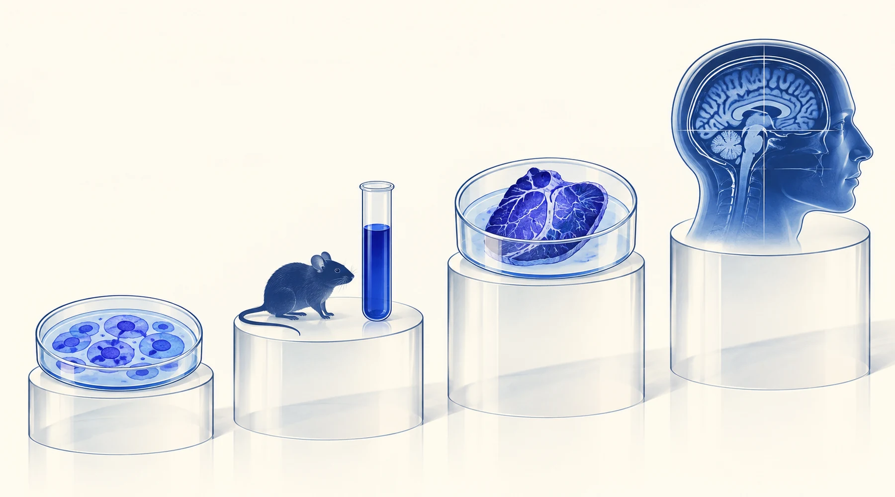
Consider two published findings. In one, a single oral dose of methylene blue increased brain activity during attention and memory tasks and improved memory retrieval by 7 percent. In the other, methylene blue reduced cerebral blood flow and brain oxygen metabolism in healthy volunteers. Same molecule, opposite directions. A reader who treats both as verdicts on the same question will conclude that one of them must be wrong.
Neither is wrong. They asked different questions, in different systems, at different doses, by different routes, measured different endpoints at different times. This article teaches you to see those differences the way a researcher does, so that the next claim you meet can be sorted before it is believed. The background on what the molecule does at the chemical level is covered in the mechanism explainer, and the question of how dose shapes effect is covered in the dosing evidence review. Here the subject is the evidence itself.
Every paper answers a narrow question
A study result is never a statement about methylene blue in general. It is a statement about methylene blue in a particular model, given by a particular route, at a particular dose, compared against a particular control, measured with a particular endpoint, at a particular time. Change any one of those and the answer can change. Seven slots, filled in for every paper you read:
- Model. Cells in a dish, a mouse, human tissue kept alive outside the body, or people. Each step up adds realism and removes control.
- Route. Swallowed, injected into a vein, injected into a joint, or pipetted onto cells. Route decides how much reaches the tissue and how fast.
- Dose units. Milligrams per kilogram for animals and people, micromolar for cells and tissue baths. These units do not convert into each other by arithmetic alone, because absorption, distribution, protein binding, and clearance sit between them.
- Comparator. Placebo, vehicle solution, untreated animals, or a before dose baseline. A result without a comparator is an observation, not a finding.
- Endpoints. What was actually measured: a brain scan signal, an enzyme level, bone density, a test score. Surrogate markers (things that stand in for an outcome) are not the outcome.
- Timing. A single dose measured after one hour is a different claim from daily dosing for a year.
- Uncertainty. Sample size, healthy volunteers versus patients, and whether the authors themselves flag limits. Small pilot studies generate hypotheses; they do not settle them.
The four examples below fill in this worksheet for real papers, including two that appear to disagree.
Example 1: a volunteer trial that found more brain activity
In a randomized, double blind, placebo controlled imaging trial, 26 healthy adults aged 22 to 62 performed sustained attention and short term memory tasks in a scanner, then swallowed either 280 mg of pharmaceutical grade methylene blue (about 4 mg/kg) or a blue food dye placebo, and repeated the tasks one hour later. The dye group showed stronger responses in the insular cortex during attention and across prefrontal, parietal, and occipital networks during memory, plus 7 percent more correct answers at retrieval. A carbon dioxide challenge showed no change in vascular reactivity, which the authors read as evidence that the signal reflected oxygen use rather than blood vessel effects.
Worksheet reading: model, healthy people. Route, oral capsule. Dose, single 4 mg/kg. Comparator, placebo with blinding protected by asking subjects not to urinate mid session (blue urine would unblind the study). Endpoints, scan signal plus task scores. Timing, one hour after one dose. Uncertainty, 26 people, healthy, one session, imaging surrogate rather than a clinical outcome.
What this proves is narrow and real: one low oral dose changed task related brain activity in healthy adults that day. It does not test treatment of any condition, long term use, or any other dose. Claims about memory and cognitive performance should be weighed with that boundary in mind.
Example 2: the contradiction that is not one
A later team measured blood flow and metabolism directly after intravenous methylene blue, in eight healthy volunteers (0.5 and 1 mg/kg) and in rats (2 and 4 mg/kg). Both species showed reduced global cerebral blood flow; human oxygen metabolism fell and rat glucose use fell. The authors' own explanation was dose behavior: methylene blue is biphasic, stimulatory at low exposure and inhibitory at higher exposure, with the low range usually described around 0.5 to 4 mg/kg and reversal above roughly 7 to 10 mg/kg, depending on the model.
Put the two worksheets side by side and the conflict dissolves. Different route (intravenous versus oral), different doses, different endpoints (metabolic rate versus task evoked activity), different timing (infusion 30 minutes before scanning versus capsule one hour before). An intravenous dose that arrives all at once is not the same exposure as a capsule absorbed through the gut, and a resting metabolic rate is not the same measurement as activity during a memory task. Contradicting papers are often just papers answering different questions, and the dose response story that connects them is laid out in the hormesis and dosing overview.
Example 3: human tissue studied outside the body
A pilot study incubated fat samples taken during heart surgery with methylene blue. The samples came from 25 heart failure patients: epicardial and perivascular adipose tissue, the fat deposits around the heart and vessels that contribute to oxidative stress. Incubated for 24 hours with 0.1 micromolar methylene blue, the tissue showed lower expression of monoamine oxidase enzymes (mostly MAO-A) and less reactive oxygen species. Adding serotonin, the MAO-A substrate, raised oxidative stress again, and methylene blue partly reversed that rise.
This is human tissue and a real biological effect, but it is not a treatment trial in people. Nobody was dosed. There was no circulation, no metabolism by the liver, no clinical outcome, and the concentration in the dish says nothing by itself about what oral dose would reproduce it. Ex vivo work (tissue studied outside the living body) sits between cell culture and clinical research: it shows an effect is possible in human material under controlled conditions, and it cannot establish efficacy, safety, or dosing. When you read coverage of tissue findings, check whether the headline describes what happened in the dish or leaps to what would happen in a patient. The broader context for this kind of redox finding is in the oxidative stress evidence review.
Example 4: the mouse study where a cell result did not survive
A mouse aging study tested long term methylene blue against age related bone loss. Aged female mice drank water containing 250 micromolar methylene blue for 6 or 12 months, and genetically diverse mice were treated from adulthood to old age. In cell culture the compound had reduced osteoclast differentiation, the kind of result that launches headlines. In the living animals it changed nothing: cortical and trabecular bone morphology aged exactly as in untreated mice, and a parallel MitoQ arm failed the same way.
This is the translation gap in its purest form. A dish holds concentration constant, removes every organ system, and measures one cell behavior. An animal adds gut, liver, kidneys, hormones, and months of compensation. A null result like this is not a failure of science; it is science working, ruling out a hypothesis (antioxidant treatment alone protects aging bone) before anyone repeats it in people. Animal findings support or weaken a mechanism. They do not prescribe human use, a boundary that matters whenever longevity and aging claims cite mouse data.
The worksheet, ready to copy
For the next paper or article about a paper, fill this in before deciding what you think:
- Model: cells, animal (which species, healthy or diseased), ex vivo human tissue, healthy volunteers, or patients?
- Route: oral, intravenous, local injection, or applied directly to cells or tissue?
- Dose units: mg/kg (body) or micromolar (dish)? Never convert one to the other by molecular weight alone.
- Comparator: placebo, vehicle, untreated group, or pre dose baseline? Was blinding protected?
- Endpoints: measured directly or surrogate? A scan signal, an enzyme level, and a disease outcome are three different strengths of claim.
- Timing: single dose or chronic? Measured after minutes, hours, or months?
- Uncertainty: how many subjects, and what did the authors themselves say was limited? Pilot, healthy volunteer, and single dose studies generate hypotheses.
Two habits make the worksheet fast. First, read the methods before the conclusion; the conclusion tells you what the authors believe, the methods tell you what they are entitled to believe. Second, when two studies disagree, compare worksheets before comparing verdicts. Model, route, dose, and endpoint differences resolve most apparent contradictions without anyone being wrong.