
Article
Methylene blue dye tests for urinary leaks and fistulas: what a blue swab proves
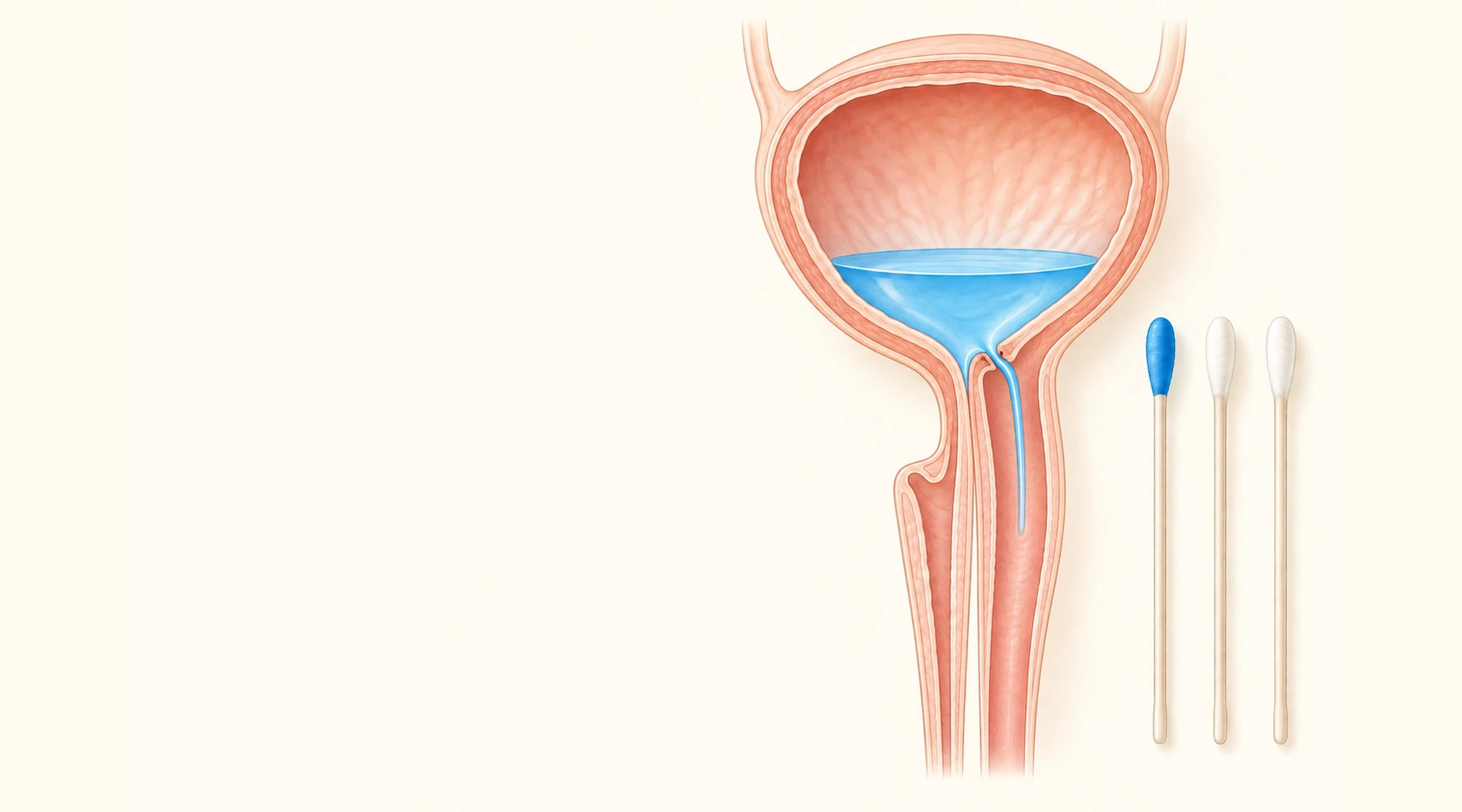
A few weeks after pelvic surgery, a patient notices she is leaking urine continuously. She assumes it is a bladder infection, finishes a course of antibiotics, and is confused when the leakage continues unchanged. The antibiotics were never going to work, because the mental model was wrong. An infection is an invasion of tissue by microbes. A fistula is a hole: an abnormal channel connecting two spaces that should be separate, in this case the bladder and the vagina. No antibiotic closes a hole.
This is the situation methylene blue dye testing was designed for. The test does not treat anything, does not detect infection, and does not produce a diagnosis by itself. It answers one narrow question: is fluid in the vagina arriving from the dye-filled bladder? Everything about the procedure, including its failure modes, follows from that single purpose. Our overview of methylene blue in medicine places this diagnostic use alongside the compound's authorized and investigational roles.
The core idea: controlled blue fluid as a tracer
The principle is deliberately simple. The bladder is filled retrogradely through a catheter with sterile irrigant containing dilute methylene blue. The vagina should contain no blue fluid. If blue fluid then appears in the vagina at an abnormal site, a communication exists between the bladder or urethra and the vagina. The dye is a tracer, and the test is a localization maneuver.
Two consequences follow immediately. First, the test can only find leaks downstream of where the dye is placed. Blue dye sitting in the bladder can never reveal a ureterovaginal fistula, because urine from the affected ureter reaches the vagina without ever entering the bladder. Second, a positive result localizes a leak but says nothing about tissue quality, fistula size, or how to repair it. That is why surgical and diagnostic procedure planning always goes beyond the dye test.
The three-swab test, step by step
The examination begins before any dye is used. A speculum examination looks for pooling of clear urine and, if visible, the vaginal opening of the fistula. History and focused examination carry a strong recommendation in the European urological guidance on urinary fistula, and no dye result overrides what the eyes can see directly.
The formal triple-swab procedure, as defined in the ICS/IUGA terminology report, runs as follows. Three dry sponge swabs are placed sequentially in the vagina: one upper, one middle, one lower. Variants use gauze packs, cotton balls, or a tampon, but the three separate swabs are the standardized form. The bladder is then filled through the catheter with dilute blue irrigant, and the clinician watches for blue fluid emerging from a vaginal opening during filling. Placing the swabs before filling matters: inserting a tampon after dye has been spilled around the entrance guarantees contamination and a meaningless result.
There is no single universal volume, concentration, or dwell time, and an honest article should say so. The FIGO fistula surgery training manual describes 60 to 100 mL of diluted dye with immediate inspection, and, if nothing is seen, walking and drinking for 30 to 60 minutes with the dye still in the bladder. One postsurgical protocol used 250 to 300 mL and specifically warned that inadequate filling causes false negatives in small fistulas. The ICS/IUGA definition removes the swabs after 10 minutes but allows up to 30 minutes for a tiny or twisting tract. The defensible instruction is to fill adequately with dilute solution according to the local protocol, not to prescribe one universal recipe.
Reading the swabs
Each swab is removed individually and documented for wetness and color. The standard interpretation, following the ICS/IUGA definitions:
| Finding | Interpretation | Caveat |
|---|---|---|
| Upper or middle swab wet and blue | Supports a bladder-to-vagina communication | Height is approximate when several tracts coexist |
| Only the lowest swab blue | Suggests a low urethral leak, or contamination | Not specific: urethral incontinence and backflow from the meatus do the same |
| Upper swab wet but not blue | Suggests an upper-tract, ureterovaginal source | Requires upper-tract imaging to confirm |
| Nothing wet or stained | No leakage demonstrated in that test | Does not exclude a small, intermittent, or underfilled fistula |
The most dangerous row is the second. Blue on the lowest swab alone is the finding most easily produced without any fistula at all, by ordinary urethral leakage or by blue fluid spilling backward from the meatus. A distal blue swab alone never closes the case.
The double-dye test: telling bladder from ureter
When vaginal urine keeps appearing but the bladder dye never shows in it, the suspicion shifts upstream to the ureter. The double-dye test makes the two sources visually distinct. The patient takes oral phenazopyridine until the urine turns bright orange (the formal definition uses 200 mg three times daily for one to two days, though some office protocols use a single dose), and then the bladder is filled with blue irrigant as before.
The logic is a small exercise in routing. Blue vaginal staining means the fluid came from the bladder or urethra. Orange staining without blue means the fluid arrived from the ureters above the bladder. Both colors suggest communications in both places. Clear upper-vaginal urine despite a blue bladder points the same upstream direction as orange. This is a clinician-directed diagnostic regimen, not a home experiment.
Cystoscopy and the operating room
Dye testing also works from the other side. During pelvic surgery, the bladder can be filled retrogradely with blue-tinted irrigant while the operative field is inspected; blue fluid welling into the field identifies a bladder defect, and the maneuver can be repeated after repair to check that the closure looks watertight. One reported technique used up to 600 mL for this purpose, which should be read as a surgical report rather than a standard every bladder must receive.
Cystoscopy defines what the swabs cannot: the number, size, and position of bladder defects and their relationship to the trigone and the ureteral orifices. European guidance recommends cystoscopy with retrograde colored filling for confirming fistula, while noting the evidence base is limited. A related but distinct maneuver checks ureteral patency during cystoscopy by watching for dye jets from the ureteral orifices. Here the marker must arrive from above, through the bloodstream or the urine, because dye placed inside the bladder is already downstream of the ureters and cannot test them. Confusing these two dye routes is exactly the kind of conceptual error the swab table is meant to prevent. Details of these operative techniques belong to our companion guide on methylene blue in surgery and diagnosis.
Why the test fails in both directions
A false negative has a short list of usual causes: too little bladder filling, a tiny or twisting tract that needed more than ten minutes, an intermittent or position-dependent leak, a ureterovaginal fistula the bladder dye could never reach, or a second fistula hiding behind the first one found. In one retrospective series of 56 postoperative fistula patients, 10 of 13 dye tests performed before referral were positive. That 10-of-13 figure is sometimes quoted as though it were sensitivity. It is not: the cohort was highly selected and the study was never designed as a diagnostic-accuracy trial. The 2020 postsurgical series supports dye testing as a rapid first-line maneuver, not as a measured sensitivity number.
False positives and misleading results are mostly contamination stories: dye spilled during tampon placement, urine wicking along packing, voiding during the test, or ordinary stress or urge incontinence staining the lowest swab. None of these indicate a fistula, which is why direct observation during filling and watching the meatus for backflow are part of the procedure rather than optional extras.
A positive dye test is not a UTI result, and vice versa
A urinary infection does not create a channel between bladder and vagina, so infection alone can never produce controlled extra-meatal blue leakage. The two conditions overlap in symptoms and can coexist, which is precisely why they get confused. Urgency and urge incontinence from infection exit through the normal urethra; fistula leakage bypasses it. Urinalysis and culture still matter when infection is suspected, and creatinine measurement in collected vaginal fluid can confirm the fluid is urine, though it cannot by itself localize the leak. Note one more confounder: some combination urinary medicines that contain methylene blue discolor the urine themselves, which is worth mentioning to the clinician before any dye-based assessment.
What still has to happen after the swabs
Dye testing opens the work-up; it does not complete it. Pelvic examination assesses tissue quality, scarring, and fistula position. Cystoscopy and urethroscopy map the internal openings before any repair. When a ureteral source is suspected or the anatomy is unclear, contrast CT with a late excretory phase or MRI shows the upper tract, and retrograde pyelography can define an uncertain ureter directly. Ultrasound comparisons exist: in a small twelve-patient feasibility study, contrast-enhanced color Doppler classified 11 of 12 cases correctly, but that result belongs to the ultrasound method, not to the dye test (Volkmer and colleagues, 2000).
The dye test also returns after treatment. FIGO's good-practice recommendations advise a dye check 10 to 14 days after fistula repair, before the catheter is removed, with the patient coughing under observation (Browning and colleagues, 2025). A negative test at that visit means no fistulous leakage was demonstrated then; it does not guarantee that later leakage from incontinence, retention, infection, or a missed ureteric source will never occur.
Methylene blue has one more diagnostic role worth keeping separate from fistula work: it is also used to check whether the fallopian tubes are open, a procedure covered in our guide to fertility testing and tubal patency assessment. Same dye, different question, different anatomy. Keeping the question attached to the result is the entire discipline of dye testing. A blue swab proves a route exists. Everything else, the size, the repairability, the prognosis, still has to be examined.