
Article
Methylene blue and permanganate diffusion: what the agar experiment shows
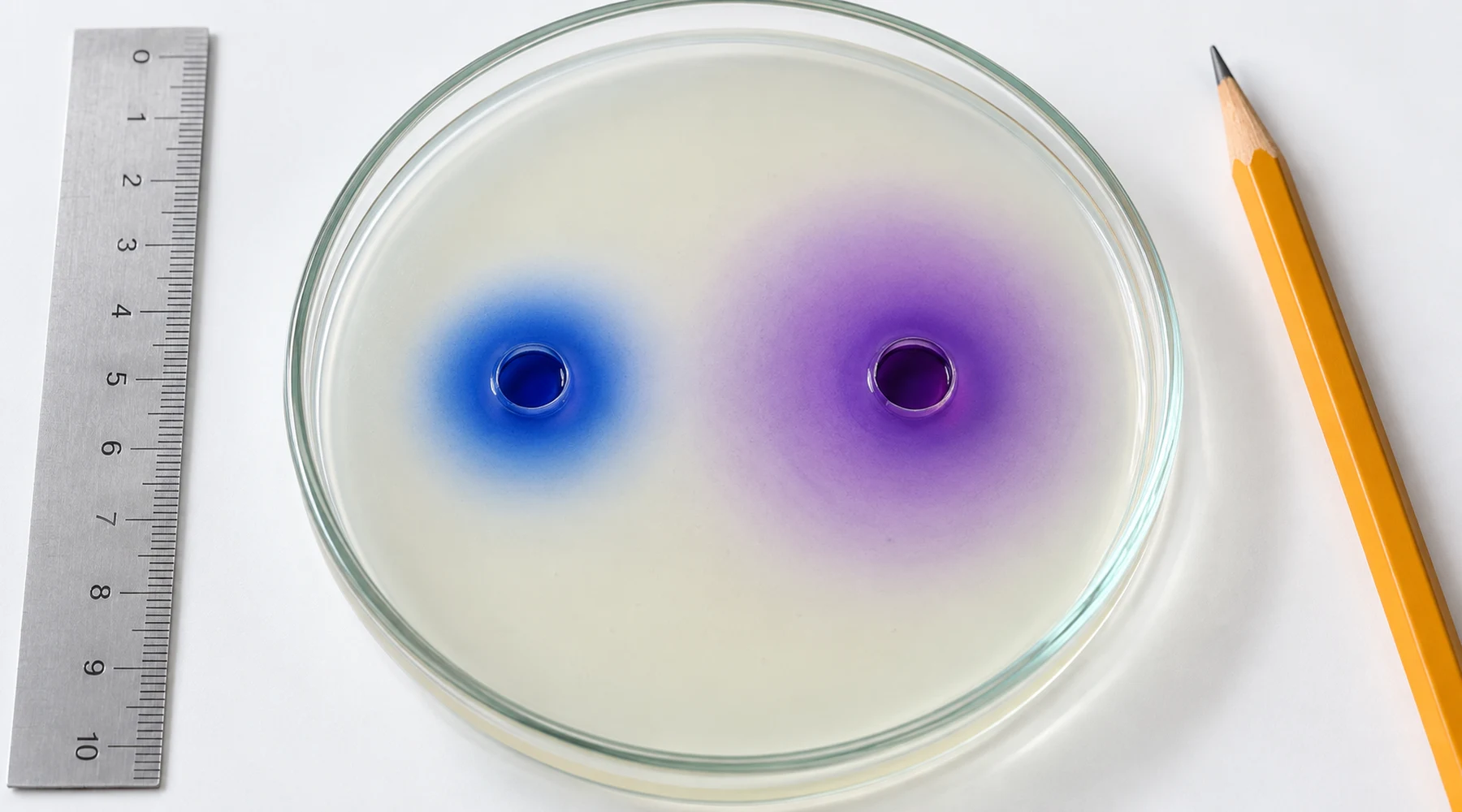
A teacher cuts two wells in an agar plate, fills one with blue dye and one with purple solution, and tells the class to watch. An hour later the purple ring is wider. The class writes down that potassium permanganate diffuses faster than methylene blue because it is lighter.
That sentence is half right and half misleading, and the misleading half is the interesting part. The demonstration is a standard diffusion lesson, described for example in this agar diffusion lab exercise, but what students see is not molecular weight in action. It is the movement of a visible color edge through a gel, shaped by ion size, hydration, dye binding, concentration, temperature, and the way the edge is measured.
The colored edge is not the diffusion coefficient
Diffusion is the spreading of dissolved particles from high concentration to low concentration. The diffusion coefficient, usually written D, describes how fast that spreading happens for a given solute in a given medium at a given temperature.
A classroom plate does not display D directly. It displays a boundary where the color becomes too faint to see. That boundary depends on dye absorptivity, lighting, camera exposure, the concentration placed in the well, and the judgment of the person holding the ruler, as well as on diffusion itself.
Call the classroom reading what it is: the movement of a visible diffusion front. Formal treatments relate mean squared displacement to D through relations of the form found in Fickian diffusion analysis, but fitting D requires concentration profiles over time, not a single diameter at the end of class. One published classroom study extracted a methylene blue diffusivity near 4.5 times 10^-10 m^2/s in gel by plotting squared distance against time rather than comparing final spot sizes. A single snapshot cannot do that work.
This is the concept behind the demo, and everything else is a detail of how faithfully the plate represents it.
Two colored ions, not two molecules
Neither solute travels through agar as the formula printed on the bottle. Both are strong electrolytes that dissociate in water.
Potassium permanganate (KMnO4, formula mass about 158.03 g/mol) separates into potassium ions and permanganate ions. The purple traveler is the permanganate ion MnO4^-, mass about 118.94 g/mol, surrounded by a shell of water. Methylene blue chloride (anhydrous salt C16H18ClN3S, about 319.85 g/mol) separates into chloride and the blue organic cation MB^+, mass about 284.41 g/mol, also hydrated. The NIST methylene blue record and the ChEBI methylene blue entry document these forms, and the chemistry and structure reference explains why the cation, the anhydrous salt, and the hydrates need separate names.
Three consequences follow.
First, comparing 158 with 320 understates the real size gap between the colored ions, since the purple species at about 119 g/mol is much smaller than the blue cation at about 284 g/mol, and both carry hydration shells that add effective size.
Second, methylene blue cations stick to things. They form dimers and larger aggregates as concentration rises, and they adsorb to agar, with both monomer and dimer binding reported in agar adsorption studies. Adsorbed dye sits still. It colors the gel without advancing the front.
Third, the common reagent powder is a hydrate, so a weighed gram contains water in the crystal. The trihydrate versus anhydrous comparison shows how that changes molarity. For a fair classroom comparison, prepare solutions at equal molar concentrations of the salts and record which solid form was used.
Why lighter does not mean faster here
In dilute liquids, a better first model than mass is the Stokes-Einstein picture, in which diffusivity scales with temperature divided by viscosity and hydrodynamic radius. Smaller hydrated size wins. Lower mass often correlates with smaller size inside one chemical family, so lighter can look like an explanation. It is a correlation, not the mechanism.
Graham's law, which states that gas effusion rates scale with the inverse square root of molar mass, does not govern this plate. Its derivation belongs to the kinetic theory of gases, summarized in this OpenStax treatment of effusion and diffusion of gases. Liquids add viscous drag, hydration, shape effects, and crowding. Gels add pore structure, tortuosity, adsorption, and charge interactions with the polymer.
The numbers expose the gap. Graham's law applied to 158 versus 320 predicts a rate ratio of about 1.42. Measured dilute-water diffusivities tell a different story: permanganate near 1.6 to 1.8 times 10^-9 m^2/s against methylene blue near 4.5 to 5 times 10^-10 m^2/s, a ratio closer to 3 or 4. Mass alone cannot produce that ratio. Hydrated size, aggregation, and gel binding do the remaining work.
Treat lighter-faster as a model with assumptions: spherical particles, no binding, no aggregation, identical conditions. The plate violates at least two of them.
How to run a fair comparison
The demonstration fails the way software fails: the underlying idea is sound, but the mapping from idea to measurement is full of traps. Each of these changes the answer without changing any molecular property.
Use one batch of agar poured to one depth, one well cutter, equal liquid volumes, equal molar concentrations, and low enough concentrations to limit methylene blue aggregation and keep the color unsaturated. Cut at least three replicate wells per solute, spaced so fronts never meet. Cover the dishes, keep them level, and hold temperature constant, since agar dye work shows strong temperature and concentration dependence. Define time zero as the moment of filling, and define the edge before measuring: a fixed photographic threshold beats an eyeballed boundary, because blue and purple have different visual detection limits. Record two perpendicular diameters per spot and convert to mean radius, since confusing radius with diameter corrupts any squared-distance plot by a factor of four.
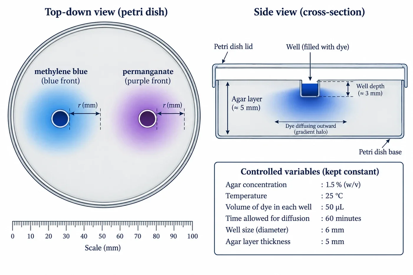
Labels: left panel shows top-down radius measured from the well edge to the color edge for each front; right panel shows the agar cross-section with well depth and outward gradient halos; the legend lists the controlled variables (agar percentage, thickness, dye molarity, volume, well size, temperature, time zero, lighting).
Watch for impostors. Liquid spilling across the agar surface looks like fast diffusion and is a documented artifact of small agar wells. Permanganate is a strong oxidant and can be consumed by traces of organic material, which stalls or bleaches its front. Methylene blue is redox active and can fade under light or reducing conditions; the redox chemistry of methylene blue and its colorless reduced form explains that behavior.
A short solvent note: both wells in the standard demo are aqueous. If a protocol ever changes the solvent, check the solubility and solution preparation guide first, because solubility and aggregation change with the medium.
Classroom observation worksheet
Copy this table into notebooks or print one row per well. One table per dish keeps replicates honest.
| Well | Solute and stock molarity | Volume added | Time zero | Elapsed time | Temperature | Agar % and thickness | Well diameter | Spot diameter 1 | Spot diameter 2 | Mean radius | Notes (edge sharpness, spill, fading, asymmetry) |
|---|---|---|---|---|---|---|---|---|---|---|---|
| A1 | |||||||||||
| A2 | |||||||||||
| A3 | |||||||||||
| B1 | |||||||||||
| B2 | |||||||||||
| B3 |
For analysis, plot mean radius squared against elapsed time for each well and compare the slopes, stating the edge definition alongside the graph. If all six curves are shown with their spread, the class sees variation as data rather than as embarrassment.
Safety is part of the protocol: wear gloves and eye protection, since methylene blue stains skin and clothing and permanganate is an oxidant that stains and irritates. Keep solutions out of the mouth, work over a tray, and dispose of plates and liquids through the school's chemical waste route rather than the sink.
The sentence worth writing down
Under these agar, concentration, and temperature conditions, the visible permanganate front usually spreads faster than the methylene blue front. That is consistent with permanganate having the larger effective diffusivity, largely because its hydrated ion is much smaller and it does not aggregate or bind agar the way methylene blue does. Molecular mass correlates with that size difference here, but mass alone does not set diffusion rates in liquids or gels.
A class that writes that sentence has learned two things: a fact about two colorful ions, and a habit of asking what a demonstration measures before trusting what it seems to say.