
Article
Methylene blue and heart health: tissue studies, animal models, and clinical context
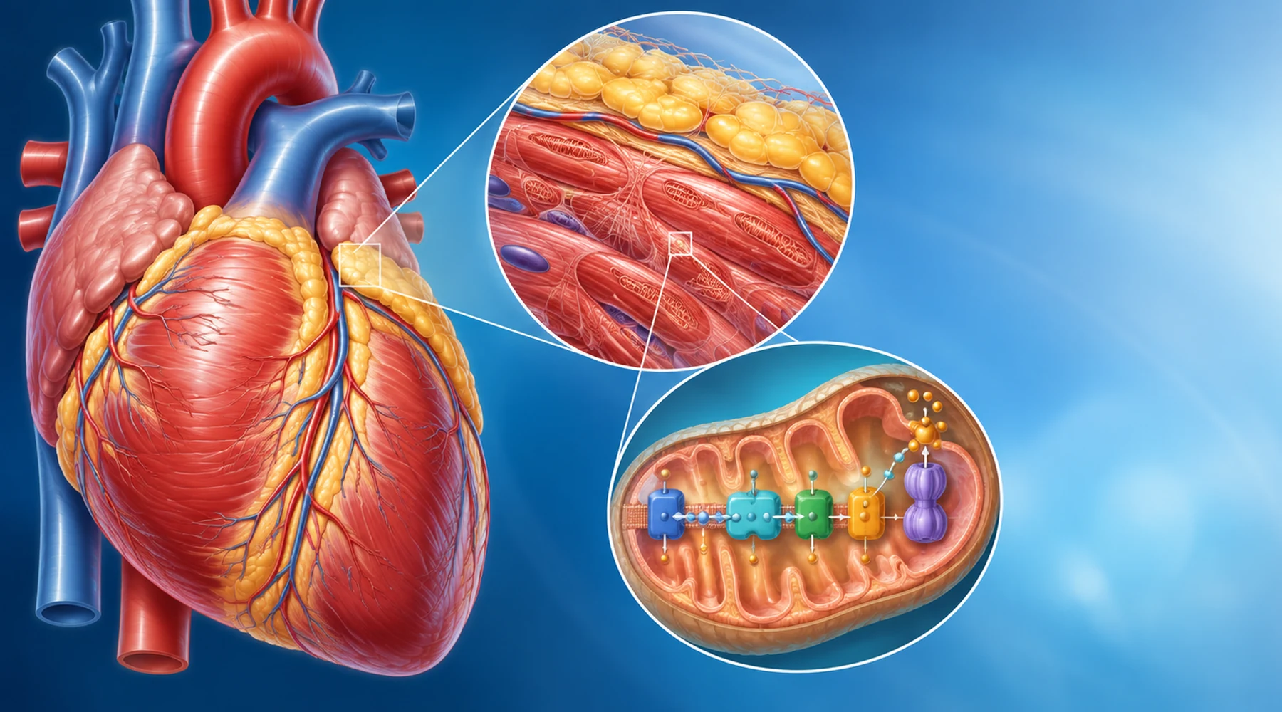
A reader finds two statements on the same week. One says methylene blue improves mitochondrial respiration in diabetic rat hearts. Another says it is used in hospitals around cardiac surgery. The natural inference is that methylene blue offers methylene blue heart benefits for heart failure or general cardiac protection.
That inference mixes three separate concepts. The first is a biochemical effect inside isolated mitochondria. The second is a change in tissue chemistry outside the body. The third is a clinical outcome in a patient, such as fewer symptoms, fewer hospital visits, or longer survival. Each has its own operational principle. A result at one level does not automatically transfer to the next.
This article keeps those levels apart. It reviews cardiac mitochondrial work, human cardiovascular tissue work, intact animal work, and the distinct hospital setting where methylene blue affects circulation.
Cardiac evidence matrix
The table is the core map for this topic. It separates isolated tissue preparations, intact animals, and clinical settings because they answer different questions.
| Evidence level | What was tested | What changed | What it does not establish |
|---|---|---|---|
| Isolated cardiac mitochondria, diabetic and healthy rats | Acute 0.1 micromolar methylene blue added to mitochondria supplied with complex I or complex II substrates | Oxygen consumption rose with both substrate types. Hydrogen peroxide release rose with complex I substrates and fell with succinate, a complex II substrate. Calcium retention did not change. | It does not show improved pumping, symptoms, or survival in a living patient. The preparation has no coronary flow, blood pressure control, kidneys, nerves, or drug interactions. |
| Intact diabetic rat heart, chronic dosing | Methylene blue at 10 mg per kg per day with echocardiography, hemodynamic study, respirometry, and proteomics | NADH oxidation increased, NAD rose, sirtuin 3 activity rose, and lysine acetylation fell at 83 sites across 34 mitochondrial proteins. Fatty acid oxidation shifted and cardiac function improved in diabetic animals. The diabetes related complex I defect itself was not corrected. | It does not establish methylene blue heart failure treatment in people. Species, diabetes model, dose, and surrogate endpoints prevent direct transfer to human heart failure. |
| Human cardiovascular fat, ex vivo | Epicardial and perivascular fat from 25 heart failure patients undergoing cardiac surgery, incubated with 0.1 micromolar methylene blue for 24 hours | Both monoamine oxidase isoforms were present, mainly MAO-A. Methylene blue reduced MAO expression and reactive oxygen species. Added serotonin increased hydrogen peroxide through MAO-A, and methylene blue partly reversed that rise. | Tissue outside the body is not a treatment trial. It does not show that taking methylene blue improves ventricular function or clinical outcomes. |
| Intact conscious dogs during exercise | Loading dose plus continuous infusion of new methylene blue during treadmill exercise, compared with saline | Arterial lactate to pyruvate ratio fell, showing a redox shift, while cardiac output, heart rate, contractility, arterial pressure, and catecholamine responses during exercise were unchanged. | It does not support improved exercise capacity or cardiac performance from a redox shift. The dogs did not have clinical heart failure. |
| Hospital hemodynamic settings | IV methylene blue for vasoplegia after cardiac surgery, septic shock adjunct care, and licensed treatment of acquired methemoglobinemia | Mean arterial pressure and vascular resistance can rise and vasopressor need can fall in selected vasodilatory shock. Methemoglobin falls in methemoglobinemia, restoring oxygen carriage. | Acute IV support for pressure or oxygen carriage is not evidence for chronic oral treatment of coronary disease, atherosclerosis, or heart failure. Route, dose, duration, and disease differ. |
Read across the last column first. That column contains the mental model correction most coverage of methylene blue heart claims omits.
What isolated heart mitochondria show
The diabetic heart study tested mitochondria removed from healthy rats and from rats with streptozotocin induced diabetes. High resolution respirometry measured oxygen use. Fluorescent assays measured hydrogen peroxide and calcium handling.
The useful surprise is the substrate dependence. With glutamate plus malate, which feed complex I, methylene blue raised respiration but also raised hydrogen peroxide release. With succinate, which feeds complex II, it raised respiration and lowered hydrogen peroxide. So the label antioxidant does not describe the full behavior. Whether reactive oxygen species rise or fall depends on which respiratory path is active and on the redox state of coenzyme Q.
A related diabetic rat study gives the mechanism more structure. Methylene blue acts as an alternate electron carrier. It helps oxidize NADH to NAD, which raises the activity of the mitochondrial deacetylase sirtuin 3. That enzyme removes acetyl groups from lysine residues on mitochondrial proteins. Proteomics found lower acetylation across fatty acid transport and oxidation, tricarboxylic acid cycle, electron transport, amino acid metabolism, and transport proteins. Cardiac function improved in treated diabetic rats even though the underlying complex I defect remained.
Think of this as rerouting electrons rather than simply scavenging radicals. The guide to mitochondrial electron transport places this rerouting idea in the larger chain. It remains a tissue level explanation, not a prescription.
What human heart tissue adds, and what it cannot add
The human study moves one step closer to patients without becoming a treatment trial. Surgeons collected epicardial fat, which sits directly on the heart, and perivascular fat, which surrounds vessels, from patients with heart failure. In the laboratory, samples were incubated with a low concentration of methylene blue for one day.
Both tissues expressed monoamine oxidase enzymes, mostly the MAO-A form. These enzymes can generate reactive oxygen species locally. Methylene blue reduced MAO expression and lowered oxidative stress signals. When researchers added serotonin, a MAO-A substrate, hydrogen peroxide rose. Methylene blue partly reversed that rise.
This matters because fat around the heart and vessels is now viewed as an active source of inflammatory and oxidative signals in cardiometabolic disease, not inert packing. The study locates a plausible methylene blue effect in that compartment in actual human tissue.
The boundary still holds. Twenty five surgical samples, treated outside the body for 24 hours, cannot show that swallowed methylene blue reaches that compartment at an effective concentration, stays there safely, changes heart function, or alters outcomes. For the general rules behind that boundary, see how to read methylene blue research.
What the intact animal exercise study shows
The dog study is valuable mainly as a negative result. Conscious dogs ran on a treadmill at two workloads. One group received saline. One received a loading dose of methylene blue plus continuous infusion. A third received methylene blue without exercise.
Methylene blue did lower the arterial lactate to pyruvate ratio, a marker tied to the NADH to NAD balance. Resting cardiac output, heart rate, pressure, and contractility measures changed little. During exercise, cardiac output doubled and whole body oxygen use rose three to fourfold in both the saline and methylene blue groups. The relation between cardiac output and oxygen consumption was unchanged by methylene blue.
In concept terms, the redox concept moved while the pumping concept did not. A biochemical shift need not change an integrative response such as exercise cardiac output. That separation is directly relevant to claims that better mitochondrial numbers must mean a stronger heart during activity.
Hospital use is a different concept from chronic treatment
Hospitals use IV methylene blue in three settings that sound cardiac but test different questions.
First, vasoplegia after cardiac surgery means severe loss of vascular tone despite adequate pumping. Trials typically used about 1.5 to 2 mg per kg IV as a single infusion. The repeated finding is higher mean pressure and vascular resistance with lower vasopressor need, without a consistent rise in cardiac output. The detailed trial review is in the vasoplegia and septic shock evidence page.
Second, septic shock adjunct care uses varied IV regimens, including a 2023 regimen of 100 mg infused over six hours daily for three doses. Median time to stopping vasopressors fell from 94 to 69 hours, while mortality and ventilation time were similar. Later pooled analyses suggest a possible short term mortality signal but rate the certainty as low and sensitive to trial selection. The stronger claim is vasopressor sparing, not proven survival gain.
Third, acquired methemoglobinemia is the licensed IV indication, usually 1 mg per kg IV with one repeat dose if needed. Here methylene blue restores hemoglobin function and oxygen delivery. Chest symptoms improve because oxygen carriage improves, not because methylene blue remodels the heart.
None of these settings tests daily oral methylene blue for chronic heart failure, coronary disease, or prevention. Current heart failure and coronary disease pathways do not list it as a recommended chronic therapy. The blood pressure evidence makes the same route and setting distinction for vascular tone: an IV pressure response in shock does not predict a home cuff response from an oral product.
Any future synthesis of chronic disease claims would need randomized data on symptoms, function, hospitalization, and safety with the actual oral regimen, not another mitochondrial assay.
Safety questions cardiac readers should settle first
Heart patients often take several interacting drugs. Three safety checks belong before any mechanistic optimism.
First, methylene blue is a potent monoamine oxidase inhibitor. The IV label carries a boxed warning for serotonin syndrome with SSRIs, SNRIs, MAO inhibitors, and several opioids. Symptoms include agitation, confusion, fever, sweating, unstable heart rate or pressure, tremor, rigidity, and clonus. Cardiac patients should bring a full medicine list to a clinician or pharmacist and should not stop antidepressants or pain medicines on their own.
Second, glucose-6-phosphate dehydrogenase deficiency is a contraindication because methylene blue can cause severe hemolysis. A normal pulse oximeter reading cannot exclude this risk. Methylene blue can also distort oximeter readings and some anesthesia monitors, which is one reason hospital use stays monitored.
Third, pathway reasoning is not a dosing guide. Nitrates act through nitric oxide and cyclic GMP. Methylene blue can blunt nitric oxide mediated vessel relaxation in experiments, but that does not show it can safely counteract a nitrate or replace prescribed heart medicine. Blood pressure effects reported with IV methylene blue include both hypertension and hypotension plus tachycardia and arrhythmias, so monitoring matters.
The practical rule is simple. Tissue and animal findings are reasons to run careful trials, not reasons to self treat a heart condition. Anyone with heart failure, coronary disease, arrhythmia, or a cardiac device should discuss any methylene blue exposure with the cardiology team, with product, concentration, amount, route, timing, and symptoms recorded.