
Article
How methylene blue enters cells: reduction, reoxidation, and membrane partitioning
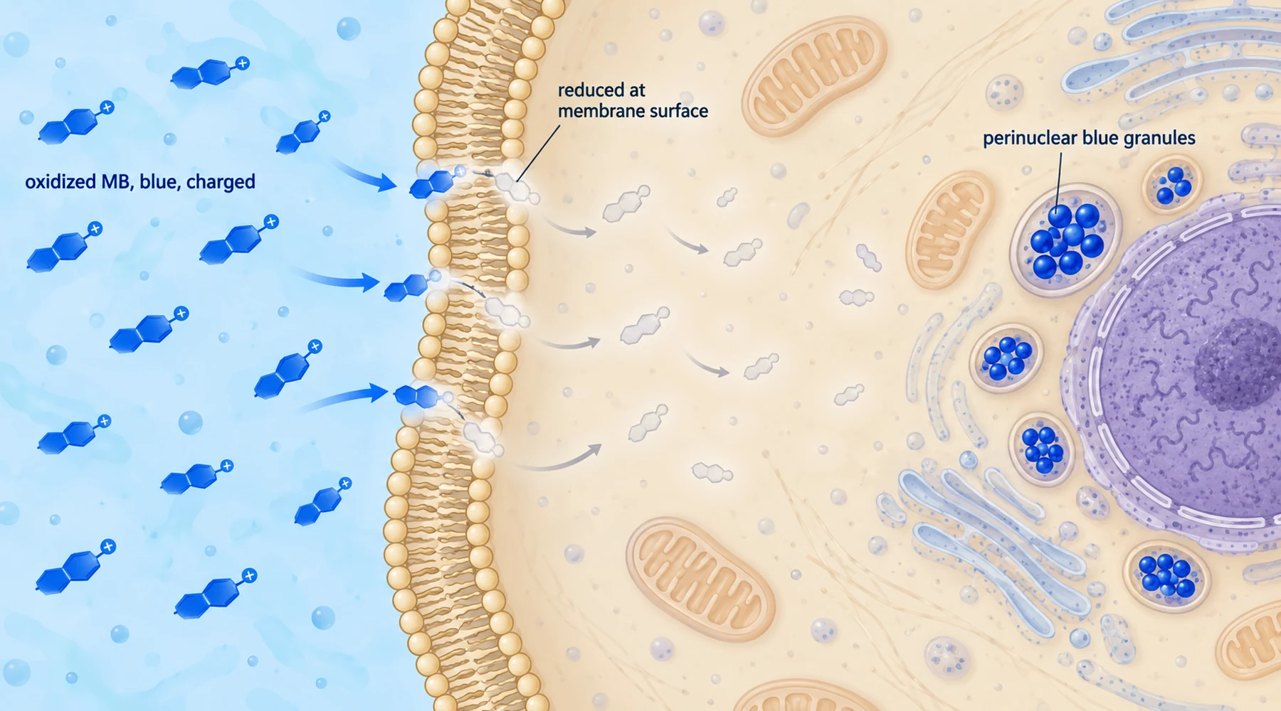
Watch a dish of endothelial cells take up methylene blue and the obvious reading is also the wrong one. The medium loses its blue color, the cells gain blue granules, so the dye must have walked through the membrane as the blue molecule. The experiments say otherwise. The blue form barely crosses. What crosses is the colorless form, and the blue you see inside is made inside, after arrival.
That inversion matters because it decides what staining can and cannot tell you. Blue granules report oxidized dye that was reoxidized and trapped in organelles. They do not report total dye in the cell, they do not report the reduced pool, and they do not measure biological effect. To see why, follow the three steps: reduction at the surface, diffusion of the reduced form, reoxidation and trapping inside.
The two forms behave like different molecules
Oxidized methylene blue is a flat, positively charged ring system. It dissolves in water, absorbs light strongly around 600 to 700 nanometers, and looks blue. The charge is the point. A charged, water loving molecule does not slip through a lipid membrane easily, so the careful phrase in the literature is relatively membrane impermeant, not impermeant.
Reduced methylene blue, called leucomethylene blue, is colorless, uncharged, and fat soluble. Remove the charge and the membrane stops being a wall. The reduced molecule diffuses across readily, and once inside a more oxidizing compartment it can lose electrons again and return to the blue, charged, trapped form. The redox chemistry of the dye is therefore also its transport mechanism. Change the electron count and you change the permeability.
The electrons for the first step come from the cell itself. Mammalian cells run electron transport across the plasma membrane, moving reducing equivalents from intracellular donors outward to acceptors at the surface. This trans plasma membrane activity is a normal cell function, found in most cell types, and the endothelial surface reductase that handles thiazine dyes behaves like others in one telling respect: it keeps working in the presence of cyanide and azide at doses that shut down the later steps. That separation is the hinge of the whole story.
Partitioning came first, and it almost explained uptake
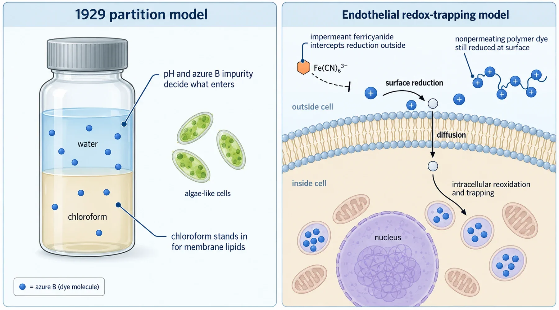
In 1929, Marian Irwin at the Rockefeller Institute asked a simpler question. Perhaps dye entry is just partitioning between water and the fatty layers of the cell, and a chloroform layer can stand in for those fatty layers on the bench. Her spectrophotometric penetration studies shook aqueous dye solutions with chloroform and compared what moved into the organic phase with what entered living algae cells.
Two findings from that work still deserve attention. First, commercial methylene blue solutions contained trimethyl thionine, now called azure B, and the impurity penetrated faster than methylene blue itself, especially at alkaline pH where the lipid soluble free base form dominates. Some apparent methylene blue entry was really selective entry of azure B. Second, pH set the outcome by setting the ionization state, and injured cells admitted far more dye than intact ones. Partition coefficients and charge state governed what the artificial system and the living cell each absorbed, and the resemblance between the two supported the idea of non aqueous layers in the cell boundary.
The model was prescient about lipids and ionization but it could not distinguish a dye that partitions as itself from a dye that is chemically converted at the surface, crosses in disguise, and is converted back inside. That distinction needed a different experiment, and it arrived seventy years later in lung endothelium.
Reduction outside, trapping inside
Merker and colleagues worked with cultured bovine pulmonary arterial endothelial cells and proposed a three step sequence: the oxidized dye is reduced on the cell surface to its colorless lipophilic form, the reduced form diffuses in, and intracellular reoxidation regenerates the membrane impermeant oxidized form, which accumulates. Their endothelial thiazine uptake study then tested the sequence by trying to break it in specific places.
The first tool was ferricyanide, an electron acceptor that cannot enter the cell. When surface enzymes reduce blue dye to its colorless form outside the cell, ferricyanide can steal those electrons back, regenerating blue dye in the medium before it ever crosses. As long as ferricyanide was present in excess (10 micromolar dye against 50 to 500 micromolar ferricyanide), dye uptake nearly stopped, and only after the ferricyanide was consumed did uptake resume at its normal rate. The kinetics told the same story in a second way. Ferricyanide disappeared at a roughly constant rate, consistent with a steady surface reduction flux feeding a recyclable mediator, while extracellular dye disappeared exponentially as its pool drained. A constant flux plus a draining pool means the ferricyanide signal reports continuous reduction, not dye molecules leaving solution.
The second tool removed the crossing step entirely. The group stitched toluidine blue O, a close thiazine relative, onto a large polyacrylamide chain too big to enter the cell in either oxidation state. Cells still reduced the tethered dye. Reduction therefore needs no intracellular access. The machinery is exposed at the surface or electrically coupled to it.
Then came the separation that gives the paper its title. Cyanide and azide at 2 millimolar strongly suppressed dye accumulation, and under the microscope the dark blue and purple inclusions that normally ring the nucleus nearly vanished. Yet neither inhibitor slowed extracellular reduction: ferricyanide cycling continued undiminished and the nonpermeating polymer was still reduced. The block sat downstream. Cyanide and azide were stopping intracellular reoxidation and sequestration, not surface reduction. Viability controls backed the reading, since the cells retained their cytosolic marker enzyme lactate dehydrogenase through these treatments, so the loss of uptake was not just poisoning or membrane rupture.
One caution from the paper still applies. The blue inclusions sat around but not in the nucleus, consistent with oxidation inside subcellular organelles, but the study did not identify which organelles. Similar patterns in other cell types had been called lysosomal on thin evidence. Treat the trapping as established and the address as provisional. Readers tracking where electrons go next can continue into mitochondrial electron transport and redox handling and the broader picture of absorption and distribution by route.
Staining is not exposure
A third study shows what happens when this uptake logic meets a biological readout. Scott and colleagues found that methyl beta cyclodextrin, a ring shaped sugar torus, binds methylene blue directly, with an association constant around 310 per molar for the inclusion complex against about 5846 per molar for dye dimerization. In L929 fibroblasts this binding blocked both cell staining and the stimulation of glucose uptake: preincubation with the cyclodextrin prevented blue staining, and adding it afterward accelerated destaining. As the optical blue signal left the cells, the activated glucose transport faded in tight parallel.
The authors read this correlation as favoring a direct interaction between the dye and a protein in the glucose transporter activation pathway over a long lived oxidative stress mechanism. That inference is reasonable but bounded. The experiment does not name the protein, it does not show binding to the transporter itself, and correlation between destaining and effect loss cannot exclude every redox mediated route.
The durable lesson is about measurement. Optical staining reports visible, oxidized, cell associated dye. It misses the colorless reduced pool entirely, so a pale cell can still hold dye equivalents, and an intensely stained cell reports trapped oxidized dye rather than total tissue exposure or effect size. The same caution scales up: comparing staining across cell types, or treating a dark tissue section as proof of higher exposure, repeats the original error of the dish. Uptake is a cycle of reduction, diffusion, and reoxidation, and any single snapshot of the blue form sees only one corner of it. The practical consequences for laboratory staining and handling and for interpreting dose and exposure claims follow directly.