
Article
Methylene blue and vision: what retinal research shows and where it stops

A reader finds a study in which methylene blue protected the retina, then asks the natural question: should it go in the eye? The research on methylene blue vision does not answer that question, because none of the key studies tested that use. One injected the dye into the vitreous of mice alongside a toxin. One added it to the medium bathing isolated cells in a dish. One put it in the drinking water of mutant mice. Each result belongs to its route and its model.
This article walks through three preclinical studies, two retinal cell types, and the boundary between laboratory protection and clinical eye treatment. The pattern is consistent: methylene blue can modify mitochondrial stress in retinal cells under controlled conditions, and that finding does not justify applying retail solutions to the eye.
Two cell types, two different diseases
The retina is layered, and the layer matters. Light strikes photoreceptors in the outer retina first. Their signals pass through intermediate neurons to retinal ganglion cells in the inner retina, whose axons leave through the optic nerve. The diagram below names the layers in order.
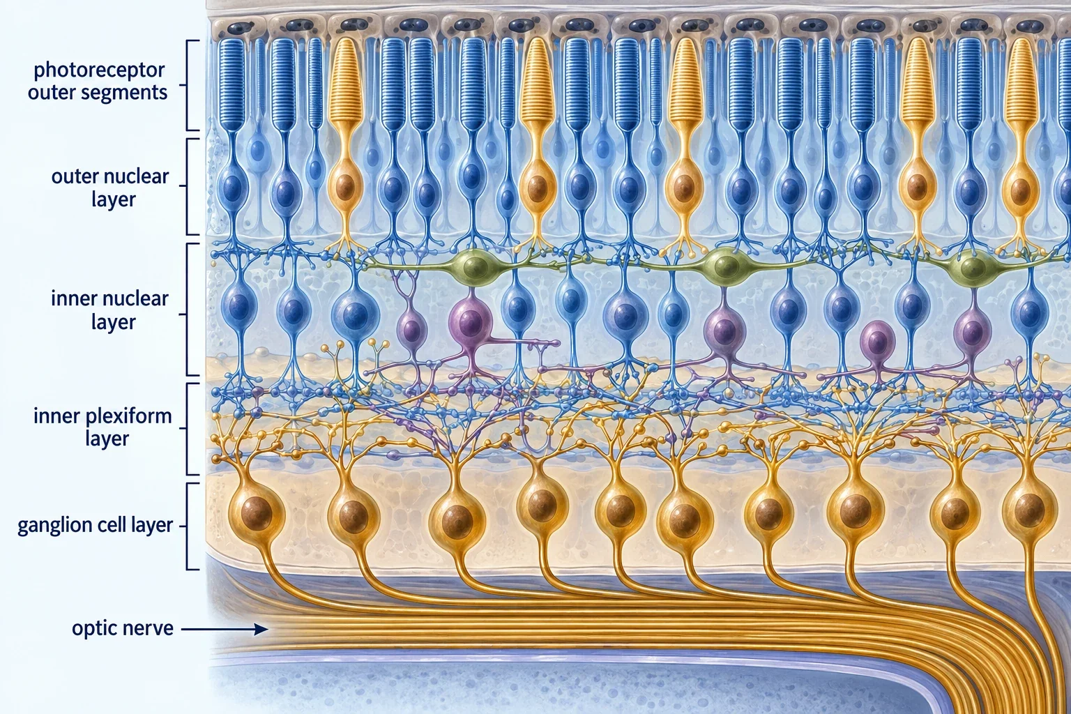
Retinal ganglion cells are the cells at stake in glaucoma and optic neuropathy. When searches mention methylene blue glaucoma, they usually mean this inner layer and its axons. Photoreceptors are the cells at stake in outer retinal degeneration. They carry the highest energy demand in the retina and run near their respiratory limit, which leaves them little reserve when mitochondrial complex I fails. A result in one cell type does not transfer to the other, and the three studies below keep them separate. The general skill of separating models from conclusions is explained in how to read methylene blue research.
Ganglion cells in the living mouse eye
In the earliest of the three studies, researchers modeled optic neuropathy by microinjecting rotenone into the vitreous of one mouse eye, using the other eye as a vehicle control. Rotenone blocks mitochondrial complex I, the entry point that passes electrons from NADH onward. Complex I dysfunction is also the feature that connects this model to Leber hereditary optic neuropathy, a disease of ganglion cells and the optic nerve.
Twenty-four hours later, the ganglion cell layer in rotenone treated eyes was thinner and held fewer cells than control eyes. When methylene blue was co-injected with rotenone, that damage was prevented in a dose dependent manner, with the highest tested dose (0.07 mg/kg, far below the mouse LD50) preserving both thickness and cell counts. Parallel tests in brain homogenates showed two effects: methylene blue reversed rotenone induced oxidative stress and reversed the drop in oxygen consumption. The authors proposed an excitotoxic mechanism, in which rotenone injures modulatory amacrine cells, ganglion cells overfire, and antioxidant action interrupts the resulting damage. The proposed chemistry behind that protection is detailed in mitochondrial mechanisms of methylene blue.
Three limits attach to this result. The dye was co-injected with the toxin directly into the eye, which is a delivery method, not a treatment schedule. The observation window was 24 hours. And the cytochrome oxidase measurements rose in both injured groups, which the authors read as overactivity rather than proof of restored respiration in the living retina.
Ganglion cells isolated in a dish
The second study removed the eye entirely. Investigators isolated primary retinal ganglion cells from young rats and exposed them to rotenone, the apoptosis inducer staurosporine, or 72 hours of low oxygen, then measured survival with a live/dead stain. Methylene blue at 1 and 10 micromolar in the culture medium improved survival against each insult, roughly 1.5 to 1.9 fold depending on the condition. A companion test showed that methylene blue preserved cytochrome c oxidase activity during a hydrogen peroxide challenge.
This is isolated cell pharmacology. The compound reached the cells by diffusion through culture medium at a defined concentration, with no cornea, tear film, lens, vitreous, blood supply, or immune system in the path. That simplicity is the point of the model: it shows that ganglion cells themselves can be protected when the compound reaches them. It says nothing about whether a drop on the corneal surface would deliver any defined concentration to the inner retina. Concentrations above 10 micromolar were avoided because of toxicity to the same cells, a reminder that dose response in a dish runs in both directions.
Photoreceptors in mutant mice and a cell line
The third study concerns the outer retina. Researchers used Harlequin mice, which carry an apoptosis inducing factor deficiency that leaves complex I incompletely assembled, and examined photoreceptor structure, outer segment markers, and mitochondrial integrity at 11 months of age. Methylene blue was given systemically in drinking water after weaning at about 10 mg/kg per day. Treated mutants kept more retinal thickness, more rhodopsin and opsin markers, and healthier mitochondrial structure than untreated mutants.
The companion cell work used 661W cells, an immortalized mouse photoreceptor-like line, exposed to rotenone with or without methylene blue for 24 hours. Survival improved at 30 micromolar, and lower concentrations normalized the NADH to NAD+ ratio and reduced oxidative stress. The striking detail is what methylene blue did not do: it did not restore ATP. Protection tracked redox relief and quieted stress pathways, including the mitochondrial unfolded protein response and mitophagy markers, rather than rescued energy supply.
That distinction matters for methylene blue retina claims. A compound can keep a stressed photoreceptor alive without fixing its energy deficit, which narrows any therapeutic story to redox stress rather than broad rejuvenation.
The three studies side by side
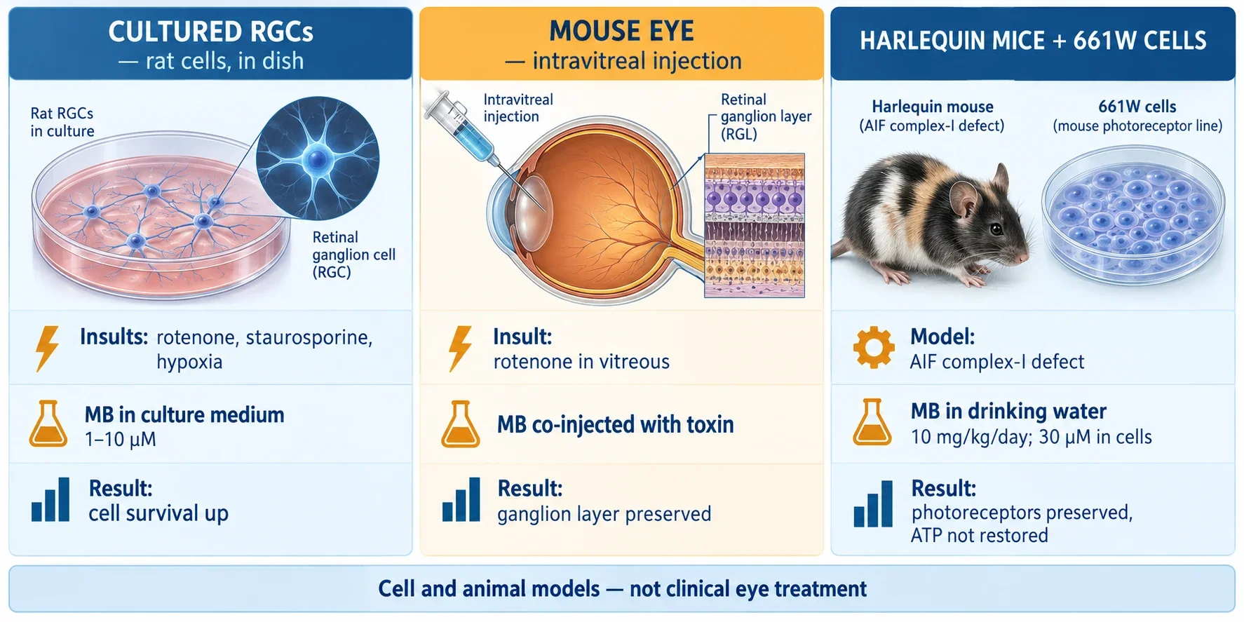
The comparison below labels each study by model and route, since those two columns determine what the result can support.
| Study | Model | Route of methylene blue | What was preserved |
|---|---|---|---|
| Zhang and colleagues, mouse optic neuropathy | Living mouse retina injured by rotenone | Co-injected into the vitreous with the toxin | Ganglion cell layer thickness and cell counts at 24 hours |
| Daudt and colleagues, primary rat ganglion cells | Isolated ganglion cells in culture | Dissolved in culture medium at 1 to 10 micromolar | Cell survival against rotenone, staurosporine, and hypoxia |
| Mekala and colleagues, Harlequin mice and 661W cells | Genetic complex I defect plus photoreceptor-like cells | Drinking water at 10 mg/kg per day; 30 micromolar in cell medium | Photoreceptor structure and mitochondrial integrity, without ATP restoration |
Every row is preclinical. No row enrolled patients, measured vision, or tested an ophthalmic formulation.
Why this is not a glaucoma treatment
Glaucoma care lowers intraocular pressure. Neuroprotection tries something different: keeping ganglion cells and their axons alive under stress. The studies above belong to the second project, not the first. None of them lowered pressure, and none compared methylene blue against pressure lowering treatment. That is why established approaches to glaucoma and pressure control remain a separate question from redox neuroprotection, and why a cell survival result cannot be read as vision saved.
Route differences reinforce the point. An intravitreal co-injection places a known amount beside the retina at the moment of injury. Drinking water dosing exposes the whole animal over months with unknown retinal concentration. Culture medium exposes bare cells directly. None of these predicts what fraction of a topical dose would cross the cornea, survive tear clearance, traverse the lens and vitreous, and arrive at either retinal layer at a helpful concentration. That delivery problem is part of the broader question of routes and distribution of methylene blue, where each route needs its own concentration measurements.
Tissue differences add a second barrier. A dye that reaches the front of the eye has not reached the back of it, and a concentration that helps one layer may injure another. Retail solutions are not manufactured, sterilized, buffered, or preserved as ophthalmic preparations, and purity, pH, osmolality, and excipients all matter for corneal and intraocular exposure. Nothing in these retinal findings validates putting such solutions into or onto the eye. Questions about preparation standards belong with safety and handling of methylene blue.
Where the evidence stops
Read together, the three papers support a narrow claim: under controlled conditions, methylene blue can relieve mitochondrial redox stress in retinal neurons and preserve their structure. The claim ends where the conditions end. Toxin and genetic complex I models are not glaucoma, drusen, or diabetic disease. Short windows and structural endpoints are not sustained vision. And a protective concentration in one compartment is not a dose for another.
A stronger case would need separate pieces: defined retinal exposure after a clinically usable route, functional vision endpoints in animals, pressure independent benefit in a glaucoma relevant model, and then human safety and efficacy data. Until those exist, methylene blue and vision remains a research hypothesis with clear contours: plausible mitochondrial pharmacology in the retina, demonstrated only in cells and animals, with no path from these studies to treating eyes.