
Article
Methylene blue and skeletal aging: why cell findings did not predict mouse bone outcomes
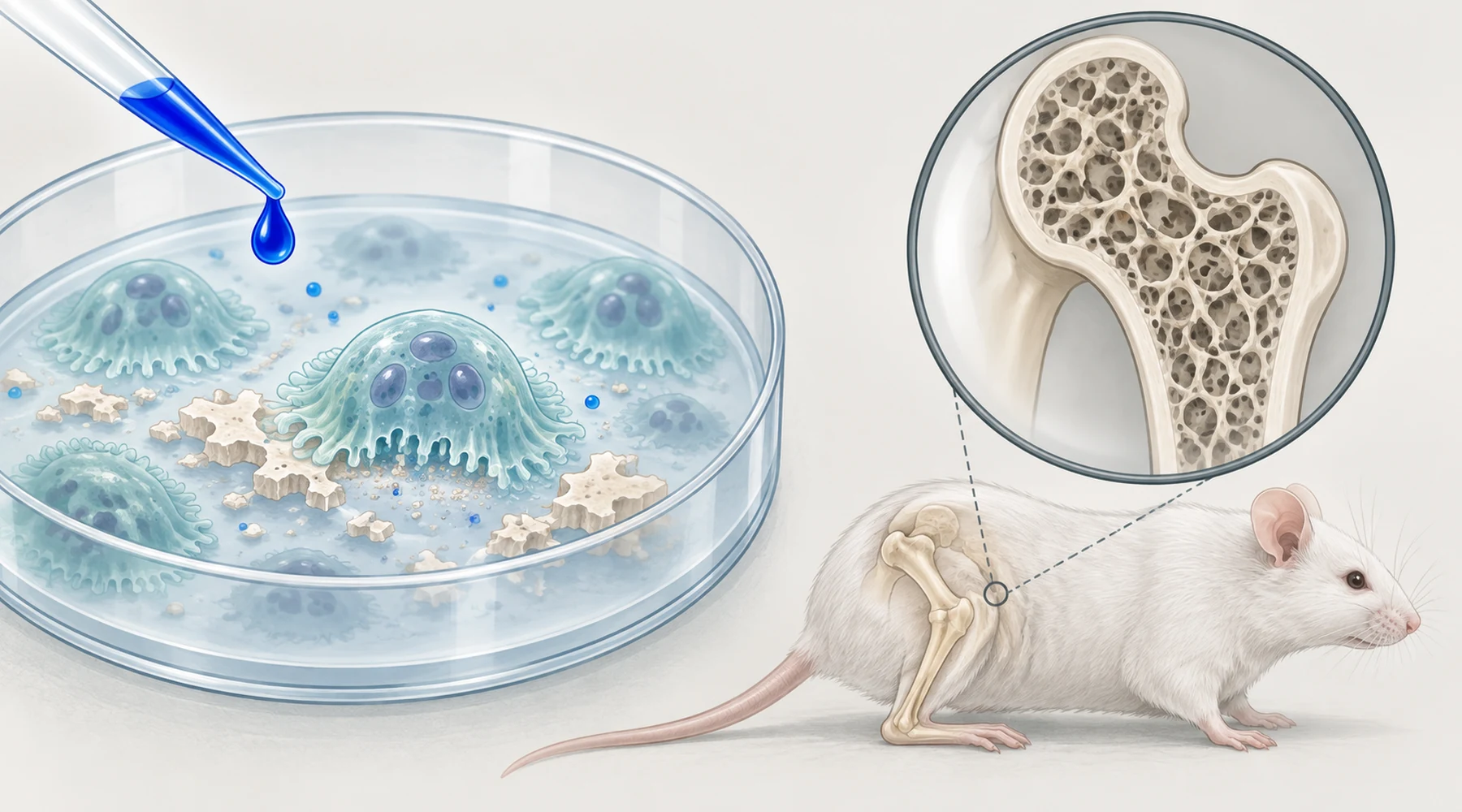
Imagine a compound that quiets the very cells that eat bone, tested in a dish and working exactly as hoped. Fewer multinucleated osteoclasts, a clean dose response, a plausible mitochondrial story behind it. Then the same compound is given to mice for most of their adult lives, their skeletons are scanned in three dimensions, and nothing is different. The aged bones look aged, treated or not.
That is the honest shape of one 2024 study, and it is worth understanding precisely, because it sits at the fault line where most supplement claims about bones break. The paper is Poudel and colleagues, published in Aging in March 2024, and it is open access, so every measurement below can be checked against the source. Its headline is a null result. Null results are not failures. They are answers, and this one answers a narrow question well: does long-term systemic antioxidant treatment with methylene blue or mitoquinone preserve the aging mouse skeleton? No.
What bone aging looks like in numbers
Bone loss with age is not one thing, so the study did not measure one thing. Using micro-computed tomography, the investigators scanned three skeletal sites: cortical bone at the femur mid-diaphysis (the dense shaft), trabecular bone at the distal femur metaphysis (the spongy network near the joint), and trabecular bone in the fifth lumbar vertebra. At each site they recorded the standard microarchitecture parameters. BV/TV is the fraction of a volume that is bone rather than marrow space. BMD is mineral density. Tb.Th, Tb.N, and Tb.Sp describe trabecular thickness, number, and spacing. Cortical thickness and cross-sectional areas describe the shaft.
Think of trabecular bone as scaffolding inside a warehouse. Aging removes some struts entirely and thins the rest, so the fraction of the space filled by structure falls, the remaining struts sit farther apart, and the whole frame carries less load. Cortical bone is the warehouse wall itself, getting thinner. A treatment that protects the skeleton should show up in these numbers. That is the test the study applied.
What happened in the dish
The cell work used marrow taken from the femurs and tibias of 6 to 7 month old UM-HET3 mice, a genetically diverse strain bred for aging research. Four findings came out, and they point in different directions, so each needs its own sentence.
First, mesenchymal stem cells, the pool that includes future bone-forming cells, stayed viable across methylene blue and mitoquinone concentrations from 0.125 to 0.5 micromolar over 48 hours. No toxicity, no boost. Second, when those stem cells were pushed to become osteoblasts in osteogenic medium and stained for alkaline phosphatase on day 18, neither compound changed the outcome at any tested concentration. The bone-building side of the dish did not respond.
Third, the bone-resorbing side did. Marrow precursors stimulated with M-CSF and RANKL, the standard recipe for making osteoclasts, formed fewer TRAP-positive multinucleated cells as the concentration rose, with the clear reduction appearing above 0.25 micromolar, in practice at the top tested concentration of 0.5. This is the finding that launches a thousand optimistic summaries, and it is real within its boundaries: direct, continuous exposure in culture suppressed osteoclast formation in a dose-dependent way.
Fourth, mature osteoblasts exposed for 48 hours and run through a mitochondrial stress assay showed no significant change in basal or maximal oxygen consumption with either compound. At 0.5 micromolar both appeared to lower respiration slightly, but the change did not reach significance. So the dish offered no mitochondrial rescue in bone-forming cells to set alongside the anti-osteoclast effect. The proposed mechanism, improved energetics through the mitochondrial electron transport chain, did not produce a measurable respiration gain where the investigators looked for it.
What happened in the mice
Two animal experiments followed, at very different scales.
The smaller one used inbred C57BL/6J females, the workhorse strain of biomedical research. Starting at 18 months of age, roughly late middle age for a mouse, half drank water containing 250 micromolar methylene blue for 6 or 12 months, until 24 or 30 months. Untreated 18-month females supplied the baseline, and 5-month females showed what young bone looked like. Aging did exactly what aging does. In the L5 vertebra, bone volume fraction fell about 35 percent, mineral density about 18 percent, and trabecular number roughly halved, regardless of treatment. Cortical thickness fell around 10 percent between 18 and 24 months and around 30 percent by 30 months. Methylene blue did not bend any of these curves.
The larger experiment ran through the National Institute on Aging Interventions Testing Program, which tests candidate compounds in UM-HET3 mice of both sexes across three sites. Methylene blue at 28 parts per million in food, and mitoquinone at 100 parts per million, each started in early adulthood and continued to about 22 to 24 months of age. The same three skeletal sites went through micro-CT. The published table reports every parameter by sex and treatment with adjusted statistics, and the treatment column reads the same throughout: no significant effect, no treatment-by-sex interaction, anywhere. Cortical or trabecular, femur or spine, male or female, methylene blue or mitoquinone. The skeleton aged on schedule.
The matched table both halves share
This is the asset the whole article rests on. Each row pairs what the dish showed with what the animal showed for the same biological process, using the study's own measurements.
| Process | Cell finding (0.125 to 0.5 µM, direct exposure) | Animal finding (months of systemic dosing) |
|---|---|---|
| Progenitor viability | No change in MSC viability at any tested concentration | Not directly tested in vivo; no downstream bone preservation that would imply a progenitor effect |
| Bone formation (osteoblasts) | No change in alkaline phosphatase positive colonies at day 18 | No preservation of BV/TV, BMD, trabecular number, or cortical thickness at any site |
| Bone resorption (osteoclasts) | Dose-dependent suppression of TRAP-positive multinucleated cells, clear above 0.25 µM | No resulting preservation of trabecular spacing, number, or volume fraction in femur or L5 |
| Osteoblast energetics | No significant change in basal or maximal oxygen consumption | Consistent with the null: no functional gain reached the tissue level |
| Whole-skeleton outcome | N/A (dishes have no skeletons) | Age-related loss proceeded identically: L5 BV/TV down ~35%, BMD down ~18%, Tb.N roughly halved; cortical thickness down ~10% by 24 mo and ~30% by 30 mo in C57BL/6J; no treatment effect in UM-HET3 of either sex |
Read row by row, the table dissolves the paradox. Only one of four cell processes responded, and it responded only near the top of the tested range under continuous direct exposure. Everything else in the dish was already null. The animal result then looks less like a contradiction and more like confirmation: three nulls plus one conditional effect in culture became an across-the-board null in the animal.
Bone structure is not lifespan
One confusion deserves its own section, because methylene blue does have a lifespan record and it is easy to misread it as a bone record. In the earlier Interventions Testing Program cohort, methylene blue at the same dietary dose raised the 90th percentile lifespan of UM-HET3 females by about 6 percent, a late-life survival effect with no change in median lifespan and no effect in males. Mitoquinone showed no lifespan effect in either sex. Those survival numbers come from the program's published longevity records, not from the bone paper.
The bone paper's contribution is to keep these ledgers separate. A compound can shift late-life survival in one sex without preserving any measured feature of the skeleton. Survival counts deaths from all causes; micro-CT counts struts in a vertebra. There is no rule that an intervention must act through bone to extend life, and this study is direct evidence that methylene blue's late-life signal in females traveled by some other route. Anyone who cites the lifespan finding as support for methylene blue and osteoporosis is spending currency from the wrong account. The general discipline of keeping these accounts apart is covered in how to read methylene blue research.
Why the dish did not predict the mouse
Five explanations fit the data, and they are not mutually exclusive.
Exposure is the first. Cells in culture were bathed continuously at known concentrations. Mice ate dosed food for months, and the study measured neither drug levels in marrow nor any marker of oxidative damage in bone. A concentration that suppresses osteoclasts when applied directly may never arrive at precursors in the marrow at that level. The paper tested one dose per compound in vivo, so dose response in the animal remains unknown.
Chemistry is the second. Reactive oxygen species do not form one well-mixed pool inside a cell. They act in small compartments on short timescales, and mitochondrial, cytosolic, and membrane redox circuits behave partly independently. Mitoquinone concentrates in mitochondria by design, while the oxidant signals that drive osteoclast formation can originate elsewhere. Lowering one pool need not reproduce what continuous bathing of the whole cell achieves.
Coupling is the third. In living bone, resorption and formation are linked: osteoclasts and their neighbors emit signals that recruit bone-forming cells to the site just resorbed. Suppressing osteoclast formation does not add an equal amount of bone, because it can also remove the signals that call in the builders. A dish containing only precursors cannot show this feedback. A skeleton cannot hide it.
Loading is the fourth. Osteocytes sense mechanical strain and set the remodeling balance for the whole bone. As animals age and move less, the mechanical signal fades, and loss follows regardless of modest pharmacological pressure on one cell type. The authors themselves name exercise and anabolic loading as the likelier lever, which is a gracious way of saying the skeleton listens more to physics than to redox chemistry.
Timing is the fifth, with a caveat. In the C57BL/6J experiment treatment began at 18 months, when substantial structural loss had already occurred, and no anti-resorptive can rebuild trabeculae that have been perforated and removed without a matching formation stimulus. But timing cannot carry the whole explanation, because the Interventions Testing Program dosing began in early adulthood and still changed nothing. Late start explains part of one experiment. It explains none of the other.
What this means for methylene blue osteoporosis claims
For a reader evaluating methylene blue for bone health or skeletal aging, the practical reading is short. The best available long-term mouse evidence reports no skeletal benefit from chronic methylene blue or mitoquinone, despite a genuine anti-osteoclast signal in culture. The lifespan literature gives methylene blue a modest late-life effect in females only, which this study shows did not pass through bone. Neither ledger supports human use for osteoporosis, and no human bone data enter the picture at all. Questions of dosing and product safety in people belong to methylene blue safety and side effects, not to this mouse skeleton.
The deeper lesson is about evidence order. A cell result is a claim about what a compound can do to one cell type under controlled exposure. A bone result is a claim about what months of systemic exposure do to an organ that couples two cell types under mechanical control. The first is where hypotheses are born. The second is where, for skeletal aging and these two antioxidants, this one died.