
Article
Methylene blue and multiple sclerosis: what the animal research tested
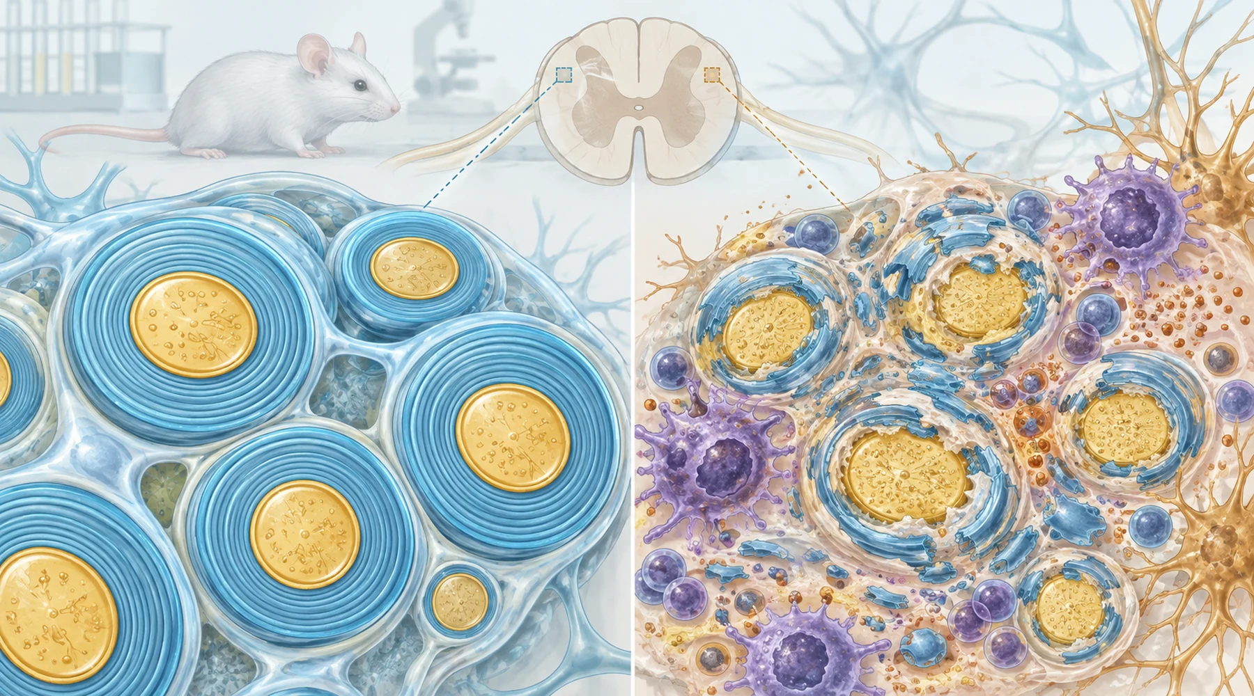
Search for methylene blue for MS and you find papers that look like they answer the same question. They do not. One paper gave methylene blue to sick mice as a treatment. The other never treated any animal with it. It used methylene blue as a blue stain on thin slices of spinal cord so a pathologist could see myelin under a microscope.
This is the concept behind the whole article. The dye has two roles in biology and they share nothing except the bottle. As a drug candidate it is dosed into a living animal and the question is whether disease changes. As a stain it is applied to dead fixed tissue after the experiment and the question is what the tissue looked like. If you miss that distinction you will read a pathology methods section as if it were a treatment result.
What EAE is, in plain terms
Experimental autoimmune encephalomyelitis, usually shortened to EAE, is the standard animal stand in for aspects of multiple sclerosis. A researcher immunizes a susceptible mouse with a fragment of a myelin protein plus a strong adjuvant. The immune system then attacks the animal's own central nervous system. The mouse develops ascending weakness that is easy to score on a 0 to 5 scale, from floppy tail to hind limb paralysis.
That scoring convenience explains why EAE is popular and also why it misleads. Human MS arises on its own, involves a wider and less defined set of targets, and produces optic, sensory, cognitive, cerebellar, and motor symptoms in relapsing or progressive courses. EAE reproduces selected mechanisms such as inflammation, demyelination, and axon injury under controlled conditions. It does not reproduce MS in miniature. Readers who want the general framework can review how methylene blue is thought to act at the cellular level before judging any single disease claim.
The two papers below also used different EAE antigens, which matters. The treatment study induced disease with MOG35-55, a short peptide from myelin oligodendrocyte glycoprotein. The pathology study induced disease with MP4, a fusion of myelin basic protein and proteolipid protein segments. Strain and antigen choices change where lesions appear and how inflammation behaves, so the two studies are not two runs of one experiment.
The map: model to question to limits
Keep this table open. It is the original reference for this article. Each row states which clinical question the model can address, what was actually measured, and where translation stops.
| Treatment study (MOG EAE) | Pathology study (MP4 EAE) | |
|---|---|---|
| Paper | MOG peptide EAE treatment study | MP4 longitudinal pathology study |
| Role of methylene blue | Intervention given to living mice | Stain applied to fixed 1 micrometer spinal cord sections |
| Clinical question it can address | Does dosing change EAE severity and immune signaling | How do demyelination and axon injury evolve over six months |
| Endpoints | Daily clinical scores, spinal cord infiltrates, demyelination, axon injury, AMPK and SIRT1 signaling, NF-kB activity, Th17 versus Treg balance | Lesion area, inflammation and edema grades, demyelinated fiber density, g-ratio by electron microscopy, axolysis, mitochondrial swelling, neurofilament spacing |
| Translation limit | One strain, one sex, short 30 day course, association not proof of mechanism | No treatment tested, cross sectional tissue sampling, lesions sampled mainly in lumbar cord |
Anyone checking the next headline can reuse the same columns with help from how to read preclinical studies: ask the species and antigen, the role of the compound, the endpoints, and whether the result is a disease course or a tissue snapshot.
The treatment result: what dosing changed
The 2016 treatment study in the Journal of Neuroimmunology used female C57BL/6 mice, about 8 to 10 weeks old, immunized with 250 micrograms of MOG35-55 per mouse. After induction the EAE animals were randomized to a methylene blue group or an untreated EAE group, and clinical scores were followed through about day 30.
Clinical scores were significantly lower in treated mice than in EAE controls from day 15 to day 30, at P less than 0.01. Spinal cord analysis pointed the same way, with less inflammatory infiltration, less demyelination, and less axonal loss in treated animals.
The distinctive part was the signaling panel. Treatment increased AMPK activation and SIRT1 expression and suppressed NF-kB activation, a central inflammatory switch. On the adaptive immune side it reduced the Th17 response and strengthened the Treg response. The proposed chain is therefore methylene blue to AMPK and SIRT1 to lower NF-kB signaling to a shift from inflammatory Th17 toward regulatory Treg to milder tissue injury to lower scores.
That chain is an interpretation, not a proven causal sequence. The study shows the drug, the signal changes, and the disease improvement moving together. It does not prove that AMPK and SIRT1 carry the full benefit, and the publicly indexed text does not expose the exact milligram per kilogram dose, route, start day, or duration with enough clarity to reproduce the regimen from the abstract alone. Treat any dosing number quoted about this paper on a sales page as unverified unless it cites the full text methods. The broader human evidence base for methylene blue is the place to check whether this early immune signal ever matured into clinical data.
The pathology result: what six months of tissue showed
The 2015 MP4 study in PLoS ONE used female C57BL/6 mice immunized with 200 micrograms of MP4 in complete Freund adjuvant plus pertussis toxin on the day of immunization and again 48 hours later. No animal received methylene blue as a drug. After fixation and embedding, one micrometer transverse spinal cord sections were stained with methylene blue for light microscopy, with electron microscopy of the ventrolateral tract for finer detail.
Tissue was sampled at three stages: acute peak with 7 mice, three months after onset with 5 mice, and six months after onset with 10 mice. Mean clinical scores at those stages looked almost identical, near 1.7 to 1.8 on the 0 to 5 scale. The tissue told a different story. Isolated myelin damage rose from about 569,000 fibers per square millimeter at acute peak to about 1,496,000 at six months, with the acute to long term comparison significant at P equal to 0.027. Combined myelin plus axonal damage roughly doubled by six months. Remyelinating fibers stayed below one percent at every stage, which is a sobering detail for repair hopes.
Axon injury followed a U shaped pattern, high at acute peak, lower at three months, highest at six months, and it correlated with mitochondrial swelling and with reduced spacing between neurofilaments. Assessment was blinded to score and stage, which strengthens the reading that clinical scores underreported ongoing structural damage.
That mismatch is the study's real contribution to an MS reader. A stable disability score did not mean stable tissue. It also shows why this paper cannot support any methylene blue and MS treatment claim. It tested time, not treatment.
Why neither study establishes benefit in patients
Think of each paper as answering a narrow question well and the patient question not at all.
First, cause differs. EAE is triggered on purpose with a defined antigen and adjuvant. Human MS develops spontaneously with a wider immune repertoire that includes T cells, B cells, innate immunity, and resident glia. A compound that shifts a MOG biased response in a mouse need not shift that wider human response.
Second, phenotype differs. Ascending tail to hind limb weakness in a mouse is a convenient readout. It compresses the heterogeneity of human MS into one motor number. Drugs routinely move short term EAE scores without changing human courses, which is why EAE is a screening and mechanism tool rather than a miniature clinic.
Third, exposure and timing are unresolved. Continuous early dosing in a 30 day mouse experiment is not chronic oral use in a person with established disease, and neither study measured drug levels in the nervous system. Without that link there is no bridge from a concentration on a plate or a dose in a mouse to a human regimen.
Fourth, the two papers do not replicate each other. Different antigens, different questions, different endpoints, and in one case no treatment at all. Citing them together as two positive MS treatment studies is a category error.
What this means for readers weighing the evidence
There is no established methylene blue protocol for MS in these papers. The treatment study supports further research on AMPK, SIRT1, NF-kB, and Th17 to Treg balance in MOG EAE. The pathology study is a reminder that tissue injury can progress while scores look flat, and that methylene blue in a methods section sometimes means a stain jar rather than a medicine cabinet. Readers following ongoing multiple sclerosis research should keep that stain versus drug distinction in mind. Safety questions, including blood and interaction risks, belong in a clinician conversation alongside general methylene blue safety data before anyone acts on laboratory headlines.
The bottom line fits in one sentence. One cited study dosed methylene blue into MOG EAE mice and reported milder disease with an anti inflammatory immune shift, while the other cited study dosed nothing and used methylene blue only to stain spinal cord while documenting six months of accumulating myelin and axon damage, so the pair does not establish that methylene blue helps human MS.