
Article
Methylene blue and ferroptosis: iron, lipid peroxidation, and cell death
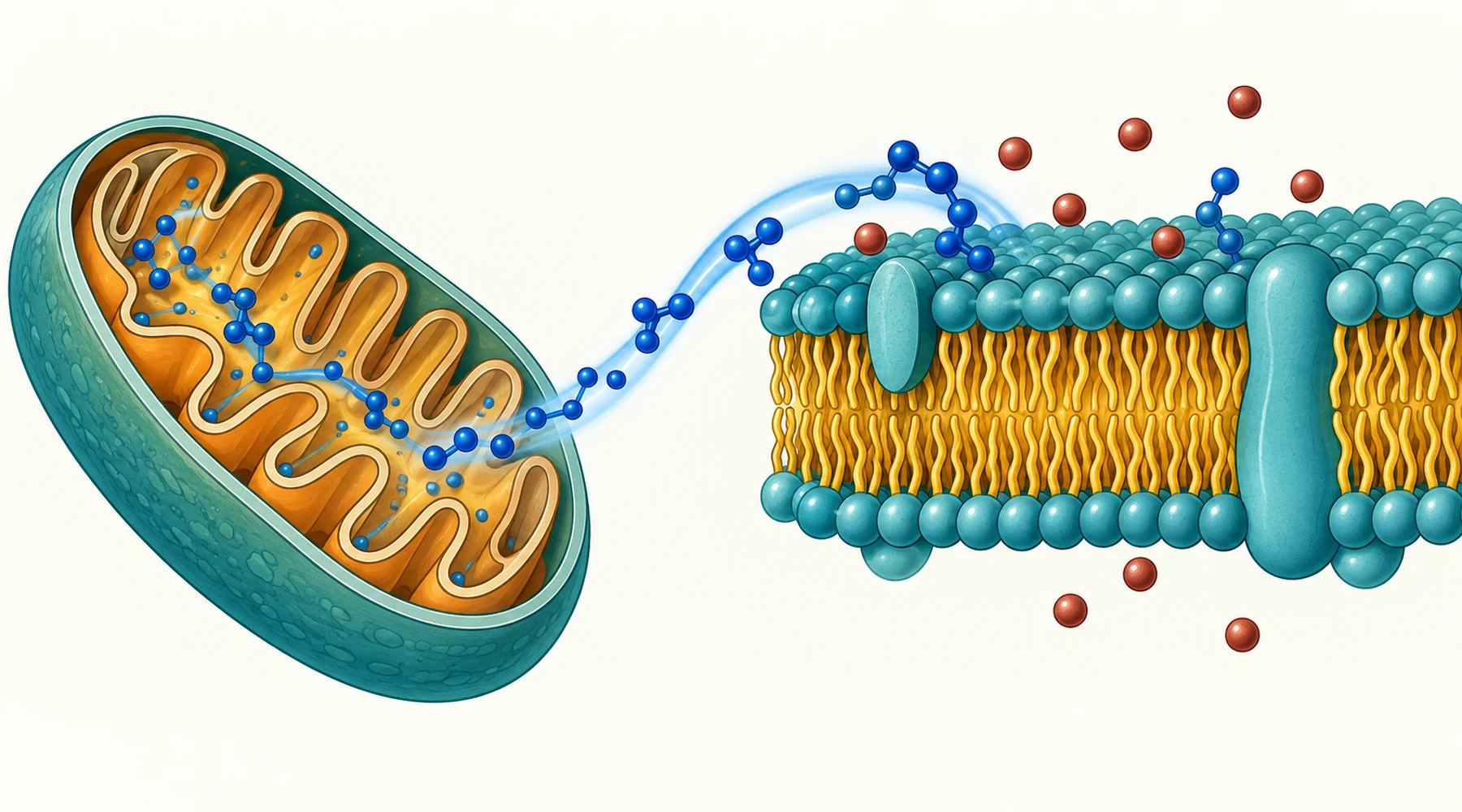
Ferroptosis is cell death by membrane damage. Polyunsaturated fats in membranes oxidize in a chain reaction, the membrane fails, and the cell dies. Iron is required for the chemistry. That sentence is the whole concept, and everything in this article is a test of one part of it.
The question here is narrower: what does methylene blue do to that chain in cell experiments, and what does that result actually show. The short answer is that in two laboratory studies, nanomolar methylene blue reduced mitochondrial lipid peroxidation and kept cells alive after triggers that otherwise cause ferroptosis. The longer answer matters more, because each link in the chain was tested with a different intervention, and each intervention has limits.
Ferroptosis in one paragraph
A membrane phospholipid that carries a polyunsaturated fatty acid tail has a weak spot: hydrogen atoms that are easy to steal. Once a radical steals one, oxygen adds to the lipid, a lipid peroxyl radical forms, and that radical steals from the next lipid. The reaction propagates along the membrane unless something stops it.
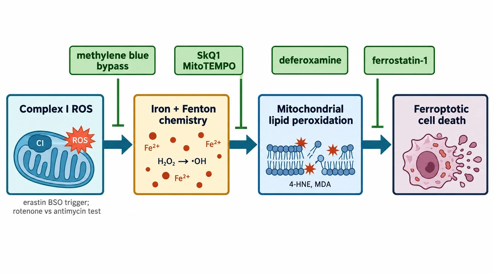
The main stop signal is an enzyme called GPX4, which uses glutathione to reduce lipid peroxides to harmless lipid alcohols. Laboratory triggers attack this defense at defined points. Erastin blocks cystine import through system xc-minus, which starves the cell of cysteine for glutathione synthesis. Buthionine sulfoximine blocks glutathione synthesis directly. RSL3 inhibits GPX4 itself. All three leave peroxides undeleted, but they do it at different steps, so a result with one trigger does not automatically generalize to the others.
Ferroptosis differs from apoptosis in the properties that define it. There is no caspase cascade and no apoptotic bodies. Iron chelators such as deferoxamine and radical-trapping agents such as ferrostatin-1 suppress it, which is the operational test researchers use to call a death ferroptotic rather than merely necrotic. The broader landscape of regulated cell death pathways puts this distinction in context, and how cells handle labile iron explains why chelation is such a decisive control.
Where mitochondria fit, and why that was disputed
Early work reported that cells without mitochondrial DNA still undergo ferroptosis, which suggested mitochondria are optional. Later work found that depleting mitochondria, or blocking respiration, can suppress ferroptosis from cysteine starvation but not from direct GPX4 inhibition. Both results can be true if the mitochondrial contribution depends on the trigger, the cell type, and where the decisive peroxidation happens.
The study that anchors this article approached the dispute with a compartment-specific probe. MitoCLox is a mitochondria-targeted version of a lipid peroxidation reporter: it shifts fluorescence when it meets lipid peroxyl radicals inside mitochondria. In fibroblasts treated with erastin, mitochondrial probe oxidation appeared in still-living cells before death, and it was heterogeneous across the population. That timing matters. Peroxidation that precedes death is a candidate cause. Peroxidation measured only in dead cells is debris. A companion guide to lipid peroxidation assays and what each probe reports covers why probe location decides what a signal means.
The same pattern appeared in a second model. Fibroblasts from a patient with Leber hereditary optic neuropathy, which carry a complex I mutation and higher baseline oxidative stress, died by ferroptosis when glutathione was depleted, while control fibroblasts resisted. Mitochondrial probe oxidation again came first. Details are in the fibroblast study of mitochondrial lipid peroxidation in ferroptosis.
What methylene blue changed in these experiments
Methylene blue is a redox carrier. Its oxidized form accepts electrons from NADH-linked chemistry at complex I and its reduced form donates them to cytochrome c, which feeds complex IV. That creates a partial bypass around the segment of the chain where electrons normally leak to oxygen as superoxide. Readers who want the full relay picture should start with how methylene blue moves electrons through mitochondria.
In erastin-treated lung fibroblasts, methylene blue at 250 nanomolar preserved viability in a cell survival readout, alongside the mitochondria-targeted antioxidants SkQ1 and MitoTEMPO. The controls define the claim. Ferrostatin-1, alpha-tocopherol, and deferoxamine also protected, which confirms the death was ferroptotic. The SkQ1 analog without an antioxidant group did not protect, which points to redox chemistry rather than nonspecific accumulation. Protection faded above roughly 200 nanomolar SkQ1, consistent with the known pro-oxidant behavior of quinone antioxidants at higher load. Methylene blue has the same two-faced dose behavior, protective at low nanomolar levels in these dishes and capable of generating hydrogen peroxide by redox cycling when pushed higher or lit strongly. Dose context of this kind is covered in reactive oxygen species in mitochondria.
The isolated-mitochondria experiments then separated the ROS sources. With ferrous iron present, substrates feeding complex I plus rotenone stimulated probe oxidation. Rotenone blocks downstream of the flavin site, so upstream carriers stay reduced and leak electrons to oxygen from the matrix-facing flavin. Substrates feeding complex III plus antimycin A produced abundant hydrogen peroxide yet did not drive lipid peroxidation. Same bulk ROS signal, different location, different outcome. SkQ1 and methylene blue each suppressed the complex I driven peroxidation without suppressing hydrogen peroxide production itself, which places their action after ROS formation, at the lipid peroxidation step.
The iron overload arm and the lipofuscin clue
A second study asked the question from the iron side. H9c2 cardiomyocytes exposed to submillimolar ferric ammonium citrate developed mitochondrial ROS, mitochondrial lipid peroxidation, then ferroptosis, followed by accumulation of a lipofuscin-like pigment within 24 hours. Lipofuscin is the autofluorescent, iron-containing residue of oxidized lipids and proteins that builds up with age, and its rapid appearance here connects acute iron-driven peroxidation to a classic aging marker. Nanomolar methylene blue and SkQ1 each prevented the peroxidation, the death, and the pigment accumulation. Isolated cardiac mitochondria confirmed the pattern: rotenone plus ferrous iron drove peroxidation and pigment-like signal, and both agents blocked it. The full sequence is reported in the iron overload study in H9c2 cardiomyocytes. The pigment side of the story belongs with lipofuscin and cellular aging markers.
What cell survival assays do not show
A viability readout such as CellTiterBlue reports how many cells in a dish remain metabolically active after a trigger plus a candidate protector. That is a test of a mechanism in a defined system. It is not a treatment outcome. None of these experiments dosed a patient, measured a disease endpoint, or established a therapeutic window in a body.
Four limits deserve explicit statement. First, the cell types are narrow: transformed lung fibroblasts, primary skin fibroblasts including one mitochondrial disease line, and a rat cardiomyocyte line, plus isolated rat heart mitochondria. Sensitivity varied by line, and the LHON line was unusually vulnerable by design. Second, the triggers are laboratory extremes: full transport blockade or synthesis blockade, and iron loads no tissue would meet acutely. Third, concentration does not translate by multiplication. Nanomolar in culture medium is not a human dose, and methylene blue distribution, reduction to its colorless form in blood and tissue, light exposure, and membrane potential all sit between a dish and a person. Fourth, the probes report their own compartment. MitoCLox oxidation shows mitochondrial lipid radicals formed; it does not show that plasma membrane rupture, the likely terminal event, was prevented directly rather than avoided upstream.
So the honest claim is this: in these models, complex I derived ROS plus iron drive mitochondrial lipid peroxidation that precedes ferroptosis, and methylene blue at low concentrations interrupts that segment. Anything beyond that segment needs different experiments in animals and then in people.