
Article
Measuring reactive oxygen species in methylene blue experiments
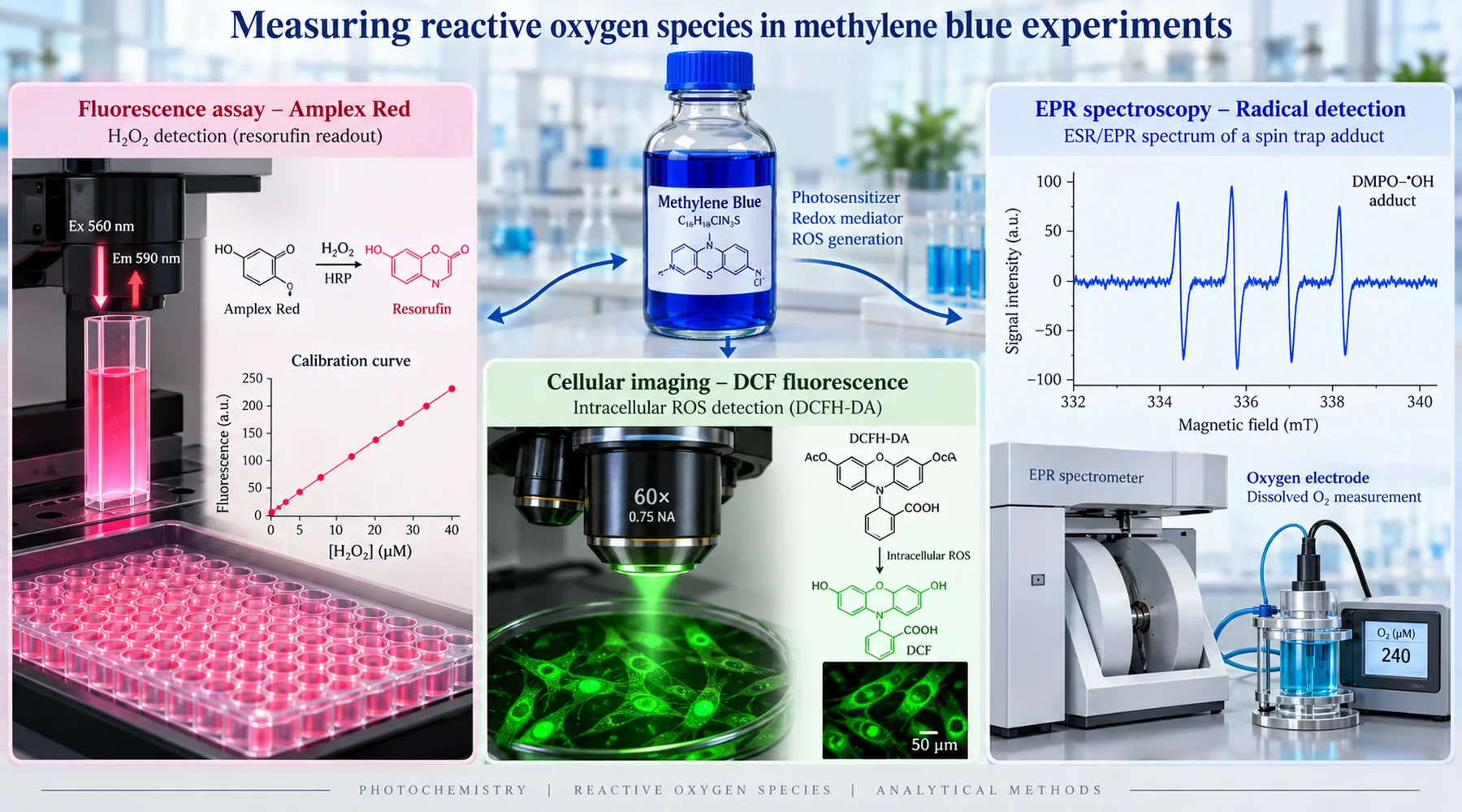
Two laboratories run what each calls a methylene blue antioxidant experiment. One reports that the dye suppresses reactive oxygen species. The other reports that it generates them. Both show clean dose response curves. Both are sincere. The difference is not fraud or incompetence. It is that the two groups measured different things under different light, with probes that answer different questions, and then described both answers with the same word: ROS.
Methylene blue is a difficult molecule to measure precisely because it participates in the chemistry it is supposed to observe. In the dark it shuttles electrons between donors and acceptors and returns to its starting form, ready to cycle again. Under light it becomes a photosensitiser, reaching an excited triplet state from which it can hand energy to oxygen to make singlet oxygen or transfer electrons to make radicals and hydrogen peroxide. Any probe placed into that system can be oxidised, reduced, bleached, or optically masked by the dye itself. So the first question of any ROS readout is never what the number is. It is what the assay actually detects, and what the dye does to the assay.
Amplex Red: a calibrated hydrogen peroxide assay
The methylene blue Amplex Red combination is the most defensible pairing in this literature, when it is set up correctly. The chemistry is specific: horseradish peroxidase uses hydrogen peroxide to oxidise the Amplex substrate to a fluorescent product, resorufin, in roughly one to one stoichiometry. Peroxide from the sample becomes fluorescence in the detector. Nothing else in the canonical ROS family drives that reaction directly.
The decisive advantage is calibration. A careful worker runs known peroxide standards in the same buffer, with the same peroxidase and substrate concentrations, the same incubation time, and the same optical settings, then reads the samples against that curve. In the isolated brain mitochondria study that anchors this method, the authors did exactly that, adding known quantities of peroxide at the end of each run, and the numbers are reported as rates in picomoles per minute per milligram of protein rather than arbitrary fluorescence units. That is what turns a glow into a measurement.
Three details from that study deserve imitation. First, the authors verified the signal with catalase, which destroys peroxide. The methylene blue induced fluorescence rise stopped when catalase was added, confirming the signal was peroxide rather than an optical artefact. Second, they tested illumination directly by shuttering the excitation beam so samples received one eighth of the normal light dose, and the signal fell by only nine percent. More than ninety percent of the peroxide they reported was independent of the measurement light. Third, they measured peroxide elimination separately with an electrode and found the dye both raised production and slowed clearance. A probe that only reports net fluorescence would have conflated those two effects.
The assay still has failure modes, and methylene blue finds several of them. The dye absorbs broadly across the same red region where resorufin emits, so at high dye concentrations the sample can mask its own signal through inner filter effects. Standards should therefore contain the same dye concentration as the samples. Amplex Red can also photooxidise on its own, and its product resorufin can enter photoredox cycling that generates superoxide and peroxide, which matters greatly when the experiment also illuminates a photosensitiser. The practical fix is to irradiate the methylene blue samples first and assay aliquots with Amplex reagents in the dark, rather than lighting dye and probe together. Finally, superoxide dismutase does not simply suppress an Amplex signal: converting superoxide to peroxide can raise it. An SOD control that increases fluorescence has not failed. It has identified superoxide as an upstream species.
DCFDA: a nonspecific oxidation indicator
The methylene blue DCFDA pairing is the source of most of the confusion. DCFH-DA is a cell loading precursor. Esterases inside the cell strip its acetate groups to leave nonfluorescent DCFH, and oxidation of DCFH yields fluorescent DCF. What oxidises it is a long list: hydroxyl radicals, peroxidase and heme intermediates, nitrogen dioxide, hypochlorous acid, and peroxynitrite derived radicals. Hydrogen peroxide alone does not oxidise it directly at any useful rate; peroxide dependent fluorescence generally needs iron, heme proteins, or peroxidase chemistry in the loop.
The critique that every methods section should cite comes from the analysis of DCFH as a probe by Kalyanaraman and colleagues, as summarised in the brain mitochondria study that rejected DCFDA for this reason, who showed that the dye reacts slowly, with uncharacterised stoichiometry, without valid calibration, and with non ROS compounds. The probe also amplifies what it measures: one electron oxidation of DCFH produces a DCF radical that reacts with oxygen to make superoxide, which dismutates to additional peroxide. The readout partly manufactures the chemistry it reports.
None of this makes DCFDA useless. It makes it a screen, not an identification. A rise in DCF fluorescence means oxidant dependent probe oxidation increased. It does not mean peroxide increased, and it certainly does not mean total ROS increased, a quantity no single probe measures. Earlier methylene blue work that relied on DCF oxidation above one micromolar reported prooxidant effects, while other systems in the same era reported protection against peroxide induced cell death. Both observations can be true of the same dye in different redox contexts, and the DCF result alone cannot say which oxidant carried the signal. Where the earlier brain mitochondria work matters is that it replaced this screen with a calibrated peroxide measurement and found the burden directly: even one hundred nanomolar dye raised peroxide release several fold across every substrate and respiratory state tested.
DCFDA carries one more methylene blue specific trap. The dye is intensely coloured and redox active, so it can bleach fluorescence, quench emission, or oxidise the probe chemistry independently of cellular ROS. A cell free control with dye plus probe and no cells is not optional decoration. It is the experiment that tells you whether the dye talks to the probe.
Spin trapping and oxygen uptake: identity and flux
Where the fluorescent probes report that something oxidisable appeared, electron spin resonance with spin traps reports something closer to what it was. The classic methylene blue photochemistry study used ESR to detect the ascorbate radical from dye, ascorbate and light, then showed peroxide formation in oxygen uptake experiments and iron catalysed hydroxyl radical formation in spin trapping runs. Each technique answered the question the others could not: ESR gave radical identity, oxygen uptake gave reaction flux independent of any fluorescent probe, and the combination showed both singlet oxygen and radical pathways operating in the same illuminated system.
That 1984 result still reads as a warning label. With illumination and a reducing agent present, the dye generated peroxide, and with iron present the system produced the hydroxyl radical, the most reactive species in the set. The authors connected this to ascorbate in the eye and questioned casual ophthalmic use. Modern readers should note the conditions: light plus reductant plus iron. Remove any one of those and the pathway weakens or vanishes.
Oxygen electrodes earn their place differently. A Clark type electrode measures net oxygen consumption, which establishes that dye driven redox chemistry is consuming molecular oxygen and gives kinetics no fluorescence artefact can fake. It does not identify the product. Peroxide, superoxide, water, and singlet oxygen decay all consume oxygen. Flux without identity is as incomplete as identity without flux, which is why the strong papers pair the electrode with a calibrated peroxide assay or a trap.
Why antioxidant results conflict
Put the three readouts side by side and the apparent contradictions in the methylene blue literature resolve into method differences plus three biological facts.
First, the dye is both an electron shuttle and a peroxide source. By diverting electrons away from the respiratory sites where oxygen would otherwise become superoxide, it can lower one ROS while its reduced form reduces oxygen directly to peroxide, raising another. A laboratory facing mitochondrial redox chemistry and its bypass role should therefore expect probe dependent answers: a superoxide sensitive readout falls while a peroxide readout rises in the same cuvette.
Second, peroxide at low doses is a signal, not just damage. The mouse brain mitochondria study that followed the earlier work found peroxide generation from as little as fifty nanomolar dye without mitochondrial DNA lesions at therapeutic levels, with protection against rotenone induced lesions after pretreatment. Only at one hundred micromolar, far above the therapeutic range, did the dye damage selected DNA fragments, and red light plus twenty micromolar dye produced singlet oxygen that damaged all fragments tested. The proposed mechanism is hormetic: mild peroxide production triggers Nrf2 linked antioxidant defences and mitochondrial biogenesis, so a short term prooxidant reading precedes a longer term antioxidant outcome. An assay read at one hour and an assay read at one day can disagree because the biology changed between them.
Third, light decides which molecule is even present. Illumination demethylates the dye toward Azure B and related products, and the microscope lamp over a dish is enough to photosensitise it. Dark and illuminated experiments are different experiments, and the controls for illumination and dye handling section of any protocol should treat them that way.
A minimum control set, and how to read any claim
The comparison below is the original reference asset for this article. Before accepting any methylene blue ROS result, check which row the authors filled and which they skipped.
| Control | What it rules out | How to run it |
|---|---|---|
| Dark versus defined illumination | Confounds ground state redox effects with photochemistry | Same dye, same matrix, one arm in darkness, one under measured light dose |
| Probe plus light, no dye | Probe photochemistry | Illuminate the probe alone; any signal is artefact baseline |
| Dye plus light, no cells or substrate | Cellular versus dye intrinsic ROS | Cell free matrix; establishes what the dye generates by itself |
| Catalase | Non peroxide signal in an Amplex run | Signal should fall; note illuminated dye can damage catalase itself |
| SOD | Superoxide participation | A rise in Amplex signal implicates superoxide upstream, not assay failure |
| Iron chelator such as deferoxamine | Fenton type hydroxyl chemistry | Tests whether redox active iron carries the DCFH or trap signal |
| Peroxide standards in full matrix | Uncalibrated fluorescence | Known peroxide additions with identical dye concentration and optics |
A practical reading rule follows. Amplex with peroxidase, calibrated, with a catalase check, is evidence about peroxide. DCFDA is evidence about nonspecific probe oxidation and nothing more. Spin trapping with scavenger controls is evidence about radical identity. Oxygen uptake is evidence about flux. A sentence that claims more than its assay can support, however confidently written, is a sentence about the probe, not about the dye. Related discussions of dose dependence and the hormetic range, mitochondrial electron flow, assay interference in stained samples, and microscopy illumination artefacts extend each of these points, and each remains valid once its placeholder reference is replaced.