
Article
When methylene blue interferes with the experiment used to measure it
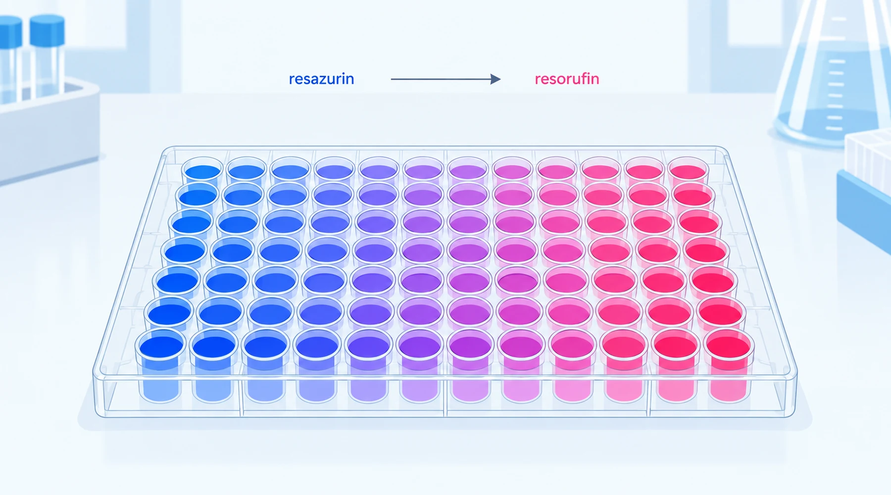
One laboratory rears zebrafish embryos in methylene blue at the standard husbandry concentration, then measures metabolism two ways. On day one, a Seahorse analyzer reports oxygen consumption going up. On days four and five, an Alamar Blue plate reports energy expenditure going down. Same dye, same fish line, opposite verdicts. Then comes the quiet control: wells containing methylene blue plus Alamar Blue reagent and no fish at all also read dimmer than reagent alone. Part of the result was biology. Part of it was the dye grading its own exam. That split, documented in a 2025 Communications Biology study of methylene blue in developing zebrafish, is the normal shape of methylene blue assay interference, and it is why redox-active dyes demand controls that ordinary test compounds do not.
What a resazurin assay actually reports
Alamar Blue is a brand name for a resazurin solution, so methylene blue Alamar Blue interference and methylene blue resazurin assay interference describe the same collision. Oxidized resazurin is blue and barely fluorescent. Metabolically active cells reduce it to resorufin, which is pink and strongly fluorescent near excitation 560 nm and emission 590 nm, with usable ranges around 530 to 570 nm excitation and 580 to 610 nm emission per the manufacturer microplate protocol. Push reduction further and resorufin converts to dihydroresorufin, which is weakly fluorescent, so overloading the well with cells or time bends the calibration curve downward at the top end.
The electrons come mostly from NADH and NADPH through cellular reductases and diaphorases in cytosol, microsomes, and mitochondria. That makes the readout an integrated reducing capacity, not a viability count and not a mitochondrial measurement. A higher signal can mean more cells, more reducing flux per cell, or both. A compound that reroutes NADH changes the signal without killing a single cell. Keep that sentence nearby, because methylene blue reroutes NADH for a living.
Two interference pathways, one lowered signal
Methylene blue bends a resazurin readout through two independent pathways, and either one alone lowers apparent fluorescence.
Pathway 1 is optical. Methylene blue absorbs strongly in the red, with its monomer band near 660 to 665 nm and a dimer shoulder near 607 to 610 nm whose tail reaches back toward the resorufin emission around 590 nm. Dye in the light path absorbs some excitation light and reabsorbs some emitted fluorescence, an inner-filter effect that dims the well even when resorufin production is unchanged. The spectrum also shifts with the dye state: a factor analysis of methylene blue spectra in water and cyclodextrin solutions resolves monomer, dimer, and host-guest complex as three species with distinct extinction coefficients from 500 to 700 nm, so the size of the optical error moves with concentration, aggregation, and binding. Experimentally, distinguish plain optical attenuation from molecular quenching with a spike recovery test described below, since the fix differs.
Pathway 2 is redox competition. Oxidized methylene blue accepts electrons from NAD(P)H to form colorless leuco methylene blue, which molecular oxygen re-oxidizes back to the blue form while producing reduced oxygen products including hydrogen peroxide. Each turn of that cycle consumes reducing equivalents that would otherwise reduce resazurin, and it consumes oxygen while generating peroxide on the side. A lowered Alamar Blue signal can therefore mean electrons traveled through the methylene blue cycle instead of into resorufin, which reads as depressed metabolism on the plate regardless of what the cells did. The same chemistry explains a mirror-image trap in oxygen instruments: reduced dye re-oxidized by oxygen drives chemical oxygen consumption with no biology present, so dye blanks belong in respirometry too, as the comparison of oxygen, ATP, and membrane potential readouts lays out for the mitochondrial case.
Altered signal and altered biology can both be true
The zebrafish study is valuable precisely because it refuses the easy either-or. The biology was real: basal oxygen consumption rose in 1-day embryos at both husbandry concentrations, ATP-linked respiration rose at the lower one, membrane potential measured with the JC-10 probe rose at day two, and mitochondrial complex transcripts fell at days one and four. The artifact was also real: fish washed three times before the Alamar Blue step still carried visible blue stain in the head, mouth, and olfactory region, and cell-free wells with dye plus reagent lost a small but significant amount of fluorescence on their own. The organismal Alamar Blue decreases at days four and five were larger than the cell-free effect, so the authors read the outcome as genuine metabolic change viewed through a slightly warped lens. That is the correct mental model. A redox cycler that is also a blue absorber will usually perturb the pathway under study and the reporter chemistry at the same time, and the job of the controls is to size each contribution, not to declare one of them imaginary. A related lesson comes from the cyclodextrin binding work: when a host molecule sequestered the dye, staining and glucose-uptake activation faded together, which argued the metabolic effect needed direct dye interaction rather than diffuse redox noise. Correlating a physical dye readout with a functional readout is exactly the move that separates mechanism from artifact, and the guide to reading dye-based metabolic claims develops that habit further.
The controls that separate them
Run these before interpreting any methylene blue result from a resazurin plate. Concentrations, medium, incubation time, and illumination must match the cellular experiment exactly, because every pathway above depends on them.
| # | Control | What it detects | Pass criterion | If it fails |
|---|---|---|---|---|
| 1 | Medium plus dye, no reagent, no cells | Dye background at assay wavelengths | Background small versus cellular signal | Subtract per-concentration blanks, never one global blank |
| 2 | Medium plus reagent, no dye, no cells | Spontaneous reagent reduction | Low stable baseline over the incubation | Shorten incubation, change medium, or accept a higher floor |
| 3 | Medium plus dye plus reagent, no cells | Direct dye-reagent chemistry | Same as control 2 within noise | Report the offset, do not attribute it to cells |
| 4 | Resorufin spike into vehicle versus dye wells | Optical attenuation or quenching | Equal recovery in both matrices | Quantify the loss per dye dose and correct, or switch readout |
| 5 | Washed versus unwashed cell-free carryover | Residual dye after your wash steps | Washing removes the cell-free effect | Internalized dye remains, so controls 3 and 4 still apply |
Controls 3 and 4 are the pair most papers skip and most methylene blue papers need. Control 3 catches chemistry, control 4 catches optics, and together they bound the artifact before any biological claim is made.
Orthogonal readouts, each with its own fine print
No single second assay certifies the first. Each orthogonal readout escapes one pathway and stays exposed to another, so pick the combination whose blind spots do not overlap. The assay-selection matrix for redox-active compounds walks through the pairing logic, and the membrane-potential probe guide covers the fluorescence caveats in detail.
Seahorse oxygen consumption follows electrons to oxygen instead of into resorufin, which sidesteps the reporter chemistry entirely, but the dye chemically consumes oxygen too, so mitochondria-free dye blanks in complete medium are mandatory. ATP luciferase assays use bioluminescence rather than resorufin fluorescence and so dodge both the optical overlap and the reductase competition, at the cost of reporting the ATP pool balance rather than synthesis flux. Membrane-potential probes such as TMRM or JC-10 answer whether the charge gradient holds, yet they fluoresce in the same visible range the dye absorbs in, so residual blue staining complicates them as well and they need dye-only blanks at each dye dose. Transcript measurements of respiratory complex subunits avoid optics altogether and integrate over hours, which complements the minutes-scale plate readouts.
The minimum credible package for a methylene blue metabolic claim is therefore: a resazurin result with controls 1 through 4, plus one non-resazurin endpoint from a different physical principle, plus a stated position on whether the change reflects cell number, reducing flux per cell, or both. Anything less leaves the reader unable to tell a quieter metabolism from a dimmer well.
Interference-control worksheet
Copy this worksheet into the lab notebook for each experiment that combines the dye with a redox reporter. It is the original asset for this article: fill one row per dye concentration.
A. Signal pathways. For each concentration, record: expected monomer versus dimer regime (low micromolar versus tens of micromolar and above), absorbance of the final well contents at 590 nm in a cell-free well, and resorufin spike recovery percentage. If recovery falls below about 90 percent, treat all fluorescence values at that dose as optically discounted, not biologically exact.
B. Chemistry check. Record cell-free dye-plus-reagent fluorescence as a percentage of reagent-only baseline at each read time. If the offset grows with time, the dye is reacting with the reporter system during the assay, and endpoint-only reads will mislead. Consider kinetic reads or a different reporter.
C. Biology cross-check. Name the orthogonal readout chosen for this run, its own dye blank, and the predicted outcome if the resazurin result is genuine. A genuine metabolic depression should move an independent endpoint in a consistent direction. If the orthogonal endpoint disagrees, trust neither result yet and redesign rather than averaging the two.
D. Verdict sentence. Write one sentence of the form: at stated dose and stage, resazurin signal changed by stated amount, of which stated amount is cell-free artifact, and the orthogonal endpoint (agrees or disagrees). If any entry is blank, the experiment is not finished.
Methylene blue remains a useful metabolic probe, and the zebrafish work shows genuine mitochondrial effects at doses many facilities pour down the drain daily. The dye simply insists on being measured with instruments it cannot touch. Give it dye-only wells, a spike recovery, and one readout built on different physics, and the interference stops being a trap and starts being a quantity with a number attached.