
Article
Can methylene blue bypass mitochondrial Complex III? Why studies disagree
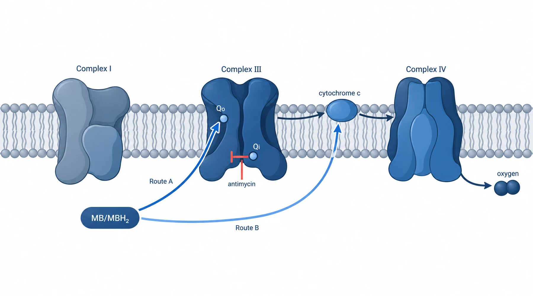
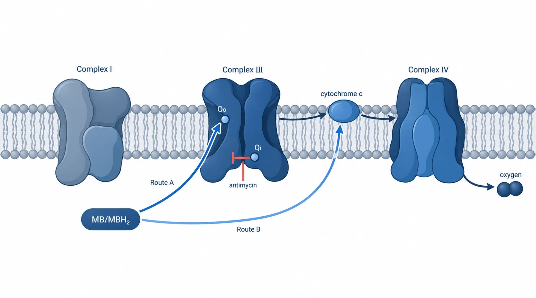
Two laboratories asked whether methylene blue can bypass a blocked mitochondrial Complex III. One answered no. The other answered yes. Both had data.
The first study, in mouse brain mitochondria, found the dye cannot. The second, in brain mitochondria from three rodent species, found it partly can. A third study, in mouse liver, is often cited nearby but tested something else. Keep it in its place and half the confusion clears.
There is no universal mechanism to announce. There are three experiments with different blockers, doses, and readouts. Read the conditions first. The conclusions follow.
What bypass means here
Electrons enter the chain through Complex I (from NADH) or Complex II (from succinate), pass through Complex III to cytochrome c, then through Complex IV to oxygen. The gradient built along the way is what the cell spends to make ATP.
A bypass claim says methylene blue picks up electrons upstream of a block and drops them off downstream. The oxidised dye takes electrons from NADH or early chain components, becoming leucomethylene blue, and the reduced form hands them to cytochrome c or back into the chain. If the handoff lands downstream of the block, respiration and membrane potential partly recover. If it lands at or upstream of the block, nothing recovers.
One detail of Complex III decides much of this dispute. The complex runs a Q cycle with two inhibitor sites. Antimycin binds the Qi site. Myxothiazol binds the Qo site. A study using only one of them has tested only half the complex.
Study one: no bypass in mouse brain mitochondria
Gureev and colleagues used cortex mitochondria from C57BL/6 mice, digitonin isolated, assayed at 0.1 mg protein per ml. Substrates spanned three regimes: pyruvate plus malate at 5 mM each, succinate at 10 mM with glutamate at 2 mM, and L-proline at 10 mM. Blockers were rotenone at 1 micromolar (Complex I) and antimycin at 1 microgram per ml (Complex III). Dye doses were 0.5, 1, and 2 micromolar, with 1 micromolar as the main concentration. Methods and traces are in the FEBS Letters communication.
Complex I bypass worked. Rotenone cut ADP stimulated respiration from about 121 to about 2 nmol per minute per mg, and 1 micromolar dye restored it to about 21. Membrane potential recovered on pyruvate plus malate and on L-proline.
Complex III bypass did not. Antimycin cut respiration to about 16, and dye moved it only to about 20. Membrane potential did not recover on any substrate, at dye doses up to 5 micromolar. Peroxide data pointed the same way: dye alone raised peroxide emission from a basal rate near 76 pmol per minute per mg to roughly 1170, 1460, and 1945 at the three doses, yet antimycin cut that dye driven signal by about 25, 88, and 96 percent respectively. If reduced dye reacted directly with oxygen, blocking Complex III should not suppress the signal. The authors inferred the reduced dye is oxidised at the Qo site of Complex III, upstream of the antimycin block. Short loop, in words: donors to dye, dye to Qo, normal route onward. Rotenone is bypassed. Antimycin is not.
Study two: partial bypass in three species, at both inhibitor sites
Svab and colleagues widened the design. Brain mitochondria from mice, rats, and guinea pigs, Percoll isolated, assayed at the same 0.1 mg per ml, on pyruvate plus malate or glutamate plus malate at 5 mM each. They used two Complex III blockers: antimycin A at 0.3 micromolar and myxothiazol at 1 micromolar. Dye was 2 micromolar in most assays, 0.5 in the cytochrome c work. All three species with matched protocols are in the open access paper.
Both blocks were partly bypassed, in all three species. In mouse mitochondria, antimycin suppressed respiration and dye raised it about 60 percent, from roughly 37 to 59 nmol per minute per mg. Myxothiazol suppressed it 88 percent, to about 4.5, and dye raised it fivefold to about 23. Membrane potential partly repolarised under either inhibitor, read with two dyes (safranin O and ratiometric TMRM) to exclude optical artefacts.
This paper also tested the handoff directly, which the first study never did. Exogenous acetylated cytochrome c at 25 micromolar was not reduced by energised mitochondria with or without inhibitors, but adding dye drove reduction in both cases. In cell free buffer, NADH plus dye reduced cytochrome c at a rate proportional to NADH. Longer jump, in words: reducing equivalents to dye, dye directly to cytochrome c, Complex IV to oxygen. That route skips Complex III entirely, so both blockers are bypassable.
Two extra readouts support that reading. NAD(P)H fluorescence fell when dye was added to myxothiazol treated mitochondria, meaning reducing equivalents drained through the new route. And dye absorbance at 660 nm tracked each addition in turn while spun supernatants showed oxidised dye enriching inside energised mitochondria over a hundredfold, then leaving when potential collapsed. The proton economics fit too: skipping the pumping steps leaves roughly 2 protons per electron pair instead of 10, so faster oxygen use with only partial repolarisation is what a real bypass looks like, not evidence against one.
The same preparation produced responses that punish casual reading. With inhibitor plus dye, ADP lowered respiration instead of raising it, and transport blockade stimulated it instead of slowing it, while membrane potential still moved in the normal directions. Oxygen consumption alone misleads in this system.
Study three: the liver study that tested Complexes I and II, not III
Lee and Boelsterli used mouse liver mitochondria and cultured hepatocytes injured by efavirenz (Complex I, IC50 near 30 micromolar) and hydrazine, the isoniazid metabolite (Complex II, IC50 near 30 micromolar). Dye at about 30 micromolar restored NADH oxidation, membrane potential, and ATP while lowering oxidants and necrotic death. The Redox Biology paper models it as dye carrying electrons around the injured Complexes I and II.
That result is settled upstream context, and it says nothing about Complex III because nothing in it was blocked there. It cannot confirm or refute either brain study.
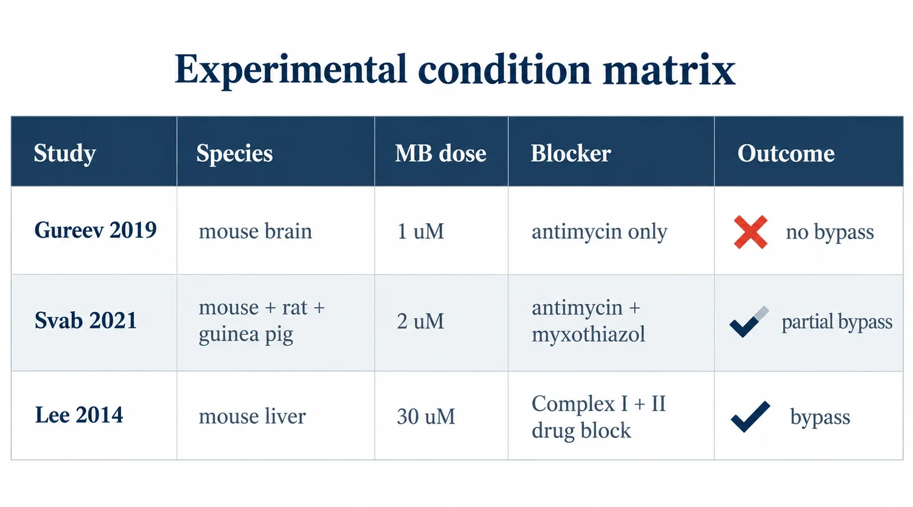
The condition matrix
| Condition | Gureev 2019 | Svab 2021 | Lee 2014 |
|---|---|---|---|
| Species and tissue | Mouse brain cortex | Mouse, rat, guinea pig brain | Mouse liver, mitochondria and cells |
| Dye dose | 0.5 to 2 micromolar, main 1 | 2 micromolar, 0.5 in cyt c assays | About 30 micromolar |
| Complex III blocker | Antimycin 1 ug per ml, Qi only | Antimycin 0.3 uM plus myxothiazol 1 uM, Qi and Qo | None |
| Substrates | PM, succinate glutamate, proline | PM or GM | NADH linked, I and II injury |
| Key endpoints | Respiration, potential, peroxide | Respiration, potential by two dyes, NAD(P)H, dye redox, cyt c reduction | Energetics, potential, ATP, oxidants, death |
| Outcome | Complex I bypass yes, III no | Partial bypass under either III blocker | Bypass around I and II injury |
Four reasons both Complex III studies can be right about their own setup
1. The blocker was not the same intervention. Antimycin alone at 1 microgram per ml against antimycin at 0.3 micromolar plus a separate Qo site blocker. A dye route feeding the Qo site fails exactly where a direct to cytochrome c route succeeds. One site cannot stand in for both.
2. The dye dose and clock differed. Main concentrations of 1 against 2 micromolar, in a redox cycler whose uptake and direct cytochrome c reduction are concentration and time dependent. Small dose gaps plus different readout timing can expose different legs of one network.
3. The preparations differed. Digitonin isolation against Percoll gradient isolation selects overlapping but not identical mitochondrial populations. Protein concentration matched at 0.1 mg per ml, so that variable drops out, but subpopulation and integrity do not. And the species suspect was cleared: the second group reproduced its phenotype in mouse too.
4. Substrate state sets what the dye can do. Three substrate regimes against NADH linked pairs. NADH supply and membrane potential jointly set how fast the dye is reduced and where it partitions, and a route needing reduced dye beside oxidised cytochrome c is hostage to that choice.
How to read the next bypass claim
Ask which complex was blocked, with which inhibitor, at which site and dose. Ask the dye concentration and when the readout was taken. Ask the tissue, species, isolation, protein concentration, and substrate. And ask whether potential, respiration, redox state, and a direct handoff assay agree, because oxygen consumption alone pointed the wrong way more than once here.
The narrow summary holds. Dye bypass around Complex I is agreed. Around Complex III, mouse mitochondria with antimycin at 1 microgram per ml and dye at 1 micromolar show no functional bypass, while three species with antimycin at 0.3 micromolar or myxothiazol at 1 micromolar and dye at 2 micromolar show partial bypass with direct dye to cytochrome c transfer. Those sentences coexist. Anything broader is extrapolation.
Related reading keeps the disputed claim inside undisputed chemistry: how methylene blue moves electrons for the relay, the Complex I bypass experiments for the settled upstream case, membrane potential readouts and their artefacts for why two dyes were needed, and laboratory handling of redox cyclers for the dosing and timing traps.