
Article
Methylene blue for brain injury: understanding the preclinical evidence
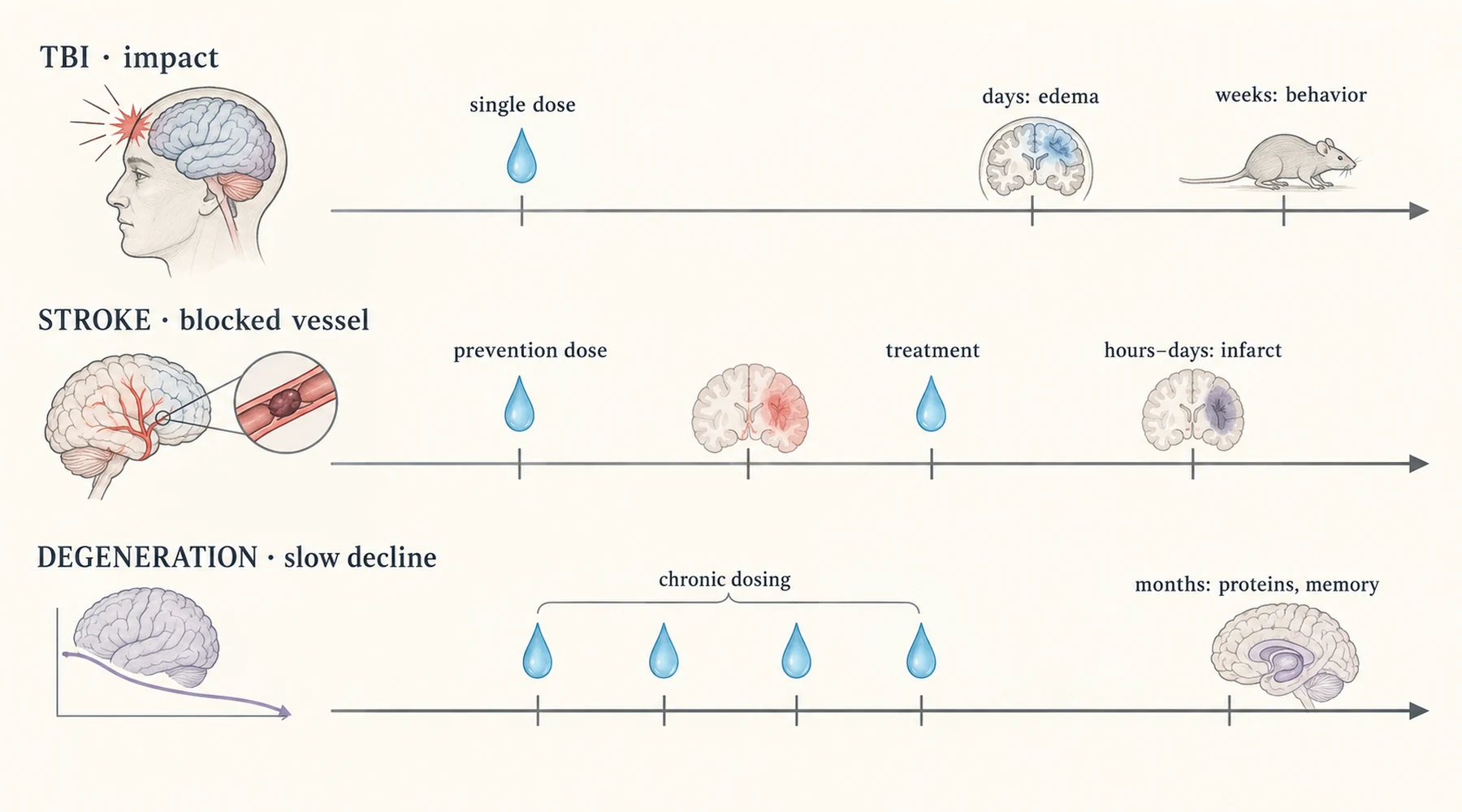
A blow to the head, a blocked artery, and a slow neurodegenerative disease can all end with the same complaint: the brain no longer has the energy to do its work. That shared endpoint is why one compound keeps appearing in all three literatures. But a shared endpoint is not a shared concept. A treatment given before an injury, a treatment given in the hour after it, and a treatment measured months later test different ideas, even when the syringe holds the same blue liquid.
This article follows the injury half of that literature. It reviews what methylene blue for brain injury means in animal experiments, separates traumatic injury from stroke and from chronic neurodegeneration, and shows why the timing of the dose explains most of the differences between studies.
Three injuries, one energy failure
The mechanism that connects these models sits inside mitochondria. Normally, electrons pass down the electron transport chain to produce ATP, and a healthy chain keeps reactive oxygen species in check. After injury, parts of the chain falter, ATP output drops, and free radical production rises. The 2018 Molecular Neurobiology review describes methylene blue as an alternative electron carrier: it accepts electrons from NADH and passes them to cytochrome c, which raises complex IV activity and restores ATP synthesis even when the early part of the chain is impaired.
The numbers reported are concrete. In cell and slice preparations, complex IV activity rose 30 to 100 percent while oxygen consumption rose in step, which indicates the extra electrons did useful work rather than leaking as radicals. In a rat stroke model, ATP production in the affected region was about 30 percent higher 24 hours after treatment than in untreated animals. The 2022 Biochemistry (Moscow) review adds the downstream consequences: less oxidative stress, less neuroinflammation, less apoptosis, plus effects on monoamine oxidase, nitric oxide synthase, mitochondrial biogenesis, autophagy, and the aggregation of misfolded proteins.
So the concept is simple. Injury chokes the normal path of electrons, and methylene blue offers a detour. Everything that follows is a question of which injury, given when, and measured how.
Traumatic injury is not stroke
In a traumatic brain injury model, force arrives all at once. The primary damage is mechanical: tissue tears, vessels rupture, and the blood brain barrier opens within minutes. What methylene blue can plausibly touch is the secondary wave that follows over hours and days, as mitochondria in surviving tissue fail, inflammation spreads, and edema builds. Endpoints in these studies reflect that sequence: lesion volume, brain water content, neuronal survival near the impact, and later behavior such as motor recovery or maze learning.
In a stroke or global ischemia model, the insult is the opposite shape. Blood flow stops, energy failure follows within minutes, and restoration of flow brings its own reperfusion injury. Here dosing synchronizes to the vessel event, often within minutes of occlusion or reperfusion, and the endpoints quantify acute tissue fate over hours to a day or two: infarct size, blood flow, and early functional recovery. One pattern in the stroke literature is pretreatment, animals dosed before vessel occlusion, which tests prevention in a way no head trauma experiment can ethically mirror in humans.
The stroke results are the strongest in the injury literature, and they include the clearest null. Rats given methylene blue at reperfusion after one hour of middle cerebral artery occlusion finished with infarcts 30 percent smaller at day 2, with more of the threatened core and penumbra salvaged (Shen, 2013). A permanent occlusion study found slower progression of threatened tissue to infarct but no reduction in final 24 hour infarct volume at either dose tested (Rodriguez, 2014). A third occlusion study cut infarct size roughly in half at the middle dose and documented mitochondrial ultrastructure and mitophagy directly in the ischemic brain, while reporting the counterintuitive detail that measured apoptosis rose as necrosis fell (Di, 2015). And in global ischemia, continuous low dose infusion restored cytochrome c oxidase activity, ATP levels, and mitochondrial membrane potential in the vulnerable CA1 hippocampal region, preserved those neurons, and protected spatial memory (Lu, 2016). That CA1 measurement is worth pausing on: direct ATP and respiratory chain data from inside an injured brain, which the TBI literature mostly infers rather than measures.
Chronic neurodegeneration differs from both. Models of Alzheimer, Parkinson, and Huntington disease dose animals for weeks or months and measure protein aggregation, dopamine handling, or gradual cognitive decline. Those endpoints have almost nothing in common with a lesion measured days after impact. Readers who carry a result from one column into another are moving a finding across a boundary the experiments never crossed. Our guide to the neurodegeneration literature keeps those long timeline studies in their own frame, and the comparison with stroke-only evidence separates vessel blockage designs from impact designs in detail.
What the TBI models actually did
No human trial of methylene blue traumatic brain injury has been published. The entire TBI record is animal work, mostly rats and mice, and it clusters around a small set of experiments that are worth naming, because their details differ in ways summaries erase.
The earliest controlled cortical impact study gave rats 1 mg/kg intravenously one hour after a mild impact, then 0.5 mg/kg at three hours (Watts, 2014). MRI lesion volume stayed lower in treated animals across two weeks of follow up. Foot faults on a walking test fell significantly on days 1, 2, and 7 but not on day 14. At the microscope on day 14, degenerating neurons in the injured cortex counted 270 versus 368 per field, and abnormal Nissl staining 41 versus 73. Early protection, in other words, with the behavioral edge fading by the second week.
A diffuse injury design used midline fluid percussion in mice and a single 2 mg/kg intravenous dose within half an hour (Fenn, 2015). Treated animals showed calmer inflammatory gene expression, fewer inflammatory microglia, less apparent edema, and no depressive-like behavior at day 7. The nulls matter as much: the injury itself raised brain water only modestly, and methylene blue did not rescue body weight loss or the motor coordination deficit. Anti-inflammatory is not the same as fully protective.
Two mouse studies used the same schedule, 1 mg/kg at one hour, 0.5 mg/kg at six hours, then daily dosing for three days, and extended the mechanism. In a cortical impact model the treatment shrank lesion volume at 24 hours, 72 hours, and day 14 while preserving neurons and raising autophagy markers (Zhao, 2016). In a fluid percussion model it reduced neuronal apoptosis, tightened a leaky blood brain barrier, and improved neurological scores across two weeks (Shen, 2019). Brain water content, though, showed no difference by 72 hours in the Zhao study, and rotarod performance showed none on days 1 and 3 in the Shen study. Lesion and cell survival endpoints moved; swelling and early motor endpoints did not always follow.
The timing standout is the delayed dosing experiment (Watts, 2016). Using the same impact model, the first dose was withheld until 24 hours after injury. By day 2 the delayed group showed lesion reductions comparable to animals treated in the first hours, and the reduction persisted at days 7 and 14. The behavioral signal was weaker: foot fault improvement in the delayed group was a nonsignificant trend. One laboratory, one model, one 24 hour window. That is genuine evidence that the window extends beyond the first hours, and it is also the entire evidence for that proposition. Note the trap in the word delayed elsewhere: a Russian series titled around delayed neuroprotection dosed at 30 minutes and used delayed to mean long lasting outcomes, not late initiation.
A smaller focal injury study in rats completes the timing picture from the other direction: dosing immediately before the injury and dosing 30 minutes after it both reduced limb dysfunction (Stelmashook, 2019). Pretreatment works in the laboratory precisely because the laboratory schedules the injury.
Dose discipline matters here. The reviews describe low doses as stimulatory to mitochondrial respiration and high doses as inhibitory, the same biphasic shape seen across methylene blue pharmacology. That is why results cannot be read as more blue, more recovery. Each study tests one point on a curve. The dose response explainer traces that curve across models, and the safety and side effect profile covers what higher doses do instead.
| Model family | When the injury happens | When methylene blue is given | What is assessed, and when |
|---|---|---|---|
| Controlled impact or percussion (TBI) | A single mechanical blow at time zero | 15 minutes to 6 hours after impact in most studies; one study first dosed at 24 hours | Evolving MRI lesion and edema over days, sensorimotor behavior over days to weeks |
| Vessel occlusion or global ischemia (stroke) | Blood flow stopped, then restored | Before occlusion in prevention designs, or after flow returns in treatment designs | Infarct size, blood flow, and early functional recovery within hours to days |
| Transgenic or toxin models (neurodegeneration) | No single onset; damage accumulates for weeks | Chronic dosing over weeks to months | Protein aggregation, transmitter handling, gradual cognitive change |
Read across that table: the same compound, three different clocks. Prevention belongs to the middle row, immediate treatment to the first two, and delayed outcomes to the first and third.
Prevention, immediate treatment, delayed outcomes
Prevention is the easiest claim to misunderstand. In ischemia models, animals dosed before vessel occlusion fare better, and that result is sometimes quoted as if methylene blue protects any brain against any future insult. It shows no such thing. It shows that mitochondria primed with an alternative electron route tolerate a scheduled loss of blood flow better. Nobody schedules a head injury.
Immediate treatment is where the TBI evidence concentrates. Dosing in the hours after impact targets the secondary injury cascade while it is still unfolding: energy failure, oxidative stress, inflammation, swelling. Lesion and edema endpoints that improve here are genuine treatment effects, but they are treatment of a process, not reversal of torn tissue.
Delayed outcomes are the third concept. Behavior measured weeks after injury, or protein handling measured months into a degenerative model, answers whether early protection persisted or whether slow processes changed course. A smaller lesion at 24 hours, a nonsignificant behavioral trend after a 24 hour delayed dose, and a maze improvement months into chronic dosing are three different claims with three different burdens of proof, and only the first currently clears its bar more than once.
What this evidence cannot show
Every result above comes with the same boundary. Rodent mitochondria respond to methylene blue in ways human mitochondria may share, but a rat given an intravenous dose minutes after a calibrated impact is not a person arriving at an emergency department hours after a fall. Anesthesia, controlled injury severity, young healthy animals, and short follow up all narrow what transfers. No blast injury study of methylene blue exists at all, so the injury type most relevant to military readers is the one with the least evidence.
There is also no dose translation on offer. The animal literature uses intravenous and intraperitoneal routes at doses chosen for experimental control, not the oral drops sold at retail. Nothing in these studies sets a human regimen, and nothing here tests methylene blue alongside standard head injury care.
So the honest summary fits in one paragraph. In rodents, methylene blue given around the time of traumatic or ischemic brain injury preserves mitochondrial energy output, and that preservation is followed by smaller lesions and better recovery scores. One impact study kept part of the lesion effect with a 24 hour delayed first dose. Whether any of this happens in injured human brains is untested, and the TBI and stroke literatures test different timings against different endpoints. Anyone selling certainty on top of this evidence is adding an ingredient the experiments never contained.