
Article
Methylene blue in stroke and cerebral hypoperfusion research: what three models actually show
A blocked artery, a starved cell culture, and a rat with narrowed neck arteries sound like three versions of one experiment. They are not. Each asks a different question, and methylene blue gives a different answer in each. Confuse them and the literature looks contradictory. Separate them and a clear pattern appears, one governed mostly by timing.
That separation is the whole point of this review. The three models are acute focal stroke, oxygen-glucose deprivation with reoxygenation in cultured cells, and chronic cerebral hypoperfusion over weeks. The proposed mechanism connecting them is mitochondrial, but the outcomes diverge because the metabolic context diverges.
The three models answer different questions
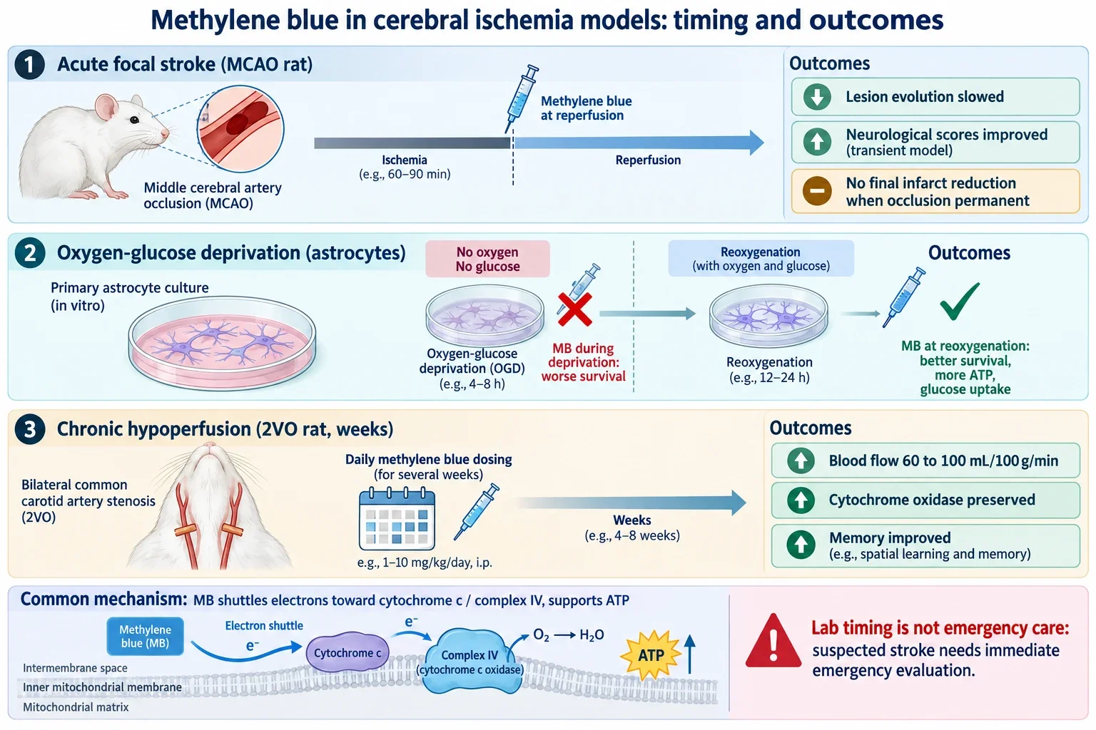
Acute focal stroke in rats is usually produced by occluding the middle cerebral artery, abbreviated MCAO. A clot-like blockage creates an ischemic core surrounded by a penumbra, tissue that is threatened but potentially salvageable if flow returns. The question here is whether an intervention slows infarct growth or preserves function around reperfusion.
Oxygen-glucose deprivation, abbreviated OGD, works at the opposite scale. Cultured brain cells are placed in near zero oxygen without glucose, then returned to normal medium. There is no clot, no collateral circulation, no blood pressure, and no immune system. The question is purely cellular: what happens to respiration, ATP, and survival when substrates vanish and then return.
Chronic cerebral hypoperfusion uses permanent bilateral carotid occlusion in rats, called the 2VO model. Blood flow to the forebrain falls and stays low for weeks. There is no sudden focal infarct and no emergency reperfusion. The question is whether weeks of metabolic support preserve vessels, mitochondria, and memory during prolonged low flow.
A memory gain after one month of treatment in the 2VO model therefore says nothing direct about rescuing human brain during an emergency thrombectomy. The distinction matters because most optimistic summaries skip it.
Acute stroke: protection at reperfusion, delay without it
The clearest acute result comes from transient MCAO with reperfusion. Rats underwent 60 minutes of occlusion, then received low dose methylene blue intravenously starting at reperfusion, with further doses at 3 hours and on day 2. MRI showed the vehicle group lesion growing by about 19 percent by day 2 while the treated group lesion shrank by about 17 percent, with roughly 30 percent smaller final infarcts and better neurological scores on days 2 and 7. Survival of both core and penumbral tissue on imaging improved. The broader neuroprotection literature places this reperfusion-timed protection alongside global ischemia and trauma findings.
The picture changes when the artery stays blocked. In a permanent MCAO experiment, methylene blue given during ongoing ischemia slowed expansion of the diffusion lesion and briefly prolonged the perfusion-diffusion mismatch, with slightly better local flow, but final infarct volume at 24 hours was not significantly different. It delayed conversion of at-risk tissue without substituting for reopening the vessel.
A third acute study adds mechanism and dose nuance. A single intraperitoneal dose immediately after occlusion showed the largest infarct reduction, about 50 percent, at an intermediate 5 mg/kg dose rather than at 1 or 10 mg/kg, alongside better sensorimotor scores. The authors linked this to preserved mitochondrial structure and increased mitophagy, the selective clearance of damaged mitochondria. Dose, route, and schedule therefore matter, and rat mg/kg values are not human stroke doses.
Cell culture: the same drug helps or harms depending on when it arrives
The astrocyte experiment is the most instructive and the most misquoted. Primary mouse cortical astrocytes underwent 6 hours of severe deprivation at 0.1 percent oxygen in medium without glucose, pyruvate, or serum, followed by 24 hours of reoxygenation with substrates restored. About half the cells died, which gives the assay room to show either direction of effect.
When methylene blue was present during the deprivation phase, survival got worse, not better. When it was introduced at reoxygenation instead, low micromolar concentrations improved viability and raised oxygen consumption, glucose uptake, and ATP production. Under normal conditions the same concentrations increased glucose uptake by roughly 89 percent at 0.1 micromolar and about 200 percent at 1 micromolar, with preserved glycogen and hexokinase activity after injury and no measured rise in superoxide.
The mechanism is easier to grasp through an analogy to a backup generator. Methylene blue acts as an alternative electron carrier that accepts reducing equivalents and passes them toward cytochrome c and complex IV, which supports electron flow and ATP synthesis while reducing upstream electron leakage. During severe deprivation that stimulation is a liability, because it raises metabolic demand when oxygen and glucose are nearly absent, which can exhaust the last reserves faster. After substrates return, the same property becomes an asset, because it helps damaged mitochondria use the restored oxygen and glucose efficiently. That is also why findings from mitochondrial electron transfer explainers need the deprivation context attached.
One caution: this timing rule belongs to that astrocyte preparation. A companion cell study with a different cell type, milder deprivation, and a lower concentration found protection even with exposure during deprivation. Cell type, residual oxygen, concentration, and protocol decide the sign of the effect.
Chronic low flow: memory, cytochrome oxidase, and vessels over weeks
The chronic experiments used daily low dose treatment for weeks, not a single emergency bolus. In the first study, rats with permanent carotid occlusion received 4 mg/kg daily for one month, timed after each daily training session to target consolidation rather than acute performance. Occluded animals learned visual discrimination problems more slowly and performed worse on a difficult memory probe, while swimming ability, motor activity, and odor recognition were unaffected. Daily treatment attenuated the learning and memory deficits without changing general motor behavior.
A follow-up mapped cytochrome oxidase, which is mitochondrial complex IV, across 44 brain regions after the same one month regimen. Occlusion lowered enzyme activity in visual, prefrontal, and perirhinal cortex plus hippocampus and amygdala, and weakened coordinated activity between visual cortex and hippocampus. Treatment preserved regional enzyme activity and strengthened those interregional metabolic correlations, and preserved activity correlated with better task performance. These correlations describe coordinated metabolic enzyme activity, not direct measures of human functional connectivity, a point that brain energy metabolism reviews help keep straight.
A separate one week study used 2 mg/kg daily and added a pathway inhibitor to test mechanism. Cerebral blood flow rose from about 60 to about 100 mL per 100 g per minute with treatment, and hippocampal vascular density rose from about 15.1 to 23 percent. Both gains were partly reversed when the PI3K/Akt survival pathway was blocked, and phosphorylated Akt rose with treatment. The blood flow, vascular density, barrier, and neuronal findings point to neurovascular protection tied to PI3K/Akt signaling rather than a pure metabolic effect.
Why none of this equals emergency stroke care
Laboratory control is the reason these results do not transfer directly. Cultured cells have no thrombus or pharmacokinetics. The 2VO model is prolonged global low flow, not a sudden focal embolus with a minutes old penumbra. Even rat MCAO uses young healthy animals, anesthesia, uniform occlusions, and precisely timed dosing, unlike older patients with varied clots, atherosclerosis, hypertension, diabetes, and unknown onset times.
No published randomized human trial has established methylene blue as an emergency treatment for acute ischemic stroke. A sometimes cited human imaging study enrolled 26 healthy adults, not stroke patients, and a single oral dose altered task related brain signals with a small retrieval gain. That shows central activity, not stroke efficacy. Current emergency guidance instead centers on rapid reperfusion with intravenous thrombolysis in eligible patients and thrombectomy for appropriate large vessel occlusions, and standard emergency stroke pathways remain the only proven route. Suspected stroke should never be delayed to administer methylene blue, and practical limits of preclinical dosing extrapolations explain why rat schedules cannot be read as clinical regimens.
Safety context reinforces the boundary. Intravenous methylene blue carries a boxed warning for serious or fatal serotonin syndrome when combined with serotonergic drugs including SSRIs, SNRIs, and MAO inhibitors, which many stroke age patients take. It is contraindicated in G6PD deficiency because of hemolysis risk. It also inhibits nitric oxide signaling, which raises vascular resistance and pressure in some settings, so it should not be described simply as a drug that increases brain blood flow. The approved 1 mg/kg intravenous regimen for acquired methemoglobinemia is not a stroke regimen.
The honest summary is narrow but real. Around reperfusion in transient focal ischemia, low dose methylene blue reduced lesion evolution in rats. Under permanent occlusion it delayed but did not prevent infarction. In astrocytes it harmed during severe deprivation and helped at reoxygenation. During chronic hypoperfusion it preserved cytochrome oxidase, vessels, and memory over weeks. One shuttle, three contexts, three answers.