
Article
Methylene blue as antioxidant and pro-oxidant: what changes the outcome?
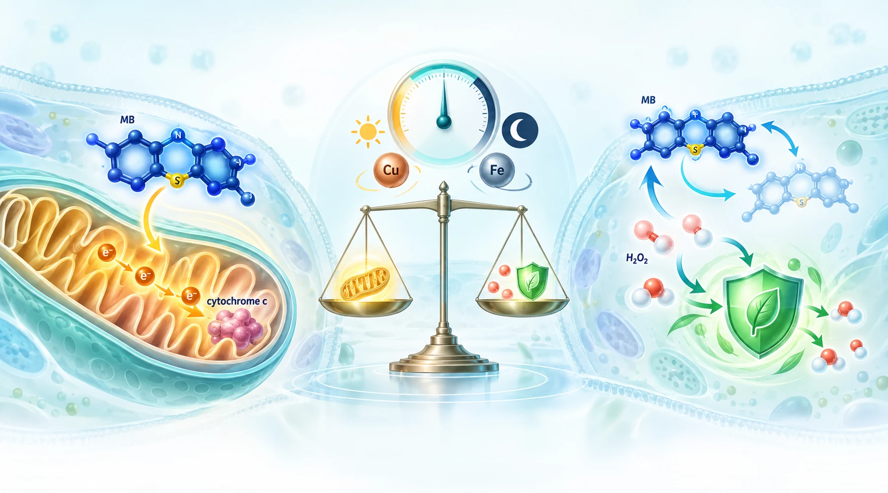
Suppose two laboratories test the same blue dye and report opposite results. In the first, isolated brain mitochondria breathe better and make more ATP. In the second, the same preparation releases far more hydrogen peroxide. Both reports are correct. They describe the same cycle seen from two ends, and the confusion starts when readers treat one end as the whole story.
The mistake is asking which label fits the molecule. Methylene blue is a relay for electrons, as the map of its cell biology targets shows in broader context. Oxidised dye accepts electrons and becomes colourless leucoMB. LeucoMB hands those electrons onward and turns blue again. What you observe depends on who donates the electrons, who receives them, and whether the system can clear the hydrogen peroxide made along the way. The question worth asking is never whether the dye is an antioxidant. It is what the conditions let the relay do.
The cycle that produces both faces
In the dark, the core loop is short. NADH, NADPH, or carriers in the respiratory chain reduce the dye. Reduced dye then has competing exits. It can pass electrons to cytochrome c and support flow around a damaged complex, which is why respiration and membrane potential partly recover under complex I or III blockade. Or it can pass electrons directly to oxygen, which regenerates the blue form and makes hydrogen peroxide. Because the dye is regenerated, a small amount can repeat this loop many times, steadily consuming reducing equivalents and steadily making peroxide.
Two further branches change the chemistry completely. Free copper or iron converts that diffusible peroxide into far more destructive local oxidants through Fenton type reactions. Red light opens a separate photochemical branch in which excited dye transfers energy to oxygen and makes singlet oxygen, a species covered in detail in the type I and type II photochemistry explainer. Dark redox cycling, metal amplified damage, and photodynamic damage are three different mechanisms that happen to share one starting molecule.
Condition to outcome matrix
The table below is the asset to keep. Each row is one variable, the direction it pushes, and the experiment behind it. Positive means a protective or improved readout. Adverse means more oxidant, more depletion, or more damage.
| Condition | Outcome direction | What the experiment showed |
|---|---|---|
| Low concentration, 0.1 to 1 uM, isolated mitochondria | Positive bioenergetics plus higher peroxide | 0.1 uM improved oxygen consumption in healthy and diabetic rat heart mitochondria; 100 nM to 1 uM partly restored ATP, membrane potential, and calcium uptake in blocked brain mitochondria, while peroxide release rose in every condition |
| Higher concentration, above 5 uM, glucose deprived cells | Adverse oxidant signal | Endothelial cells oxidised a general ROS probe and lost reduced glutathione above 5 uM when glucose was absent; adding glucose prevented both |
| Complex I substrates (glutamate plus malate) | More peroxide | Both brain and heart mitochondrial work found raised peroxide release with NADH linked substrates |
| Succinate plus rotenone, heart mitochondria, 0.1 uM | Less peroxide in that protocol | The one clearly antioxidant peroxide readout in the set; note the protocol included rotenone and a different tissue, so substrate alone does not explain it |
| Red light, 20 uM plus 630 nm | Adverse DNA damage | No significant mitochondrial DNA lesions in the dark at 20 uM; the same concentration with red light damaged every tested fragment |
| Very high dark concentration, 100 uM | Adverse selective DNA damage | Selective mitochondrial DNA lesions at purine rich sequences, a step change from the nanomolar peroxide signal |
| Copper or iron plus NADH, cell free DNA | Adverse oxidative lesions | Copper directed damage to a p53 hotspot sequence; iron EDTA caused broad damage; catalase and metal chelators blocked the respective pathways; oxidised guanine increased |
| Glucose present, intact cells | Protective buffering despite faster cycling | Glucose increased dye reduction yet lowered net stress, consistent with pentose phosphate supply of NADPH for antioxidant recycling |
| Ascorbate loaded cells | Mixed: ascorbate consumed, probe signal buffered | Dye progressively oxidised intracellular ascorbate even with glucose present, while partly suppressing probe oxidation; the defense pool paid the bill |
| Rotenone pretreated animals, dye given first | Positive tissue outcome | Dye pretreatment reduced rotenone induced mitochondrial DNA lesions in mouse midbrain despite raising peroxide in isolated mitochondria |
The pattern across rows is the point. Peroxide production and biological protection are separate axes. A treatment can score adverse on the first and positive on the second at the same time.
Why the same dose gives different answers
Concentration sets shuttle capacity, not fate. Peroxide release from brain mitochondria rose detectably from 50 nM upward in work on intact brain mitochondria, while frank mitochondrial DNA damage needed 100 uM in the dark. That is roughly a two thousand fold gap between first signal and genotoxicity, but it is not a safety margin. The endpoints, compartments, and exposure times differ, so the numbers mark distinct phenomena rather than points on one curve. The dose response discussion places this kind of low signal versus high damage gap in its wider hormesis context.
Substrate decides where electrons enter. NADH linked fuels give the dye abundant reducing equivalents for both the useful bypass and the wasteful oxygen reaction. The often quoted summary that succinate makes the dye antioxidant while glutamate makes it pro-oxidant overreads the data, because the heart study's succinate peroxide condition also contained rotenone and used a different tissue and dose than the brain work that found raised peroxide under every substrate and respiration state.
Light flips the mechanism, not just the magnitude. The cleanest contrast in the set is 20 uM in the dark doing nothing detectable to mitochondrial DNA fragments, and 20 uM under red light damaging all of them. Anyone comparing a lit experiment with a dark one is comparing photochemistry with redox cycling.
Metals convert the oxidant. Without metals, the dye's dark cycle makes peroxide, a diffusible molecule that cells also use as a signal. With accessible copper or iron, NADH reduced dye feeds metal reduction, and the reduced metal activates peroxide into hydroxyl type or copper peroxide species beside DNA. That is how cell free work with NADH and metal ions produced hotspot selective and broad DNA damage from the same starting dye. Metal handling of the compartment, not the dye alone, decides whether this branch matters.
Fuel status decides whether the cell can pay. In endothelial cells exposed to 2 to 20 uM dye, glucose removal exposed the oxidant cost while glucose supply erased it, even though glucose also sped up dye reduction. Faster cycling with funded defenses beats slower cycling with empty defenses. Treat any oxidant claim that omits glucose or NADPH status as incomplete.
Tissue decides the hardware. Endothelial cells regenerate NADPH through glucose metabolism and carry cytosolic antioxidant pools. Isolated mitochondria carry none of that. Purified DNA carries not even repair. A result in one preparation predicts little about the others, which is why the planned piece on tissue specific responses will need to keep these systems separate.
Direct measurement is not downstream defense
Much writing collapses two layers that the experiments keep apart. One layer asks how much oxidant appeared. The other asks how the cell answered.
Amplex type peroxide assays sit closest to the first layer, and even they report net flux, meaning production minus destruction minus export. The brain mitochondria study found both sides moving: more peroxide formed and slower clearance of an added peroxide bolus. Further downstream sit glutathione oxidation, ascorbate consumption, oxidised guanine, and polymerase blocking DNA lesions. Each step adds biology between the oxidant and the readout.
Nrf2 driven antioxidant enzymes and mitochondrial biogenesis sit in the second layer. They are adaptive programs that a mild peroxide pulse can trigger, and the authors of the brain mitochondria protection study propose exactly that mild stress trigger while noting the causal chain through Nrf2 rests on prior work rather than their own complete demonstration. Protection that arrives hours later through new proteins does not mean the initiating chemistry was antioxidant. It means a small pro-oxidant event bought a larger defense. The guardrails for reading such claims, including assay limits and preparation differences, are the subject of the guide to reading methylene blue research.
Why assays disagree with each other
A general probe such as dihydrofluorescein reports broad probe oxidation, not a peroxide concentration. It depends on peroxidase and heme chemistry and can miss a peroxide rise that a calibrated Amplex assay catches. The brain mitochondria study saw sharply higher peroxide with no matching superoxide signal, which is a warning against quoting any single dye based antioxidant claim. Optical overlap is a legitimate control question since the dye absorbs visible light near common excitation and emission bands, but the careful study calibrated against known peroxide, cut excitation exposure with little change in signal, and confirmed slower peroxide removal with a separate electrode. Interference is a control to run, not an automatic dismissal.
The calcium result illustrates the same need for precision. Acute dye did not change total calcium load before permeability transition in the heart study, yet it improved the rate of calcium uptake in blocked brain mitochondria in the diabetes focused heart mitochondria study. Different endpoints, different systems, no contradiction.
A checklist before trusting a verdict
Treat every antioxidant or pro-oxidant verdict as a report on a system, and ask five questions. At what concentration, and in what compartment. With which electron donor and which competing acceptor. Lit or dark, and at what wavelength and dose. With what metals available and what fuel and antioxidant pools funded. And with which assay, measuring oxidant flux or damage or adaptive response. Change any one answer and the same molecule can move across the matrix from protective to damaging without any paradox at all.