
Article
Reading E. coli and other colony appearances on EMB agar
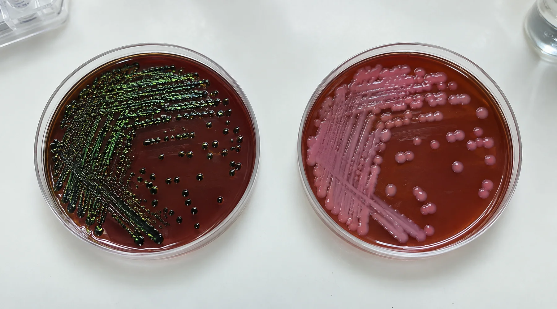
A student streaks an unknown onto eosin methylene blue agar, incubates it overnight, and returns to a plate of dark colonies with a green shimmer. The notebook entry reads "E. coli, confirmed." That entry claims more than the plate can support. The plate shows a growth and fermentation phenotype under specific conditions. Species identity is a separate claim that needs separate evidence.
This guide treats EMB agar results as an interpretation task. It explains the underlying idea, walks through an illustrative plate atlas with controls, and names the cases where the textbook picture misleads.
The idea behind the plate
EMB agar is both selective and differential. The dyes eosin Y and methylene blue suppress many Gram positive organisms, so the medium favors Gram negative growth. The same dyes act as indicators: when an organism ferments the available sugar and produces enough local acid, the colony takes up dye heavily and turns dark purple to blue black. With vigorous acid production, the colony surface can show a metallic green iridescence. The ASM protocol for eosin methylene blue agar plates describes this combination of selection and differentiation and supplies reference photographs for teaching.
One detail matters before reading any result: check which formulation is in use. The older Holt Harris Teague formulation contains lactose plus sucrose, while Levine EMB contains lactose only. An organism that ferments sucrose but not lactose, such as many Serratia marcescens isolates, can look different on the two versions. The manufacturer documentation for EMB media states this formulation difference explicitly. Record the manufacturer and formulation in the notebook alongside the result.
Prepared uninoculated EMB agar is typically red brown to reddish purple, sometimes with a greenish cast or fine precipitate. That background color is not growth. Compare every read against an uninoculated plate or the documented baseline, and read the primary plate after about 18 to 24 hours of aerobic incubation at 35 to 37 C unless the teaching protocol specifies otherwise. Extended incubation can change fermentation appearance.
How to look at the plate
Use two kinds of light. First inspect the plate under reflected or oblique light and tilt it: the metallic sheen is iridescent and can appear or vanish with viewing angle. Then examine it with transmitted light to judge colony color and dark centers. Work from well isolated colonies, since crowding and neighboring colonies change local acid accumulation and can suppress or exaggerate color. The ASM image set demonstrates this directly: E. coli in mixed culture may lose its expected sheen because surrounding organisms alter the acid environment.
Include controls whenever the exercise allows it. A common teaching set is E. coli ATCC 25922 for the blue black and metallic sheen phenotype, a nonfermenter such as Salmonella Typhimurium ATCC 14028 or Pseudomonas aeruginosa ATCC 27853 for the colorless phenotype, and a Gram positive such as Enterococcus faecalis or Staphylococcus aureus as an inhibition control, as listed in standard QC tables for EMB media. If the positive control lacks sheen, suspect incubation, crowding, or formulation before suspecting the unknown.
Illustrative plate atlas
The plates below are generated illustrations, not photographs of real cultures. They show the pattern to look for, not the exact shade of any single strain. Compare them with the ASM reference photographs and with real plates from the teaching laboratory.
Plate 1: Escherichia coli on EMB agar, vigorous lactose fermenter

E. coli on EMB agar is typically dark purple to blue black, often with a dark center and a metallic green sheen from vigorous acid production. The sheen is the famous finding students search for, but treat it as supporting evidence rather than a definition. Sheen intensity varies with strain, incubation time, colony density, and viewing angle. Absence of sheen does not exclude E. coli, and a dark colony without sheen points to several other fermenters. Pick a well isolated colony for any follow up work rather than reading from a crowded sector.
Plate 2: Klebsiella pneumoniae on EMB agar, mucoid fermenter

Klebsiella pneumoniae on EMB agar typically forms large, glistening, mucoid colonies in pink purple to brown purple shades, often with dark centers but usually without a strong metallic sheen. Similar morphology occurs with related fermenters such as Enterobacter. Mucoid texture and colony size are part of the reading: a pink colony that is small and flat carries a different weight than a large domed mucoid one. As with E. coli, the plate proposes a fermentation phenotype, and species level naming needs biochemical confirmation described below.
Plate 3: Salmonella on EMB agar and other nonfermenters, colorless colonies

Salmonella on EMB agar and related non lactose fermenters, including Shigella and many Pseudomonas isolates, typically grow as colorless to translucent or pale amber colonies. Some formulations and conditions give a faint pinkish cast, so a pale pink colony is not automatically a fermenter. EMB does not distinguish Salmonella from Shigella or from other nonfermenters; that distinction belongs to later tests. Note also that lactose positive Salmonella strains with E. coli like metallic colonies have been documented, which is one more reason a single plate cannot close the case.
Plate 4: Serratia marcescens and the formulation trap
Serratia marcescens earns its own section because students often expect red colonies and then misread the plate. Most clinical isolates do not ferment lactose, so on Levine EMB they commonly appear pale to weakly colored. Because many isolates ferment sucrose, a sucrose containing formulation may give a darker or more acid associated reaction. Do not expect the classic red prodigiosin pigment at 35 to 37 C either; pigment production is unreliable at incubator temperature. When the protocol names Serratia marcescens on EMB agar, check which formulation the statement assumes. A review of Serratia metabolism summarizes the lactose negative and sucrose variable pattern behind this trap.
Plate 5: controls and inhibited organisms

Gram positive organisms such as Staphylococcus, Enterococcus, Streptococcus, and Bacillus are generally inhibited or markedly suppressed on EMB agar, though inhibition is not absolute for every strain. A control panel makes the reading honest: the E. coli control should show good growth with blue black color and sheen, the nonfermenter control should show colorless growth, and the inhibition control should show little or no growth. If the inhibition control grows well, the selective step failed and every other reading on that batch is suspect. Staining methods answer a different question from culture media; for the slide stain side of the laboratory, see the methylene blue staining atlas.
Bounded comparison
| Observation on EMB agar | Likely meaning | What it does not establish |
|---|---|---|
| Dark blue black colony with green sheen in oblique light | Vigorous fermentation, E. coli a leading candidate | Species identity; other vigorous fermenters can sheen |
| Large mucoid pink purple colony, no sheen | Fermenter with capsular character, Klebsiella a candidate | Klebsiella pneumoniae specifically; Enterobacter looks similar |
| Colorless or translucent colony | Non lactose fermenter, for example Salmonella, Shigella, Pseudomonas | Which nonfermenter; Salmonella versus Shigella needs other tests |
| Pale colony where red was expected | Possibly Serratia on lactose only EMB at 37 C | Absence of Serratia; check formulation and temperature |
| No growth or tiny suppressed colonies | Possibly Gram positive inhibition | Sterility or identity; some strains escape inhibition |
Three sentences summarize the misleading appearances. Dark does not equal E. coli, since Klebsiella, Enterobacter, and other fermenters develop dark centers without sheen. Pink does not equal Klebsiella, since moderate or slow fermenters and some nominal nonfermenters appear pink or lavender. Sheen does not equal definitive E. coli, since other vigorous fermenters can produce it. The teaching protocol and its mixed culture figures illustrate each of these failures.
Confirmation limits
EMB agar reports a phenotype, not a species. Different organisms can share one colony appearance, and one organism can deviate from its textbook picture with strain, age, crowding, formulation, and incubation. That is why teaching and clinical workflows treat EMB morphology as presumptive.
A defensible student workflow is: describe the isolated colony (size, color, center, sheen under oblique light, mucoid character), record formulation, incubation time and temperature, and controls; then subculture a well isolated colony to purity before any confirmatory test. Common follow up includes Gram stain plus IMViC reactions for presumptive E. coli and coliform identification, TSI or KIA and organism specific biochemical panels for enteric organisms, with serologic, MALDI TOF, or other validated identification where the task requires it. For background on how EMB fits among selective and differential enteric media, see introductions to enteric culture media and Gram stain follow up. For the related question of what a blue slide stain can and cannot prove, the methylene blue staining atlas keeps observation and conclusion separate in the same spirit.
Write the observation before the conclusion. "Dark blue black colonies with angle dependent green sheen on Levine EMB at 24 hours, consistent with vigorous lactose fermentation" is defensible. "E. coli, confirmed" requires the confirmation step the plate alone cannot supply.