
Article
Methylene blue simple staining versus Gram staining: what each method can prove
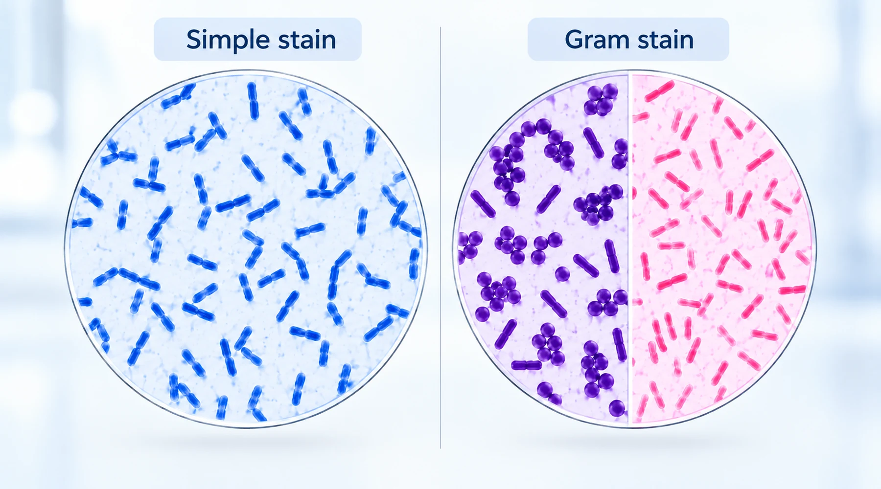
A student prepares a smear from a culture labeled E. coli, floods it with methylene blue for one minute, rinses, and looks through the oil lens. Blue rods cover the field. The lab report reads: "Gram-negative rods identified as E. coli." The observation is correct. The conclusion is not earned by that slide.
The error is conceptual, not technical. The student treated a simple stain as if it were a differential test. A simple stain and a Gram stain answer different questions because they embody different staining concepts. One asks: what shape is present. The other asks: how does the cell wall behave when challenged. Confuse the two and a correctly stained slide produces an unjustified report.
This comparison keeps the two methods separate, shows what each result licenses you to claim, and answers the two questions students ask most: can methylene blue be used in Gram staining, and what does a methylene blue stain of E. coli actually show.
The short answer
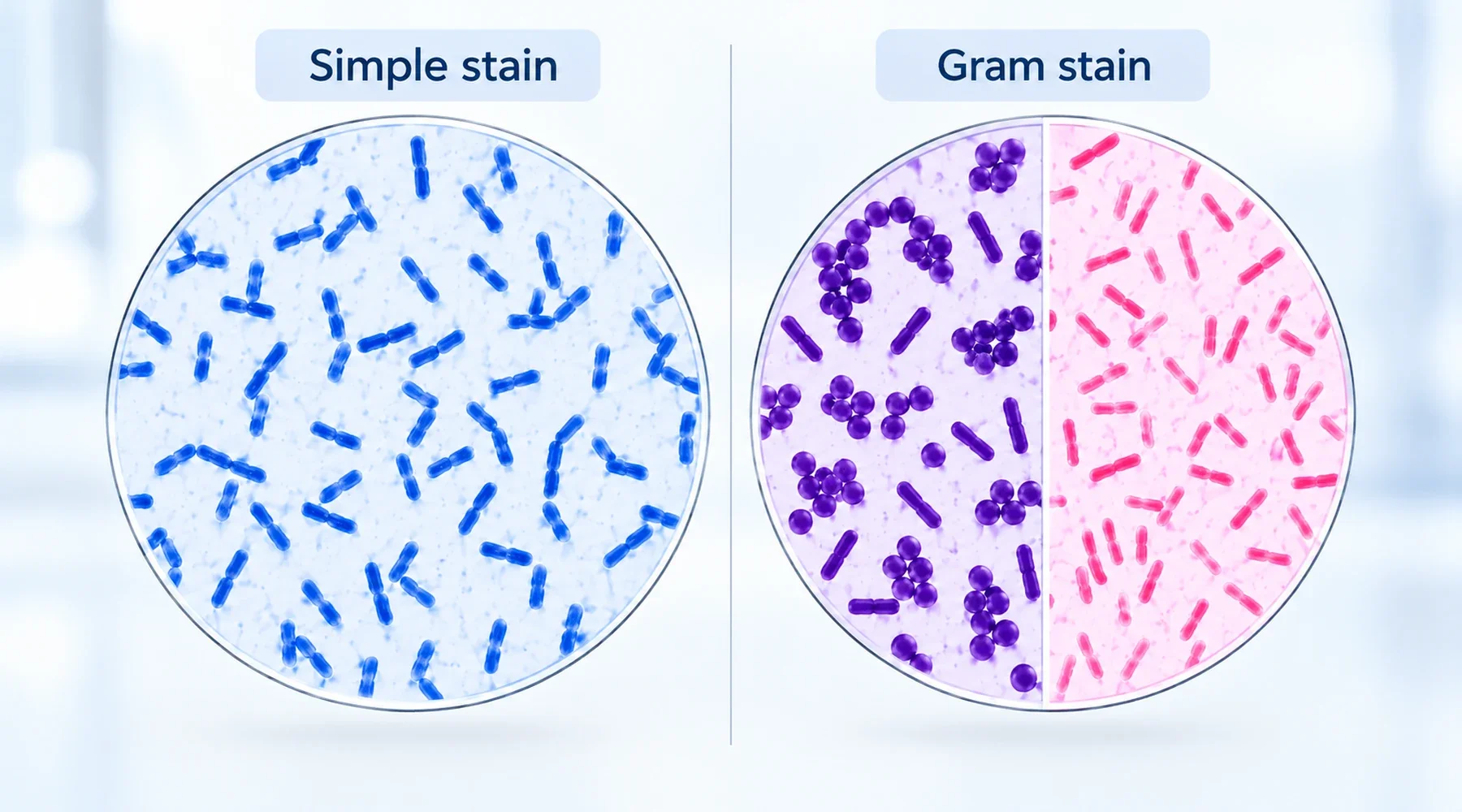
Methylene blue simple staining colors every stainable cell the same blue, so it reveals morphology (rods, spheres, spirals) and arrangement (singles, pairs, chains) and nothing about Gram category. Gram staining adds iodine, a decolorization step, and a counterstain, so it separates bacteria into Gram-positive (purple) and Gram-negative (pink or red) based on how their envelopes retain dye. Blue rods after methylene blue mean "rod-shaped cells are present." Pink rods after a full Gram sequence mean "Gram-negative rods are present." The second statement is stronger because the procedure performed an additional test.
For the wider family of one-dye bacterial methods this article belongs to, see the microscopy staining atlas.
Simple staining: one dye, one question
Methylene blue is a basic (cationic) dye. Its colored ion carries a positive charge and binds negatively charged structures in the cell, including nucleic acids and acidic surface groups, so cells appear blue against a pale background. OpenStax describes this positive-staining principle in its staining chapter. The dye cation itself is the small flat phenothiazine ion detailed in the methylene blue chemistry reference; the chloride counterion does not do the coloring.
A standard teaching protocol is deliberately plain: make a thin smear, air dry, heat fix, flood with about 1% aqueous methylene blue for roughly one minute, rinse gently with water, blot dry, and examine with oil immersion. A LibreTexts teaching protocol gives exactly this 1% one-minute form, while noting that some labs extend the time. No mordant, no decolorizer, no second dye. That absence is the point. With only one dye and no selective removal, every cell that binds dye ends up the same color.
With E. coli, the expected field is blue rods (bacilli), single or in pairs or small groupings. The blue comes from the dye. The rod shape comes from the organism. But the combination does not identify the organism, because many species are rods and many rods stain blue in a simple stain. ATCC makes this morphology-only limit explicit in its microbiology culture guide. A correct report reads: "blue bacilli observed." Anything beyond shape and arrangement needs more evidence.
Gram staining: four reagents, one behavioral test
Gram staining is a differential stain. It subjects all cells to the same sequence and watches which cells hold the first dye and which lose it. The American Society for Microbiology Gram stain protocols define the standard four-step order: crystal violet (primary stain), Gram iodine (mordant), alcohol or acetone alcohol (decolorizer), then safranin (counterstain).
| Step | Reagent | Function | Gram-positive result | Gram-negative result |
|---|---|---|---|---|
| 1 | Crystal violet | Primary stain, colors all cells purple | Purple | Purple |
| 2 | Gram iodine | Mordant, forms a complex with crystal violet | Purple | Purple |
| 3 | Decolorizer | Selectively removes the complex | Stays purple | Becomes colorless |
| 4 | Safranin | Counterstain, colors decolorized cells | Stays purple | Pink or red |
The standard teaching model explains the split through envelope structure. Gram-positive cells have a thick peptidoglycan wall that dehydrates and retains the crystal violet iodine complex during the alcohol wash. Gram-negative cells have a thin peptidoglycan layer plus a lipid-rich outer membrane that the decolorizer disrupts, so the complex washes out and the cells then take up safranin. The NCBI Bookshelf Gram stain review gives this cell wall account. Specialists add that the full physicochemistry is more complicated than wall thickness alone, but for bench interpretation the behavioral rule holds: purple means retained, pink means lost then counterstained.
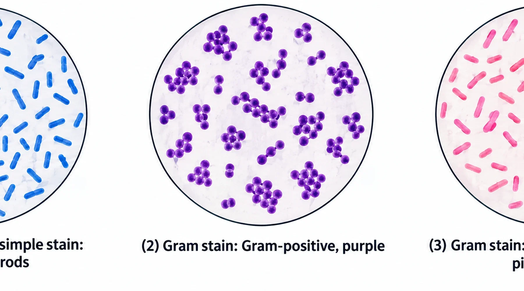
Illustrative panels. Stylized drawings for method comparison, not camera micrographs. Colors are simplified to teach the logic: one color in a simple stain, two colors after Gram decolorization.
Can methylene blue be used in Gram staining
This question has two readings, and they need different answers.
Reading one: can a single flood of methylene blue substitute for the whole Gram sequence. No. Without iodine, decolorizer, and counterstain there is no differential step, so no Gram category is tested. A blue field after methylene blue alone is a simple stain result even if the bottle sits next to the Gram reagents. Do not record it as a Gram result.
Reading two: can methylene blue replace safranin as the counterstain at step 4. Functionally it can color the decolorized cells, and a textbook exercise predicts purple Gram-positive cells and blue Gram-negative cells after such a swap. The logic is sound because correctly decolorized Gram-negative cells are nearly colorless and will take up another cationic dye. But it is not the standard protocol. ASM protocols specify safranin, with recognized alternatives such as basic fuchsin in some variants, not methylene blue. There is also a practical cost: blue against purple is harder to read than pink against purple, especially for beginners and especially under tired microscope lamps. For coursework and any result that matters, use the counterstain your protocol names, usually safranin, and treat a methylene blue counterstain as a demonstration variant rather than an equivalent standard.
The related question of how methylene blue compares with other single-dye options is covered in a comparison of simple bacterial stains.
What methylene blue stain of E. coli proves, and what it does not
E. coli is a Gram-negative rod, but that sentence combines facts from several methods: morphology from microscopy, Gram reaction from a Gram stain, identity from culture plus biochemical or molecular testing. A methylene blue smear contributes only the first part. It establishes that rod-shaped cells are present in the stained sample and how they are arranged. It does not establish the Gram reaction, because no decolorization challenge was run. It does not identify the species, because rods of many species look alike at this resolution.
This is where reports go wrong in a predictable way. The writer imports background knowledge ("this culture is supposed to be E. coli, and E. coli is Gram-negative") into the observation line, so the slide appears to confirm what was actually assumed. A cleaner report separates the layers: "Methylene blue simple stain: blue bacilli observed; morphology only, no Gram category tested." Then, if a Gram stain was run correctly with controls: "Gram stain: pink rods, consistent with Gram-negative." Species identity comes later, from the identification workflow, not from color alone.
A practical interpretation guide
Use this decision rule at the bench. If all cells are one color and the protocol used one dye with no decolorizer, you ran a simple stain. Report shape, size estimate, and arrangement. Stop there. If the protocol ran crystal violet, iodine, decolorizer, and counterstain with known positive and negative controls, you ran a Gram stain. Report color plus morphology: purple cocci in chains, pink rods, and so on.
Three mistakes account for most confusion. First, overreading blue rods as E. coli confirmation. Second, logging a methylene blue slide under the "Gram stain" heading in a notebook, which later readers will misinterpret. Third, swapping counterstains casually and then comparing colors across labs as if the palettes matched. Each mistake has the same fix: name the method that was actually performed, and claim only what that method tests.
Simple staining earns its place precisely because it claims little. It is fast, forgiving, and sufficient when the question is morphology: is anything there, and what shape is it. When the question is classification, run the differential test. The blue slide tells you what the cells look like. The purple or pink slide tells you how their walls behave. Neither slide, alone, tells you the species.