
Article
Methylene blue in wound research: dressings, light, and healing outcomes
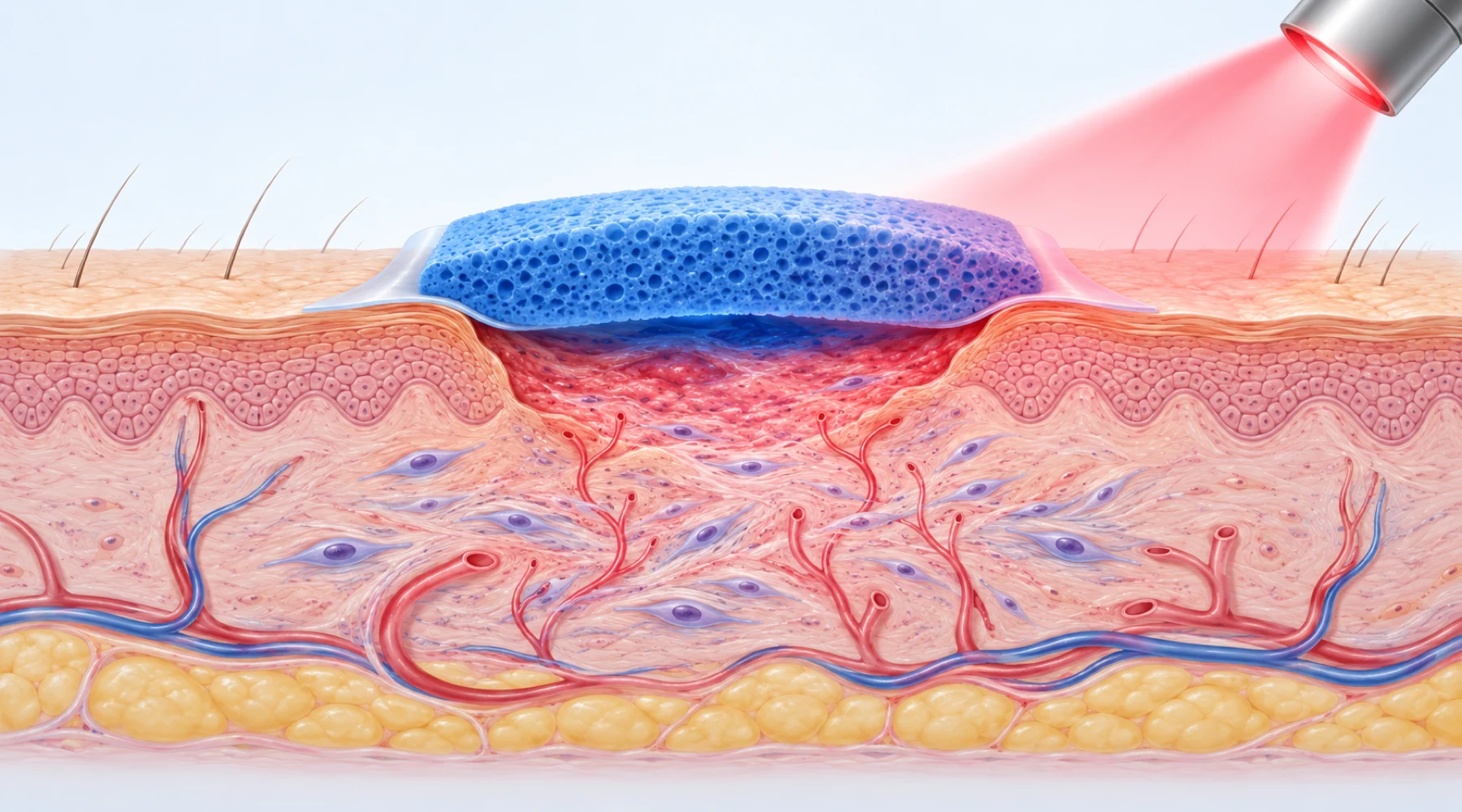
A blue-stained bandage and a dropper bottle of blue liquid look like the same intervention. In wound research they are not. One is a manufactured dressing with a fixed dye load, tested as a finished device. The other is a solution applied by hand, where concentration, volume, and contact time vary with every application. Studies of the two answer different questions, and their endpoints differ accordingly.
This review keeps the two apart. It maps what researchers applied, what they applied it to, and what they measured: wound area, closure rate, bacterial counts, or a laboratory proxy such as scratch closure in a dish.
The three settings, stated plainly
Wound research with methylene blue falls into three settings. Each has its own concept of what the dye is doing.
First, dye-containing foam dressings. Products such as polyvinyl alcohol foam impregnated with methylene blue and gentian violet (marketed as Hydrofera Blue antibacterial foam) carry the dyes bound in the dressing matrix. The stated mechanism is antibacterial activity associated with the dressing, which is intended to help manage bacterial burden in the wound environment while the foam handles exudate. Treatment decisions with such a product belong to the clinician choosing a dressing for a specific wound; the dressing's instructions for use define wear time and change frequency, not a general recipe for blue dye.
Second, methylene blue photodynamic therapy. Here a methylene blue solution is applied as a photosensitizer and then illuminated, typically with red light near the dye's absorption peak. Light and oxygen participate in generating reactive species that damage microbes. This is distinct chemistry from the antioxidant rationale used in skincare marketing, a distinction our guide to methylene blue in skincare also draws. The intervention is the combination of dye, light dose, and oxygen, and changing any one of the three changes the intervention.
Third, methylene blue without light in cells and animals. These experiments ask whether the dye itself alters fibroblast behavior, matrix proteins, or closure of experimental wounds. They are mechanism studies, not dressing trials.
Study-model map: what was tested, on what, and what changed
Use this table as the article's map. Read each row left to right: the material, the model, and the endpoint that row can support. No row borrows the endpoint of another.
| Material or intervention | Model and exposure | Measured endpoints |
|---|---|---|
| Methylene blue plus gentian violet foam dressing | Human chronic wounds (pressure, venous, diabetic) managed with the finished dressing per its instructions | Wound area change, PUSH and BWAT wound scores, clinical infection signs, pain, closure time |
| Methylene blue solution plus red light | Human infected or hard-to-heal ulcers in case series and small trials; defined dye concentration and light dose | Wound area change, BWAT scores, infection signs, healing time; occasionally microbiology |
| Methylene blue solution plus light | Infected animal wounds and biofilm models | Colony-forming-unit counts, wound contraction, histology, bacterial dissemination |
| Methylene blue without light | Cultured human fibroblasts; reconstructed skin models | Scratch-closure speed, viability, hydration and dermal thickness in the model, matrix-protein expression |
| Loose dye solution dabbed on a wound | Uncontrolled application | No interpretable endpoint without fixed concentration, contact time, and a comparator |
Dressings: a finished device, not a dye recipe
The foam dressings studied in wound care are manufactured devices. The Hydrofera Blue range described on the manufacturer's product page combines the two dyes in sterile foam: the CLASSIC polyvinyl alcohol version carries at most 0.00025 g of each dye per gram of foam, and the READY polyurethane version at most 0.00035 g per gram. Because the dyes are loaded during manufacture, each dressing carries a known quantity, and clinical reports describe the product by name, wear time, and change schedule.
These are cleared medical devices, not approved drugs. The original dressing holds FDA clearance K013462 from 2002, the polyurethane Hydrofera Blue version holds clearance K130670 from 2014, and the bordered READY version holds clearance K190268 from 2019. Clearance through the 510(k) route establishes substantial equivalence to an earlier device; it is not an efficacy approval. The antimicrobial claims in the device documentation rest substantially on activity within the dressing and in laboratory testing, which should not be translated into a claim that the dressing sterilizes infected tissue by a stated log reduction.
Published clinical experience consists largely of small uncontrolled series. In a prospective study of 29 chronic wounds by Woo and Heil, Hydrofera Blue Classic changed three times weekly for four weeks, mean wound area fell from 21.4 to 12.3 cm² (a 42.5% reduction) and PUSH scores fell from 13.3 to 10.7. A case series of 15 lower-extremity wounds by Coutts, Ryan and Sibbald found decreasing wound size at four weeks in 8 of 14 evaluable wounds, with pain improvement in some patients, particularly those with diabetic foot ulcers. A retrospective cohort of 53 wounds reported by Lullove combined the foam with an ovine collagen matrix, so its 91.3% mean area reduction at twelve weeks cannot be attributed to the foam alone. A large retrospective program analysis by Hurd reported faster healing where the dressings were used inside a standardized care bundle, which again tests the bundle rather than the dye.
One limitation runs through all of these: bacterial burden appears as clinical infection scores or semiquantitative swabs, not serial quantitative tissue cultures. The honest summary is that dual-dye foam studies report area reduction, improved wound-bed scores, and sometimes less pain, without establishing a methylene-blue-specific dose response or a measured bacterial-kill rate in tissue. And nothing in the dressing studies validates soaking gauze in a dropper solution as an equivalent. Concentration, distribution, release rate, sterility, and wear time all differ.
Light plus dye: photodynamic wound studies
Photodynamic wound research adds illumination to the protocol. Concentrations in human studies generally fall between 0.01% and 1% methylene blue, with red light near 635 to 660 nm at fluences from a few joules per square centimeter to over one hundred. Control arms vary: some studies compare against standard care, others report before-and-after bacterial counts within the same wounds.
The most consistent human signals concern wound quality and area rather than bacterial counts. In a controlled study of 12 diabetic ulcers by Carrinho and colleagues, 0.01% methylene blue with 660 nm light at 6 J/cm², three times weekly for ten sessions, produced significantly greater area reduction than the control treatment. A case series of five infected wounds by Shen and colleagues reported complete healing after a mean of four sessions of 1% dye with 635 nm light, without quantitative bacterial endpoints. The largest randomized test to date, a 94-participant double-blind trial of diabetic foot ulcers by Chagas and colleagues, found Bates-Jensen wound scores improving from the third session through thirty-day follow-up with 1% dye plus 660 nm laser, with no treatment-related adverse events, but again without a quantitative bacterial-load endpoint.
Two caveats keep this literature honest. First, the dramatic amputation result sometimes quoted from Tardivo and colleagues used methylene blue combined with toluidine blue, so it cannot be attributed to methylene blue alone. Second, healing after a light session cannot automatically be credited to antimicrobial photochemistry, because red light itself can modify repair. In a rat wound experiment by Sperandio and colleagues with dye-only, light-only, combined, and control arms, the laser-alone condition influenced healing on its own. Attribution to bacterial killing requires microbiology plus the right controls. The animal work is clearer on that point: in an infected mouse wound model by Pérez and colleagues, photodynamic therapy produced the best visible healing outcome while mupirocin produced the strongest bacterial reduction, separating the two endpoints cleanly. Our overview of methylene blue and red-light evidence places these wound protocols alongside the non-wound light settings so the shared photochemistry does not get mistaken for a shared indication.
Cells and skin models: where the healing signal starts
The laboratory root of the wound-healing claim is a 2017 study by Xiong and colleagues on human dermal fibroblasts and reconstructed skin. Fibroblasts from two donors, pretreated with 100 nM methylene blue, repopulated a scratch gap significantly faster than untreated cells at 24 hours. In full-thickness reconstructed skin, longer exposure increased measured hydration and dermal thickness, with elastin and COL2A1 upregulation among the matrix changes.
These are real wound-relevant endpoints, and they belong to their models. A scratch assay measures collective cell migration across a gap in a dish. It contains no immune response, no bacterial colonization, no exudate, and no dressing. Reconstructed skin adds tissue architecture but still lacks circulation and the full healing cascade. Neither model tests a foam dressing or a photodynamic protocol, and neither yields a concentration for home application.
Formulated methylene blue has also been tested in rodent wounds. A niosomal 0.005% gel studied by Farmoudeh and colleagues accelerated early closure of rat excisional wounds relative to free dye and vehicle, with more favorable histology and oxidative-stress measures. A 0.1 mg/mL nanoemulsion studied by Gao and colleagues in diabetic mice accelerated closure with increased angiogenesis markers and reduced oxidative and apoptotic signals. The formulation matters in both cases; neither result transfers automatically to plain aqueous dye.
And the direction of effect is not universal. In a rat anastomosis experiment by Dinc and colleagues, 1% methylene blue placed inside the abdomen reduced anastomotic bursting pressure and hydroxyproline on day three, showing impaired early healing strength at that dose and route. Concentration, tissue, formulation, and route decide the outcome.
What to ask of any wound claim
Every wound claim about methylene blue should survive four questions. First, what exactly was applied: a named manufactured dressing, a defined photosensitizer-plus-light protocol, or an unspecified solution? Second, to what: cells, an animal wound, or human wounds of a stated type? Third, against what comparator, over what duration? Fourth, with what measured endpoint: area change, closure, bacterial counts, or a laboratory proxy?
Claims that blur the first question are the most common failure. A result obtained with a dye-loaded foam does not validate painting a wound blue, and an antimicrobial signal after a photodynamic session does not validate dabbing a solution without light. The forthcoming guide to reading methylene blue wound claims will give this checklist a permanent home with worked examples; until then, the table above serves as the map.
Wounds that are deep, spreading, foul-smelling, fever-accompanied, or not improving need clinical assessment rather than a dye. The research reviewed here is worth following precisely because it is specific about materials, models, and measurements. Keep those three attached to every claim and the field reads clearly.