
Article
The methylene blue reduction test for rumen fluid
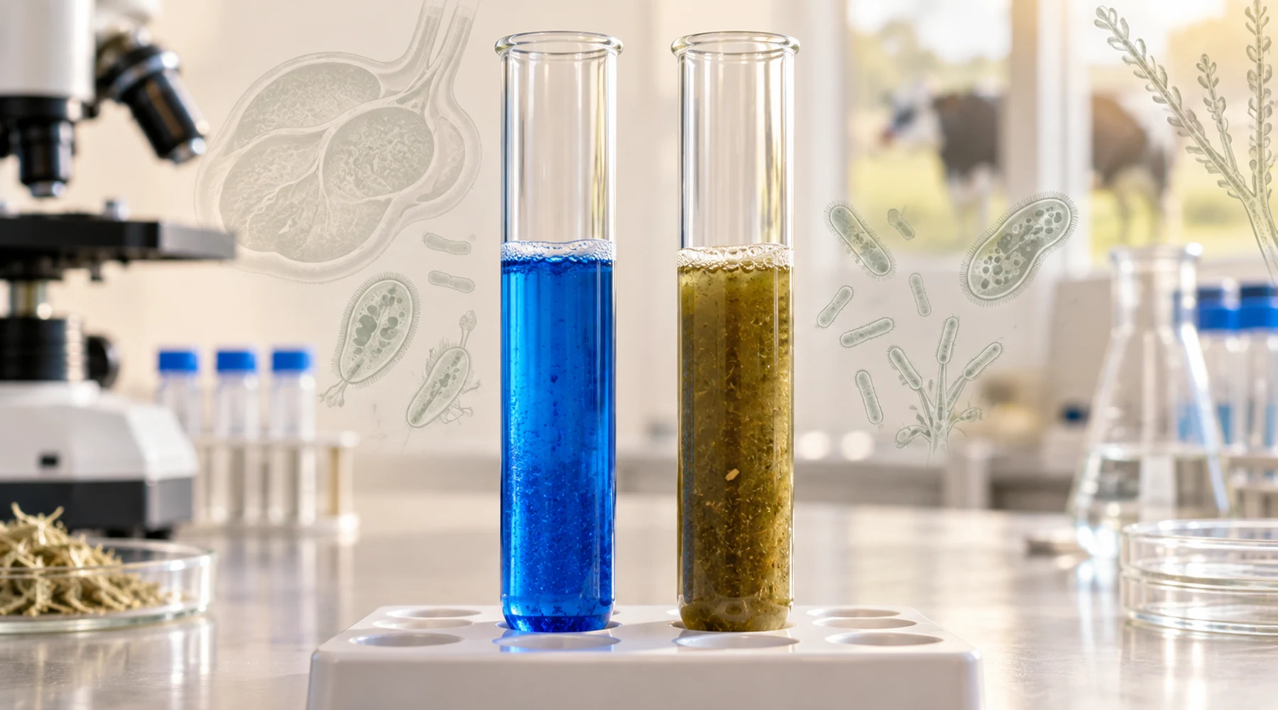
A graduate calls from a farm with a blue tube in her hand. She added methylene blue to rumen fluid ten minutes ago and the blue is still there. She asks whether she should give more dye to the cow.
That question reveals a wrong mental model. In the methylene blue reduction test for rumen fluid, the dye goes into the tube, never into the animal. The tube is the whole assay. Timing how fast the blue disappears tells you something about the microbes in that sample, not about a treatment the animal needs.
The confusion is understandable because methylene blue appears in three separate veterinary contexts. This article keeps them apart and shows how to read reduction time alongside pH, protozoa, sampling quality, and clinical signs.
What the test actually measures
Methylene blue is blue when oxidized and nearly colorless when reduced. Fresh rumen fluid from a healthy animal is strongly reducing because anaerobic bacteria are actively fermenting feed. When you mix a small amount of dye into such fluid, bacterial metabolism removes the color within a few minutes.
So the operational principle is simple: active anaerobic flora clears the blue fast, inactive or damaged flora clears it slowly. A short time means greater reducing capacity in that sample. A prolonged time points to depressed microbial activity, provided the sample was collected and handled well.
It does not measure rumen redox potential directly. A cattle methods study that compared the reduction test with direct millivolt measurements found no linear correlation between reduction time and measured redox potential, and concluded the two should not be treated as interchangeable readings of the same variable. The full comparison is summarized on PubMed. Think of the reduction test as a functional assay of the microbial population, not as a cheap electrode.
The Merck/MSD description of simple indigestion in ruminants places prolonged reduction time in the same pattern as abnormal rumen pH and reduced protozoal numbers. No single value makes the diagnosis. The pattern does.
A standard bedside procedure
Protocols vary slightly between schools, so record the exact volumes and temperature you use and compare results only within that protocol. One widely taught version is:
- Collect at least 10 to 20 mL of fresh rumen fluid with minimal saliva, air exposure, and cooling. Test promptly.
- Prepare 0.03 percent methylene blue, which is 0.3 mg per mL.
- Add dye at 5 percent of the fluid volume. That is 1.0 mL dye to 20 mL fluid, or 0.5 mL dye to 10 mL fluid. Both give the same 20 to 1 fluid to dye ratio.
- Keep a second tube of untreated fluid from the same sample as the color control.
- Mix gently, start the timer at once, and hold the tube near body temperature, around 37 to 39 degrees Celsius depending on local protocol.
- Record the time until the test tube matches the control. A thin blue ring at the air surface may persist because oxygen reoxidizes the dye there. Do not wait for that ring.
The redox chemistry of methylene blue explains why oxygen matters here. Vigorous shaking, a wide open tube, or a long delay lets oxygen in and keeps the dye blue longer than the rumen itself would. Details on clean rumen fluid collection technique matter for the same reason. Saliva dilutes the sample and raises pH, so with a stomach tube many clinicians discard the first portion when volume allows.
What different observations measure
Reduction time answers one question only. The rumen assessment below shows what each bedside observation contributes. Use the full row, not one cell.
| Observation | What it measures | Typical bedside finding | Common confounders |
|---|---|---|---|
| Methylene blue reduction time | Functional reducing activity of anaerobic bacteria in the sample | Cattle often 2 to 6 minutes; sheep and goats often faster, with 6 minutes used as a practical cutoff for adequate activity and times beyond 10 minutes markedly abnormal | Cold, delayed, aerated, or saliva diluted sample; low substrate after prolonged anorexia; oral antimicrobials; comparison across different dye ratios or temperatures |
| Rumen pH | Chemical environment, acid or alkali load | Roughage fed cattle often near 6.0 to 7.2; small ruminant texts often cite about 6.5 to 7.5; diet and time since feeding shift the value | Saliva contamination raises pH in stomach tube samples; carbon dioxide loss after collection; grain feeding lowers pH |
| Protozoal number and motility | Integrity of the ciliate population, especially large forms sensitive to disturbance | Many organisms of several sizes with vigorous directional movement in a warm fresh drop | Cooling slows movement; severe acidosis can eliminate protozoa; delay makes a normal sample look sluggish |
| Odor, color, and consistency | Fermentation character and contamination | Slightly aromatic, greenish brown, mildly viscous fluid | Putrid or sour odor with severe dysbiosis; watery or heavily salivary fluid after poor collection |
| Rumen contractions and appetite | Whole animal forestomach function and intake | Regular contractions and steady rumination in a bright animal | Recent ration change, pain, dehydration, systemic illness |
The table is the point. A prolonged reduction time plus sluggish protozoa plus abnormal pH plus weak contractions points toward genuine microbial depression. An isolated prolonged time in a bright ruminating animal with vigorous protozoa points toward the sample, not the rumen. Repeat the collection before acting.
Reading time with pH and history
Diet sets the baseline. Concentrate rich rations ferment fast and can clear the dye in about a minute, while hay diets may take several minutes, even in healthy animals. So a fast time does not by itself prove better health. It proves more fermentable substrate met active microbes.
Severe grain overload breaks that simple trend. Early carbohydrate fermentation may be intense, then acid damage suppresses the normal flora and protozoa, and reduction becomes prolonged. That is why one teaching rule states the test is not valid when rumen pH is below about 5.5. At that point the flora being tested has changed.
A practical pattern guide:
- Normal pH with reduction within about 6 minutes suggests functionally active flora.
- Normal pH with markedly prolonged reduction suggests inactivity from anorexia, antimicrobial effect, or a compromised sample.
- High pH with prolonged reduction often follows prolonged anorexia with little substrate, but consider saliva contamination from the tube.
- Low pH with reduced protozoal activity raises concern for carbohydrate related dysbiosis. Guidance on rumen pH interpretation helps here. Values below 5.5 with systemic depression point away from simple indigestion toward grain overload and severe acidosis.
Always record recent oral tetracyclines, feed ionophores, prolonged systemic antimicrobials, abrupt ration changes, duration of anorexia, and water intake. The MSD manual specifically warns that oral tetracyclines can substantially depress ruminal microflora. Ionophores intentionally shift bacterial populations. Without that history, a prolonged time is uninterpretable.
Sampling quality deserves equal weight. Controlled work found stomach tube samples higher in pH and lower in volatile fatty acids than cannula samples from the same animals, largely from saliva. Reduction time lengthened significantly by 60 minutes after collection for every volume tested, with small samples most affected. Collect at least 10 mL, test within 30 minutes, perform the reduction test early, keep the sample warm, and minimize air contact.
Three uses that must not be confused
The same dye, three different systems.
First, the rumen fluid assay described here. Specimen is collected rumen fluid. Dye is added in vitro at 0.03 percent. Endpoint is time to decolorization. Purpose is assessment of microbial reducing activity.
Second, methylene blue given to a live ruminant. The established example is intravenous therapy for nitrate or nitrite toxicosis with methemoglobinemia, where the drug restores functional hemoglobin. Dose, formulation, residue, and withdrawal rules apply. That decision has nothing to do with timing a tube. If dye is used as an in vivo tracer in a specific procedure, the principle is where the dye travels, not how fast rumen microbes reduce it.
Third, the milk reduction test. Specimen is milk. Dye is added to the milk sample. Rapid loss of blue generally indicates greater microbial activity and poorer keeping quality, while slow reduction indicates better quality. It belongs to food microbiology and milk quality testing, not rumen medicine, and it is not a bacterial count.
Keeping the names explicit prevents error: rumen fluid reduction time, systemic methylene blue therapy or tracer use, and milk reduction test. The safety rule fits in one sentence. Dye for the rumen assay stays in the laboratory tube and that tube is discarded. It is never dosed to the animal to perform the test.
A compact rule for the farm call
Standardize the dye at 0.03 percent and 1 part dye to 20 parts fluid, test fresh fluid near body temperature against an untreated control, and read the minutes with pH, protozoal motility, collection quality, diet, drugs, contractions, appetite, feces, and hydration. An isolated number never diagnoses indigestion. A concordant pattern does.