
Article
Methylene blue and progeria: lessons from a cellular aging model
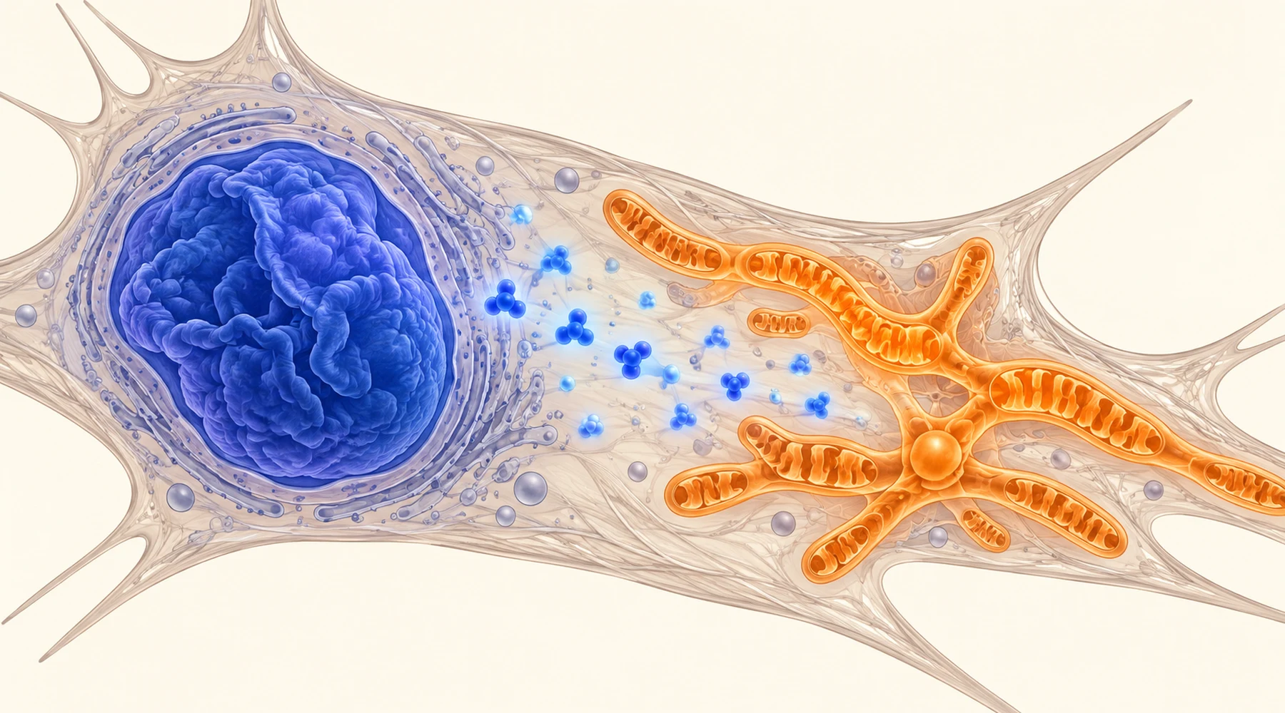
A single-letter change in one gene makes children age at terrifying speed. Hair loss, stiff joints, narrowed arteries, and a life expectancy in the teens. That disease is Hutchinson-Gilford progeria syndrome, and for years its research centered on the nucleus, where the mutant protein does its damage. A 2016 study from the University of Maryland asked a different question: what do the mitochondria look like in these cells? The answer connected two compartments of the cell, and a century-old blue dye touched both.
The study is Xiong and colleagues, published in Aging Cell in 2016. It worked entirely with cultured cells. That fact organizes everything below. The findings are genuine and specific. Their distance from any treatment is equally specific.
The model: one mutation, one sticky protein
Progeria starts with a C to T substitution at position 1824 of the LMNA gene. The change does not alter an amino acid. It activates a hidden splice site, which deletes 50 amino acids from the lamin A protein. The shortened product, called progerin, loses the cleavage site that would normally let the cell remove its farnesyl anchor. So progerin stays permanently farnesylated and stuck to the inner nuclear membrane, where normal lamin A would cycle on and off.
The consequences at the nucleus are well documented and visible under a microscope. Nuclei bleb and wrinkle instead of holding a smooth oval. Heterochromatin, the densely packed silent DNA that normally lines the nuclear edge, thins out. Thousands of genes change expression. Think of the nuclear lamina as scaffolding behind wallpaper: when one scaffold piece is welded in the wrong position, the wall bulges and the paper pattern distorts across the whole room.
The study used skin fibroblasts from progeria patients alongside passage-matched control cells, plus a second line engineered so that normal fibroblasts produced progerin directly. The engineered cells developed the same defects, which pins the blame on progerin rather than on the patients history or on time in culture. Severity tracked dose: the line expressing more progerin showed worse defects throughout.
What the mitochondria looked like
Healthy fibroblast mitochondria form a branching perinuclear network, long tubules that travel along cytoskeletal tracks. In the progeria lines that network was gone. A larger fraction of mitochondria were fragmented, many were swollen, and electron microscopy showed a roughly twofold increase in severely damaged organelles. Time-lapse imaging showed them moving shorter distances at slower speeds than controls.
Function followed form. Mitochondrial superoxide and whole-cell oxidant levels rose. More cells showed disrupted membrane potential. ATP production fell. None of this is surprising in isolation, since damaged mitochondria leak reactive oxygen species and make less energy. What was new was the proposed cause. PGC-1alpha, the transcriptional coactivator that drives mitochondrial biogenesis through downstream factors such as NRF1, TFAM, and the fusion and fission regulators MFN1, MFN2, OPA1, FIS1, and DRP1, was suppressed about eightfold at the mRNA level in progeria cells, with matching loss of protein. Progerin appeared to be pressing down on the master switch for making and maintaining mitochondria. That connection between nuclear lamina damage and energy metabolism is part of the wider account of mitochondrial energy production, and the oxidant side of the same balance is covered in oxidative stress and ROS control.

What methylene blue changed
The investigators treated cells with 100 nanomolar methylene blue, a low concentration, continuously for weeks. In cell culture terms this is a long, gentle exposure, not a brief drug pulse. Methylene blue cycles between oxidized and reduced forms and can carry electrons within mitochondria, which is why it is described as a mitochondrial-targeting redox compound rather than an ordinary antioxidant that is consumed when it neutralizes one oxidant.
Mitochondria responded in both normal and progeria cells. ATP rose, oxidant levels fell, and the fraction of severely defective mitochondria shrank. PGC-1alpha expression recovered partially, along with some of its downstream targets. So far this is a general mitochondrial support effect, not specific to progeria.
The nuclear results were the surprise, and they were specific to the diseased cells. Nuclear blebbing decreased by quantitative shape analysis. Total amounts of lamins A and C rose. Most strikingly, progerin redistributed: in untreated progeria cells only about 30 percent of progerin sat in the soluble nucleoplasmic fraction, with the rest anchored at the membrane. After treatment the soluble share rose to roughly 65 percent, near normal lamin behavior. Freed from the membrane, progerin drags less of its neighbors with it, and the soluble fractions of lamins A and C recovered too.
Downstream of that release, two nuclear phenotypes improved. Perinuclear heterochromatin, examined by electron microscopy and by staining for heterochromatin protein HP1-alpha, was visibly restored. And RNA sequencing showed the transcriptome moving back toward normal: untreated progeria cells differed from controls at over 3,200 genes, while treated progeria cells differed at about 1,670. In normal cells the same treatment altered only a few dozen genes, which argues the nuclear effect is tied to progerin rather than being a general transcriptional jolt. The principle that cell and animal preparations answer narrower questions than their headlines suggest is explained in how to read methylene blue research.
Why this is not a treatment
Four limits keep this study where it belongs: inside the laboratory.
First, everything happened in dishes. Fibroblasts in culture lack blood supply, immune context, tissue architecture, and the whole-body pharmacology that decides whether a compound reaches a target at all. No animal was dosed and no patient was treated in this paper.
Second, progeria fibroblasts are a model of one cell type. The disease kills mainly through arteries, bone, and fat, and the study checked only fibroblasts plus one smooth muscle preparation. Other tissues may behave differently.
Third, the nuclear rescue came from weeks of continuous nanomolar exposure in culture medium. Dish concentrations are not doses, and nothing in the paper converts 100 nanomolar in medium into a human regimen. Methylene blue has its own dose response and safety profile elsewhere in medicine, and this study supplies no basis for home use. General safety context belongs to methylene blue safety and side effects.
Fourth, progeria is not normal aging. It is a rare single-mutation disease whose nuclear scaffold breaks in a particular way. A compound that eases that specific breakage in cultured cells says little about aging in people without the mutation. The cell phenotype is a window into nuclear-mitochondrial crosstalk, not a longevity result.
The fair summary is therefore conditional. In progeria fibroblasts, progerin suppresses PGC-1alpha and leaves mitochondria fragmented, sluggish, oxidant-heavy, and energy-poor. Low-concentration methylene blue restored mitochondrial readouts in all cells and additionally released progerin from the nuclear membrane in diseased cells, with partial recovery of nuclear shape, heterochromatin, and gene expression. That dual action makes the study worth knowing about. It does not make methylene blue a therapy for progeria or for aging.