
Article
Methylene blue for nail and skin fungal infections: what the photodynamic trials show
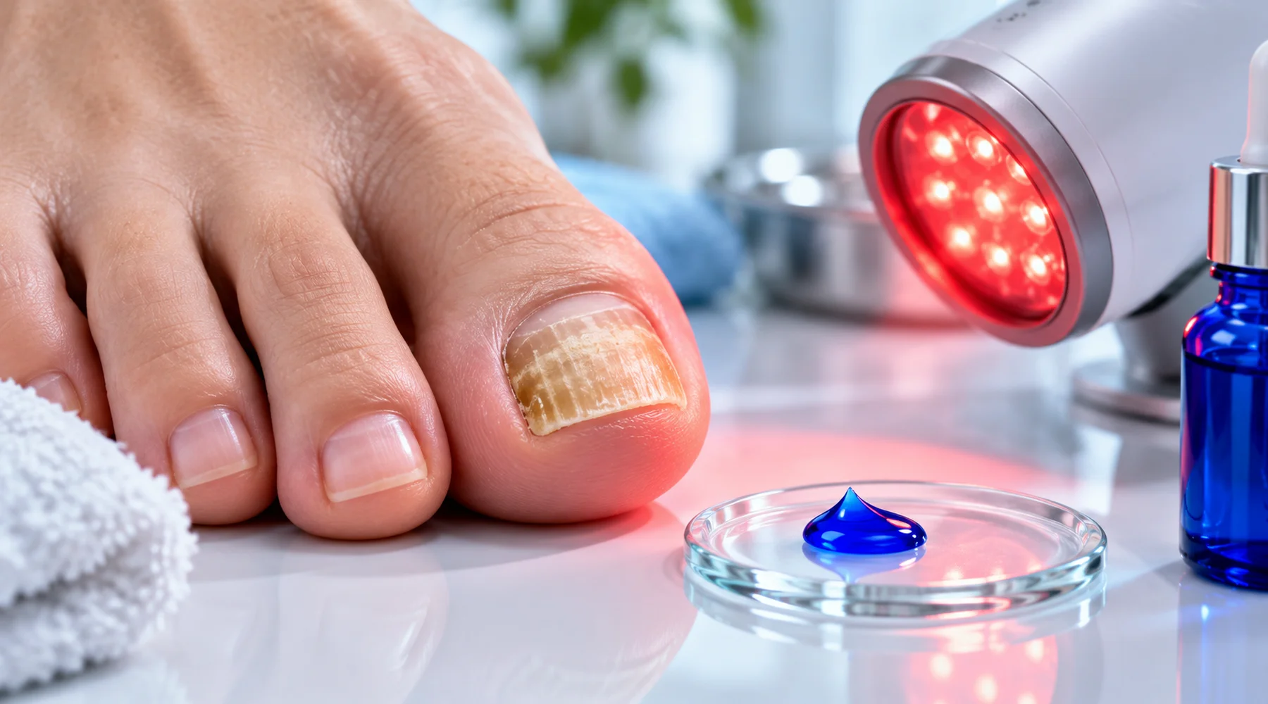
Search results for methylene blue for nail fungus tend to blur two very different things. One is a bottle of blue dye applied at home. The other is a clinic procedure in which the dye is painted on, left briefly, then activated with a measured dose of red light. Almost all of the clinical evidence belongs to the second. This article compares the three photodynamic studies directly, so you can see what was actually tested and what each one proved.
The shared mechanism is simple. Methylene blue absorbs red light strongly, and the excited molecule hands that energy to oxygen, producing singlet oxygen and other reactive species that damage fungal cells. Without light the effect is far weaker. That is why every study below pairs the dye with a specified lamp, wavelength, and dose, and why results from lit protocols say nothing about dabbing dye on a nail in daylight. The general mechanism is covered in more detail in the photodynamic therapy explainer, and the broader antifungal research context sits in the overview of methylene blue as an antimicrobial.
Toenail onychomycosis: dye plus light against weekly fluconazole
The largest trial is a randomised comparison in 80 patients with toenail onychomycosis, published in 2014. Half received photodynamic treatment and half received oral fluconazole 300 mg once weekly, each for 24 weeks.
The light protocol was specific. A 2% aqueous methylene blue solution was applied to the nail, then illuminated after a short incubation with a non-coherent red LED at 630 nm delivering 18 joules per square centimetre. Sessions repeated every 15 days, twelve times over six months. Nails with thick subungual debris, longitudinal streaks, or a dermatophytoma were mechanically abraded first, in both groups, because light and drug alike fail against a wall of keratin.
The headline result favoured the light group with statistical significance. Later extractions of the full text report roughly 90% response after the six-month course against 45% with fluconazole, falling to about 80% in the light group at twelve months after treatment ended. A stricter re-analysis that demanded all three at once, negative microscopy plus negative culture plus full regrowth of clear nail, put the final complete-cure figures at 50% against 25%. The two sets of numbers describe different endpoints, not contradictory findings, and the gap between them is the point of the next section. Treatment was reported as well tolerated, with one recorded episode of pain and burning.
Two details matter for anyone asking how to use methylene blue for toenail fungus. First, the comparator was fluconazole, a drug with modest efficacy against nail disease, not the more potent terbinafine. Second, the dye concentration was 2%, far above anything sold for home cosmetic use, applied under supervision with debridement and a calibrated lamp.
Severe toenail disease: dye plus light on top of terbinafine
The second nail study, a randomised trial in severe dermatophytic toenail infection, published in 2020, answers a narrower question. Twenty patients with more than 60% of the great toenail involved all received oral terbinafine 250 mg daily for twelve weeks plus urea softening of the plate. On top of that backbone, ten received methylene blue photodynamic treatment and ten received photodynamic treatment with a different sensitiser.
The methylene blue arm used 2% aqueous solution with about three minutes of incubation, then a 635 nm LED delivering 37 joules per square centimetre over ten minutes. Nine sessions ran at two-week intervals across sixteen weeks, with 40% urea ointment on the evenings before each session and mechanical removal of debris.
By week 52, roughly nine months after the last light session, the severity index in the methylene blue group had fallen from about 24 to under 1, nail involvement from about 65% to under 2%, and complete cure, meaning a normal-looking nail plus mycological clearance, stood at 70%. Mycological clearance itself reached 100% in the methylene blue arm against 90% with the alternative sensitiser. No local side effects from the dye and light were reported; two patients had gastrointestinal discomfort attributed to the terbinafine they were also taking.
The honest reading is that this trial cannot isolate the contribution of the dye. Every patient received a full course of the best systemic drug plus intensive nail preparation, there was no drug-only group, and the two light arms performed similarly. What it does show is that a methylene blue light protocol layered onto standard therapy produced high clearance in severe disease with long follow-up, and that the light sessions themselves added no local toxicity in this small group. Drug interactions and systemic risks of combined regimens are discussed in the safety and interaction guide.
Resistant skin infection: one patient, one stubborn fungus
The skin evidence is far thinner. A 2026 study of methylene blue light treatment against drug-resistant Trichophyton indotineae concerns a single 25-year-old patient with extensive skin lesions that had persisted for eight months despite six weeks of systemic terbinafine at 250 mg daily. Laboratory testing showed a terbinafine minimum inhibitory concentration of 8.0 micrograms per millilitre against 1.0 for methylene blue, consistent with the resistant dermatophytosis now spreading in many regions.
Lesions were treated with 1% topical methylene blue and red light at about 630 nm delivering 100 joules per square centimetre, once weekly for four weeks. After two weeks the treated lesions showed resolution of scaling with negative fungal microscopy, and by week four they were described as fully healed with only mild residual pigmentation, while lesions treated with terbinafine cream still carried visible hyphae. No severe reactions and no pain were reported.
That is a genuine mycological response in resistant disease, and it is also a single patient with four weeks of observation. There was no documented post-treatment culture conversion beyond microscopy, no recurrence follow-up at three, six, or twelve months, and no population from which a response rate can be computed. It supports further study, not a treatment recommendation. Resistant species and susceptibility testing are examined further in the guide to antifungal resistance.
The three studies side by side
| Souza 2014, toenail | Alberdi and Gomez 2020, severe toenail | Xu 2026, resistant skin lesions | |
|---|---|---|---|
| Design and size | Randomised, 40 per arm | Randomised, 10 per arm analysed | Single patient, lesions as own controls |
| Organism | Mixed dermatophytes, yeasts, moulds | T. rubrum or T. mentagrophytes | T. indotineae, terbinafine-unresponsive |
| Dye and incubation | 2% aqueous, minutes | 2% aqueous, about 3 minutes | 1% topical, light-proof cover per preprint protocol |
| Light | 630 nm LED, 18 J per cm2 | 635 nm LED, 37 J per cm2 | About 630 nm, 100 J per cm2 |
| Sessions | 12, every 15 days over 6 months | 9, every 2 weeks over 16 weeks | 4, weekly |
| Co-treatment | Nail abrasion where indicated; placebo capsule | Terbinafine 250 mg daily for 12 weeks, 40% urea, debridement | None on treated lesions; terbinafine cream on control lesions |
| Clinical outcome | About 90% response at end of treatment, about 80% at 12 months | Severity index about 24 to under 1; 70% complete cure at week 52 | Scaling resolved by week 2, healed by week 4 |
| Mycological outcome | Strict complete cure 50% vs 25% at final follow-up | 100% PAS and culture negative at week 52 | Microscopy negative at 2 weeks; no reported post-treatment culture |
| Follow-up | 12 months after treatment | About 9 months after last light session | 4 weeks, no recurrence data |
| Adverse effects | One episode of pain and burning | No local effects; terbinafine gut discomfort in 2 of 10 | No severe reactions, no pain |
Clinical cure is not mycological cure
Reading these papers requires one distinction. Clinical cure means the nail or skin looks normal. Mycological cure means microscopy and culture find no fungus. Complete cure means both at once. Nails regrow slowly, so a nail can look better months before the fungus is gone, and dead fungal material can keep a test positive after the infection is dead. The Souza trial shows the consequence directly: loose response criteria give 80 to 90%, while demanding clear nail plus negative tests gives 50%. Neither figure is wrong. They measure different things, and any claim about efficacy should say which one it cites.
What this means for treatment decisions
Three conclusions survive a strict reading. First, methylene blue with calibrated red light has produced real clearance in toenail fungus across two randomised trials, once against fluconazole alone and once layered onto terbinafine with urea preparation. Second, the one resistant-skin report is encouraging but proves almost nothing on its own; skin dermatophytosis treated with dye and light has no randomised evidence yet. Third, every positive result came from a supervised combination: pharmaceutical-grade dye at 1 to 2%, timed incubation, measured light dose, and usually debridement or a systemic drug alongside.
None of this transfers to applying retail dye at home. Home application has no measured light dose, no incubation control, no debridement, no mycology, and no follow-up, which removes every element the trials relied on. Anyone considering light-based treatment should bring these protocols to a dermatologist and ask whether supervised photodynamic treatment is available, rather than reproducing the colour while omitting the procedure.