
Article
Methylene blue and NAD+/NADH: interpreting cellular redox balance
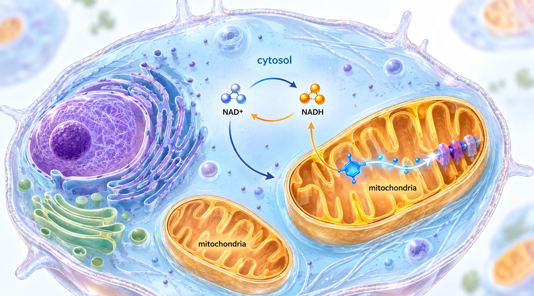
Reports on methylene blue and NAD+ tend to blur three different claims into one. The first claim is that the NAD+ to NADH ratio moved toward the oxidized state. The second is that the cell holds more total NAD. The third is that energy production recovered. These sound like one story about restoring metabolism. In the experiments they come apart regularly, and the interesting results are exactly the cases where they diverge.
The confusion is understandable because the three quantities share one vocabulary. NAD+ accepts electrons in metabolic reactions and becomes NADH, then mitochondria oxidize NADH back to NAD+ at complex I and use the released energy to power ATP synthesis. A fuller account of that electron route sits in the mitochondrial electron transport explainer, and the energy side in ATP and energy metabolism. But a ratio and a pool are different objects. The ratio says how the existing shuttle fleet is distributed between loaded and unloaded states. The pool says how many shuttle molecules exist at all. A better ratio with the same pool means redistribution, not manufacture.
Two compartments, two reporters
There is a second distinction that causes quiet misreadings. Cytosolic and mitochondrial NAD pools are largely separated by the inner mitochondrial membrane, so each compartment can run relatively oxidized or reduced on its own. A single whole cell number hides this.

Liver research made the compartment split measurable. The lactate to pyruvate equilibrium reports the cytosolic NAD+ to NADH ratio, because lactate dehydrogenase ties those two pairs together. The 3-hydroxybutyrate to acetoacetate equilibrium reports the mitochondrial ratio through a similar coupling. When both pairs are measured from freeze clamped tissue, as in the chronic ethanol feeding study in rats, the result is two redox readings from one liver, and a compound can move one more than the other.
Methylene blue acts on this system as an electron shuttle rather than as fuel. It accepts electrons from NADH near complex I, becoming reduced, then passes them downstream to carriers such as cytochrome c. The net effect is NADH oxidation without passage through the full normal route. That mechanism explains both its appeal and its ceiling: it can relieve a reductive jam, but it does not rebuild a damaged complex or synthesize new NAD molecules.
Five studies, one pattern of split results
The table below lines up the five studies by tissue, redox endpoint, energy endpoint, and what actually recovered. Read the last two columns as a pair. That pairing is the article.
| Tissue and insult | Redox endpoint with methylene blue | Energy and signaling endpoint | Functional outcome |
|---|---|---|---|
| Rat liver, chronic ethanol diet | Cytosolic and mitochondrial NAD+/NADH ratios largely restored; glycerol 3-phosphate normalized | ATP unchanged by ethanol or treatment; slight ATP dip with dye at the late time point | Fatty infiltration unchanged; lipid and triacylglycerol stayed high |
| Hepatocytes and high fat diet mouse liver | NAD+/NADH ratio and NAD+ content raised in cells and liver | SIRT1 up, PGC-1alpha deacetylated, mitochondrial DNA and oxygen use up, SIRT1 dependent AMPK activation | Hepatic fat accumulation and steatohepatitis reduced |
| Diabetic rat heart | NADH/NAD+ ratio lowered during substrate oxidation in isolated mitochondria | SIRT3 activity raised; lysine acetylation lower at 83 sites on 34 proteins | Cardiac volumes and ejection fraction improved; complex I defect itself not corrected |
| AIF deficient mouse retina and rotenone treated photoreceptor cells | NADH/NAD+ ratio normalized in cells; oxidative stress lowered | ATP deficit not corrected in cells; mitochondrial stress pathways quieted | Retinal thickness and photoreceptor markers preserved; cell survival improved |
| Human and rat Achilles tendon, oxidant and collagenase injury | NAD+ balance and total NAD chemistry restored toward control | ATP raised and ROS lowered where rescue occurred | Matrix markers improved except under direct complex I blockade, where chemistry moved but tissue did not |
Where redox correction did not rescue tissue
The oldest study in the set remains the sharpest warning. Rats fed ethanol as 36 percent of energy developed lower cytosolic and mitochondrial NAD+/NADH ratios, measured through both reporter pairs, and methylene blue at about 42 mg per kg per day in the diet largely prevented those shifts. Yet hepatic triacylglycerol roughly tripled by the late time point whether or not the dye was present, and histology showed the same centrilobular fat. The authors drew the uncomfortable inference directly: the redox shift that accompanies ethanol oxidation is not by itself what produces the fatty liver. A reader who treats a corrected ratio as a corrected organ would misreport this paper. The full design, including pair fed controls and midnight sampling at peak intoxication, is laid out in the ethanol liver study.
Contrast that with the high fat diet liver work, where the same kind of ratio movement coincided with real tissue improvement. In hepatocytes and in mouse liver after oral dosing, methylene blue raised the NAD+/NADH ratio, and the signaling chain was traced step by step: SIRT1 up, PGC-1alpha deacetylated, mitochondrial biogenesis markers and oxygen consumption higher, then AMPK, CPT-1, and PPARalpha engaged, with AMPK activation shown to depend on SIRT1. Mice on the high fat diet accumulated less hepatic fat. The proposed logic is that the redox shift here unlocked a gene regulatory axis rather than merely rebalancing chemistry, as detailed in the SIRT1 and AMPK liver study. Same measurement family as the ethanol study, opposite tissue verdict. The difference is the downstream pathway, not the ratio alone.
Heart and retina: sirtuins, acetylation, and ATP free rescue
The diabetic heart study connects the redox shift to protein acetylation through a different sirtuin. Diabetic mitochondria carry excess lysine acetylation on metabolic enzymes, which tracks substrate inflexibility. Methylene blue at 10 mg per kg per day facilitated NADH oxidation in a dose dependent way across 6 to 18 micromolar in isolated mitochondria, raised SIRT3 activity, and lowered acetylation broadly, while cardiac volumes and ejection fraction improved even though the underlying complex I impairment stayed. The paper is explicit about dose behavior elsewhere in the literature: low concentrations favor the reductive shuttle role while higher ones can reroute electrons unproductively, so the effect is hormetic rather than monotonic. The proteomic survey and cardiac measurements are reported in the diabetic heart acetylation study. The oxidative stress framing behind this sits in oxidative stress and ROS control.
The retina study pushes the separation furthest. In Harlequin mice with AIF deficiency, a complex I assembly defect, methylene blue preserved retinal thickness, photoreceptor outer segments, and markers for rods, cones, Muller, and ganglion cells. In rotenone treated 661W photoreceptor cells, the dye normalized the NADH to NAD+ ratio and lowered reactive oxygen species at 15 micromolar, yet ATP stayed depressed at the concentrations that promoted survival. Structure and survival were preserved without energy rescue, alongside quieting of the mitochondrial unfolded protein response and mitophagy. That result reframes the dye as a reliever of reductive and oxidative stress rather than an energy restorer, documented in the retinal photoreceptor protection study.
Tendon: the experiment where chemistry moved and tissue did not
The Achilles tendon work contains both patterns inside one paper and is worth reading slowly. The full branch by branch breakdown is in the 2026 Achilles tendinopathy analysis. Diseased tendon showed NAD+ depletion, a low NAD+ to NADH ratio, weak complex I markers, low ATP, and disordered collagen. Methylene blue restored NAD chemistry and ATP and improved matrix markers across rat, human explant, and cell preparations.
Then came the two perturbations. Under rotenone blockade of complex I, the dye still protected NAD+ chemistry and ATP related readouts but failed to relieve oxidant load, restore collagen balance, or preserve viability. Under AIF knockdown, where complex I machinery was underbuilt rather than pharmacologically barred, the dye rescued broadly: NAD balance, oxidative stress, membrane potential, and collagen markers. The lesson is precise. A better NAD number can be supported even when complex I is blocked, but tissue rescue in this preparation depended on a workable complex I pathway. Ratio movement is necessary in this story without being sufficient, a point developed further in the tendon redox study.
How to read the next redox claim
Three checks handle most misreadings. First, ask which compartment the ratio belongs to and which reporter pair produced it. A cytosolic lactate to pyruvate result says nothing direct about the mitochondrial matrix. Second, ask whether the paper reports a ratio shift, a larger total pool, or ATP recovery, and treat each as a separate finding. Third, ask what stayed broken: uncorrected fat, an uncorrected complex I defect, or an uncorrected ATP deficit all appear in these papers next to genuine redox improvement.
None of this is a dosing guide. All five studies are animal, cell, or tissue preparation work with controlled insults: liquid ethanol diets, streptozotocin diabetes, genetic complex I defects, rotenone, peroxide, collagenase. Concentrations in dishes do not convert to oral doses by inspection, and no human in these papers took methylene blue for redox balance. The through line for redox pharmacology and dosing is narrower and more useful than any protocol: methylene blue oxidizes NADH and shifts the measured ratio, sirtuins translate that shift into deacetylation and metabolic reprogramming in some tissues, and whether the tissue follows depends on machinery the dye cannot replace.