
Article
Does greater oxygen consumption mean more ATP? Lessons from methylene blue studies
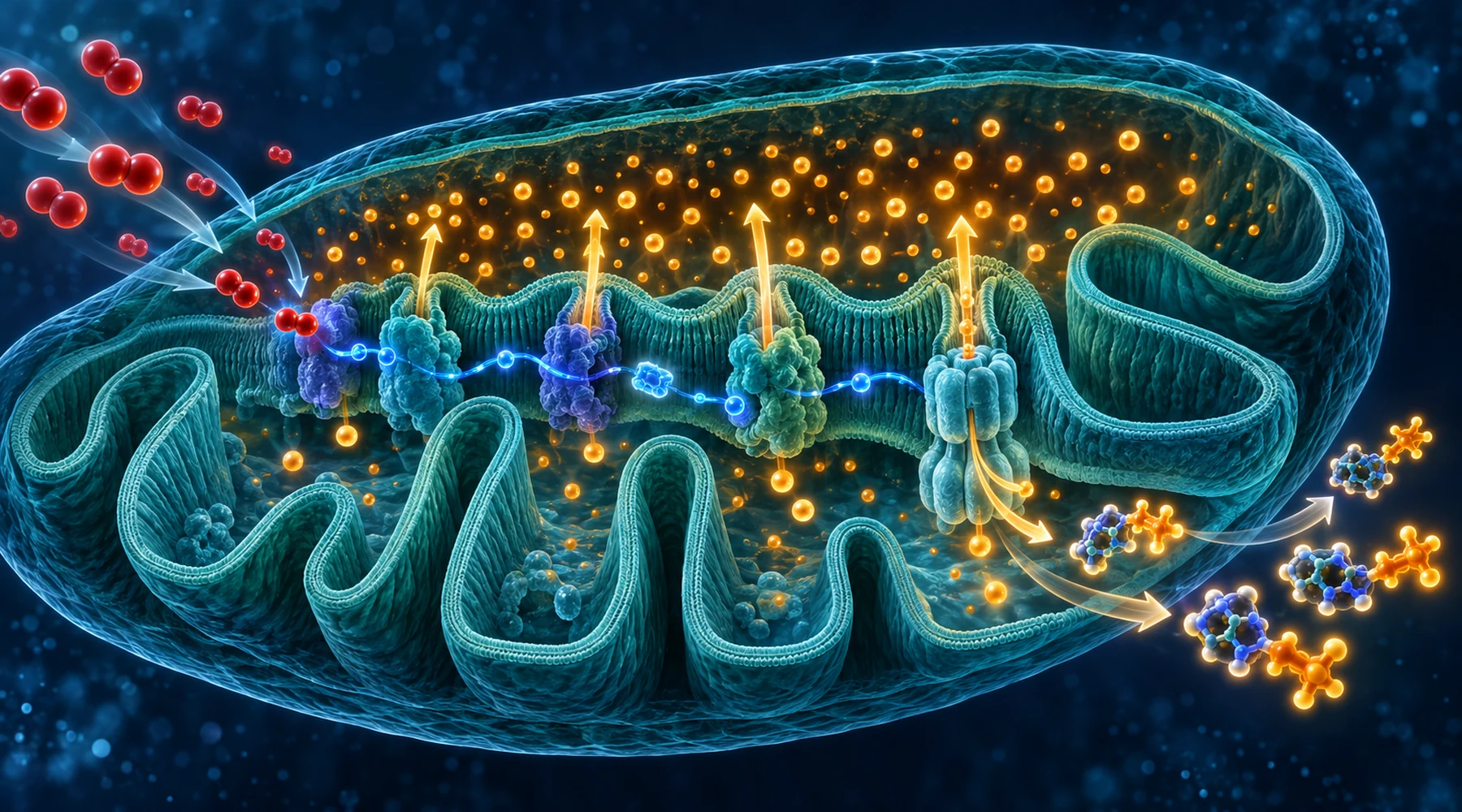
An isolated mitochondrion sits in a chamber with fuel but no ADP. It consumes oxygen slowly. Add ADP and oxygen consumption jumps several fold. Wait until the ADP runs out and oxygen consumption falls back. The fuel never changed. Only the demand for ATP did.
That small sequence contains the whole distinction this article is about. Oxygen consumption measures how fast electrons flow through the respiratory chain to oxygen. ATP production measures how much of that flow gets captured as usable energy. The two share a middle step, the proton gradient across the inner mitochondrial membrane, and anything that changes how tightly the gradient is coupled to ATP synthesis can move the two endpoints in different directions. Methylene blue is a useful teacher here because it has been caught doing both: raising oxygen use alongside ATP in some preparations, and raising oxygen use while ATP stays flat or falls in others.
Readers who want the broader relay picture first can start with how methylene blue shuttles electrons through the respiratory chain, and readers who want the vocabulary of the early oxygen experiments can read what the first respiration measurements actually showed.
Respiration, ATP, and the gradient between them
Electrons from NADH and FADH2 enter the chain at complexes I and II, pass through complex III to cytochrome c, then through complex IV to oxygen. The energy released at complexes I, III, and IV pumps protons out of the matrix into the intermembrane space. That separation of charge and acidity is the proton motive force, and its electrical part, the membrane potential, is usually the dominant part.
ATP synthase lets protons flow back into the matrix and uses the released energy to join ADP and phosphate into ATP. So the chain is:
oxygen consumption, then proton pumping, then gradient, then ATP synthesis.
The catch is that protons can return by other routes. Any return that bypasses ATP synthase is proton leak. Leak forces the chain to pump the same protons again to hold the membrane potential steady, which consumes more oxygen without making more ATP. A strong enough leak is uncoupling: oxygen use rises while ATP stays flat or drops, and the wasted energy leaves as heat.
State 3, state 4, and the respiratory control ratio
The classical vocabulary for this comes from isolated mitochondria. State 3 is respiration with substrate plus ADP present: ATP synthase drains the gradient, the membrane potential dips, the chain accelerates to rebuild it, and oxygen use is high because ATP is being made. State 4 is respiration after the ADP runs out: ATP synthase idles, the gradient rises, the high membrane potential resists further pumping, and oxygen use falls to whatever is needed to compensate for leak.
The respiratory control ratio, state 3 divided by state 4, therefore reports how tightly the preparation is coupled. A high ratio means ADP controls respiration strongly. A falling ratio with rising state 4 means leak is taking over. This is the lens through which the methylene blue results below make sense.
One mental model is worth keeping. High membrane potential slows the chain. Dissipating the potential speeds it up. The chain responds to how hard pumping has become, not directly to whether ATP is being made.
When the endpoints move together
The clearest coupled example comes from isolated guinea pig brain mitochondria with blocked respiratory complexes. At 100 nanomolar to 1 micromolar methylene blue, resting oxygen consumption rose with glutamate plus malate, succinate, or glycerophosphate as fuel, while ADP stimulated respiration was unchanged. In mitochondria poisoned at complex I or complex III, the dye modestly increased ATP production, restored membrane potential, and improved calcium uptake. The proposed route is a shortcut: the dye accepts electrons upstream of the block and hands them to cytochrome c, bypassing the damaged complex.
A second coupled case uses a different ATP source entirely. In isolated brain mitochondria with ATP synthase inhibited, methylene blue still stimulated respiration on every substrate tested. But ATP synthesis rose only on alpha ketoglutarate and glutamate, the two substrates that form succinyl CoA. With oligomycin blocking ATP synthase, the extra ATP came from substrate level phosphorylation by succinyl CoA ligase in the citric acid cycle, not from oxidative phosphorylation. The dye also restored membrane potential when complex I and ATP synthase were inhibited together. Here oxygen use and ATP moved together, but only where the substrate permitted the second ATP pathway. On succinate or malate, respiration rose without extra ATP. Same dye, same chamber, different fuel, different answer.
When the endpoints diverge
The divergence cases are just as published and just as instructive. In an isolated rat liver mitochondria study from 1997, methylene blue accelerated state 4 respiration in tightly coupled succinate respiring mitochondria, which drove the respiratory control ratio down. The effect was visible from 0.5 micromolar and strong at 5 micromolar, a concentration close to plasma levels after a 100 milligram intravenous bolus. At 5 micromolar the mitochondria also swelled, though the coupling defect appeared at lower concentrations first, which argues that the leak came before the structural damage rather than from it. In uncoupled mitochondria the dye stimulated respiration in a saturable, concentration dependent way. Oxygen use rose in both preparations. ATP synthesis per oxygen consumed did not. That is textbook uncoupling plus electron shuttling, in the same molecule.
The most striking split comes from retina. In AIF deficient mice and rotenone treated photoreceptor cells, methylene blue preserved retinal thickness, photoreceptor markers, and mitochondrial structure, and it lowered the NADH to NAD ratio and quieted stress pathways including the unfolded protein response and mitophagy. It did so without correcting the ATP shortfall. Protection without restored energy means the benefit ran through redox balance and mitochondrial integrity, not through extra ATP. Anyone equating survival of the tissue with restored energy output would misread that experiment completely.
The brain mitochondria work adds a cost column to the same ledger. The same low concentrations that improved bioenergetics also raised hydrogen peroxide generation under every condition tested and slowed its elimination, measured with a calibrated Amplex assay rather than a nonspecific dye. More ATP and more oxidant came as a pair.
A practical reading checklist
Four questions separate a coupled result from an uncoupled one.
First, which endpoint was actually measured. Oxygen consumption alone cannot report ATP. ATP synthesis, membrane potential, and the respiratory control ratio each add one piece the oxygen electrode cannot see. The guide to reading methylene blue research claims walks through this endpoint discipline in detail.
Second, which substrate fed the mitochondria. The succinyl CoA result shows that fuel choice can decide whether extra respiration can become extra ATP at all.
Third, which concentration was used. Nanomolar to low micromolar methylene blue in brain mitochondria gave modest bioenergetic gains with an oxidant cost; half a micromolar and above in liver mitochondria eroded coupling; 5 micromolar added swelling. Concentration is part of the mechanism, not background detail.
Fourth, which model carried the result. Isolated mitochondria reveal the wiring, but intact cells and tissues add transport, reduction to the colourless form, and competing sinks for electrons. Laboratory dosing work, including how concentration and protocol shape methylene blue effects, is the bridge between chamber numbers and any claim about a person.
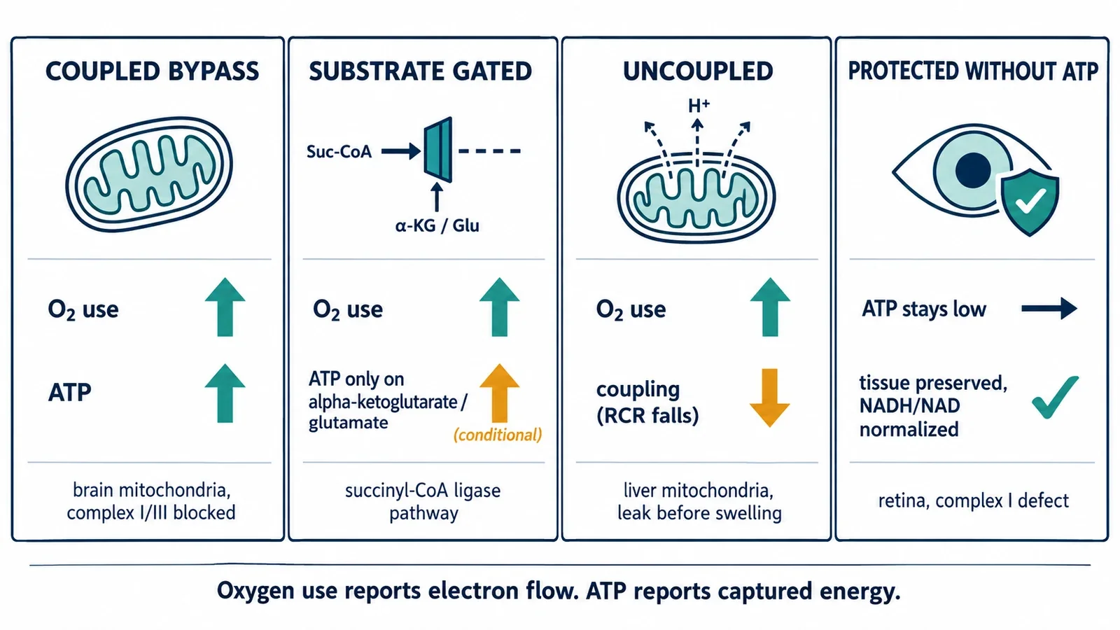
Oxygen consumption reports electron flow. ATP reports how much of that flow was captured. Methylene blue can change either one, and the experiments above show all four combinations that follow: both rise, respiration rises only where the substrate allows, respiration rises while coupling falls, and tissue is preserved while ATP stays flat. The number on the oxygen electrode is the start of the interpretation, not the end of it.