
Article
Methylene blue and inflammatory signaling: inflammasomes, IL-6, and STAT3
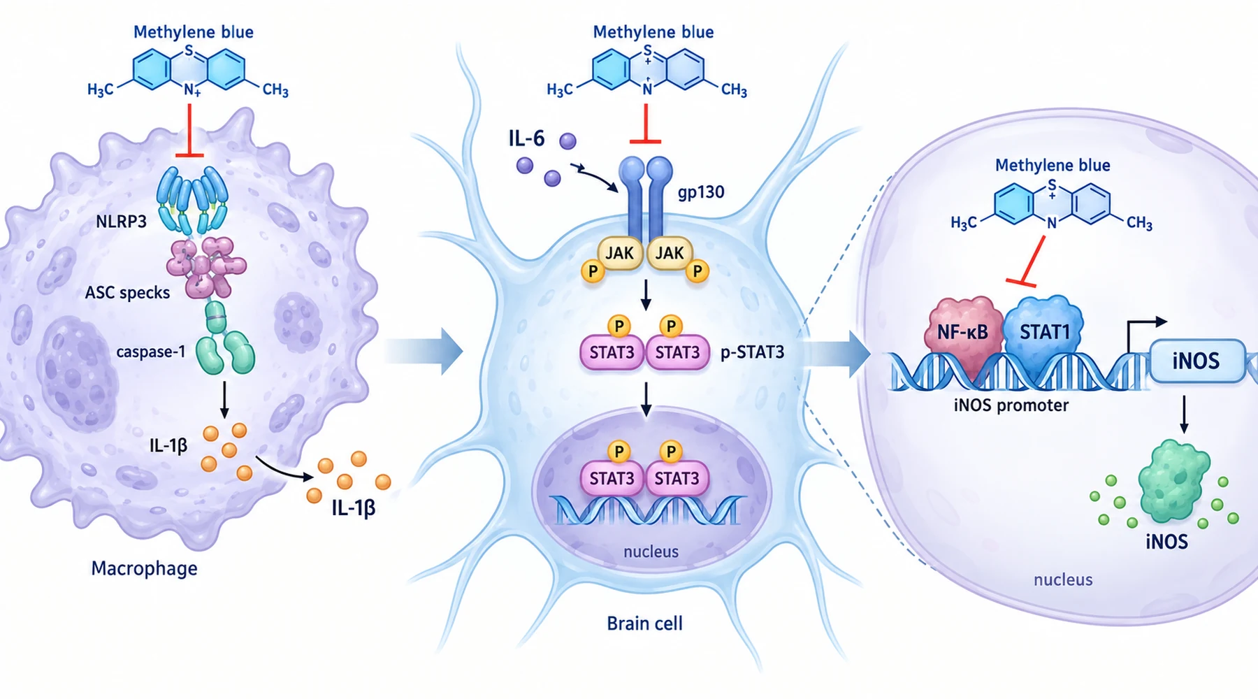
Suppose three labs report that the same blue compound is anti-inflammatory. One shows less IL-1beta from macrophages. One shows less iNOS enzyme in the same cell type. One shows less IL-6 in blood and less STAT3 activity in brain. The temptation is to merge the three into one claim. That merge hides the finding.
Each result belongs to a different signaling concept with its own trigger, assembly step, and readout. Methylene blue meets each concept at a different point. Whether any of this improves a health outcome is a separate question, handled in our overview of methylene blue and inflammation. This page stays with the pathways.
Pathway 1: inflammasome assembly and caspase-1
An inflammasome is a protein scaffold built on demand inside a myeloid cell. The parts are a sensor, an adaptor called ASC, and an effector enzyme called caspase-1. Once assembled, caspase-1 cuts pro-IL-1beta and pro-IL-18 into their secreted forms and can trigger pyroptosis, an inflammatory form of cell death. The sensor defines the type: NLRP3 responds to crystals and pore forming toxins such as nigericin and ATP, NLRC4 responds to bacterial flagellin, AIM2 responds to cytosolic double stranded DNA, and the non-canonical pathway responds to intracellular lipopolysaccharide.
Activation needs two signals. Signal one is priming: a Toll like receptor ligand drives NF-kB dependent transcription of NLRP3 and pro-IL-1beta so the parts exist. Signal two is assembly: a second trigger causes ASC to oligomerize into specks, caspase-1 to be cut into its p20 fragment, and mature IL-1beta p17 to be released. Mitochondrial reactive oxygen species and crystal uptake are known contributors to signal two for NLRP3.
The 2017 macrophage study of broad inflammasome inhibition tested methylene blue at both signals. In lipopolysaccharide primed mouse bone marrow macrophages, nigericin, ATP, and monosodium urate crystals each induced IL-1beta and caspase-1 release plus ASC speck formation, and methylene blue at 20 to 200 micromolar reduced all three in a dose dependent way without killing the cells. Flagellin and Salmonella for NLRC4, double stranded DNA and Listeria for AIM2, and transfected lipopolysaccharide or live E. coli for the non-canonical pathway showed the same pattern. The compound alone did not trigger release, and control plates showed no antibacterial effect, so the result sits with the host response.
Upstream, three interventions were measured. Rotenone induced mitochondrial superoxide fell with methylene blue, and rotenone driven IL-1beta release fell with it. Uptake of fluorescent beads fell, consistent with reduced phagocytosis before crystal sensing. A reporter driven by the NLRP3 promoter with two NF-kB sites showed lower activity with methylene blue, while a truncated promoter without those sites did not respond. In a cell free tube, methylene blue also directly lowered recombinant human caspase-1 activity. Adding the compound only at priming or only at activation still reduced IL-1beta, IL-18, and caspase-1 release, so both steps are involved.
The mouse tests matched this selectivity. A lethal lipopolysaccharide dose killed controls within hours while methylene blue improved survival; peritoneal IL-1beta fell but IL-6 in the same fluid did not. In a Listeria peritonitis model, peritoneal IL-1beta fell while total cell counts were unchanged. Human THP-1 monocytes behaved like mouse macrophages across all four inflammasome types. An inflammasome result explains IL-1beta and IL-18 maturation, not every cytokine, which is also why brain immune cell shape in microglia and astrocyte experiments answers a different question from scaffold assembly.
Pathway 2: iNOS transcription through NF-kB and STAT1 binding
Inducible nitric oxide synthase, or iNOS, is barely present at rest and is transcribed on demand. Lipopolysaccharide drives it mainly through IkB-alpha degradation followed by NF-kB phosphorylation and nuclear entry. Interferon-gamma drives it mainly through Janus kinase phosphorylation of STAT1 followed by STAT1 nuclear entry. Each factor then binds its site on the iNOS promoter. Enzyme activity and gene induction are separate control points, and a drug can act at either or both.
The 2015 study of iNOS induction in macrophages and endotoxemic mice placed the effect at the message. In RAW 264.7 cells and primary mouse macrophages, 1 micromolar methylene blue given 30 minutes before lipopolysaccharide, interferon-gamma, or both reduced iNOS protein and mRNA across 8 to 24 hours, with lower nitrite in the medium and no viability loss. In mice given 25 mg per kg lipopolysaccharide, a single 5 mg per kg dose one hour earlier reduced iNOS protein and mRNA in lung, liver, and heart.
The distinctive detail is what did not change. IkB-alpha degradation, NF-kB phosphorylation, STAT1 phosphorylation, and nuclear accumulation of either factor stayed intact. The break came one step later: nuclear extracts showed less NF-kB binding to its DNA element after lipopolysaccharide and less STAT1 binding after interferon-gamma, and chromatin immunoprecipitation confirmed less occupancy of either factor at the iNOS promoter inside cells. Tumor necrosis factor alpha, whose promoter also uses NF-kB, fell as well. The factors were activated and arrived, but engaged their target DNA poorly.
Note the contrast with pathway 1. This concerns factor to DNA engagement, uses STAT1 rather than STAT3, and reads out mRNA, protein, and nitrite rather than ASC specks and caspase-1 fragments. Our map of methylene blue targets across cell models keeps this transcriptional level separate from direct enzyme and mitochondrial measurements.
Pathway 3: serum IL-6 and tissue STAT3 activation
IL-6 signals through the gp130 receptor family into the Janus kinase to STAT3 cascade. Phosphorylated STAT3 dimerizes, enters the nucleus, and drives inflammatory programs, while IL-6 itself is an NF-kB target, so the two form a feedback loop often called the IL-6 to STAT3 axis. The standard readouts are serum IL-6 upstream, the ratio of phosphorylated to total STAT3, and effector enzymes such as iNOS and COX2 downstream.
The 2023 mouse study of IL-6 and STAT3 after lipopolysaccharide measured the axis across compartments. Male C57BL/6 mice received 1 mg per kg lipopolysaccharide daily for three days, with 5 or 10 mg per kg methylene blue 30 minutes after each dose. On day three, serum IL-6 was lower in both methylene blue groups than with lipopolysaccharide alone, while most other cytokines in the panel did not move with treatment. Phosphorylated to total STAT3 ratios were lower in cortex, hippocampus, and ear skin, with lower Iba-1 microglial staining, lower iNOS and COX2, and partly attenuated weight loss.
Two cautions belong here. Lower serum IL-6 plus lower tissue STAT3 phosphorylation fits axis modulation but does not isolate the primary node, since less upstream cytokine, less receptor signaling, or altered phosphatase activity could each produce the same ratio. And weight over three days of acute challenge is a whole animal readout, not a symptom or remission score. It adds context beyond a blot without converting a signaling result into a treatment claim. The separate chemistry of mitochondrial redox handling runs in parallel to this cytokine account and should not be merged with it.
Pathway comparison: intervention points and readouts

| Pathway | Trigger in these papers | Where methylene blue acts | Readout that changed |
|---|---|---|---|
| Inflammasome assembly | Nigericin, ATP, urate crystals, flagellin, DNA, cytosolic lipopolysaccharide | Less priming transcription, less mitochondrial ROS, less phagocytosis, less ASC assembly, direct caspase-1 inhibition | Less caspase-1 p20, less mature IL-1beta and IL-18, fewer ASC specks |
| iNOS transcription | Lipopolysaccharide via NF-kB; interferon-gamma via STAT1 | Less NF-kB and STAT1 binding to promoter DNA despite normal activation and nuclear entry | Less iNOS mRNA and protein, less nitrite |
| IL-6 to STAT3 axis | Three day lipopolysaccharide in mice | Lower serum IL-6 with lower tissue STAT3 phosphorylation | Lower pSTAT3 to STAT3 ratio in cortex, hippocampus, skin; lower iNOS, COX2, Iba-1 |
The table blocks three invalid transfers. An ASC speck result does not prove a STAT3 result. A STAT1 to iNOS promoter result does not prove a STAT3 result, even though both proteins are STAT family members. A serum IL-6 result does not prove reduced inflammasome assembly: in the peritoneal experiment IL-1beta fell while IL-6 did not. Each row needs its own trigger and readout, which is why reading cell, animal, and clinical methylene blue studies insists on matching each claim to its measured level.
None of this establishes dosing, safety, or benefit in people. Lipopolysaccharide models are controlled triggers for testing a mechanism, not replicas of chronic illness, and dosing alongside the trigger tests prevention of an induced change rather than reversal of established disease. Keep this mechanism account separate from patient outcomes, and judge any health claim by the patient study behind it.