
Article
Methylene blue in G6PD laboratory tests: what the dye is actually doing
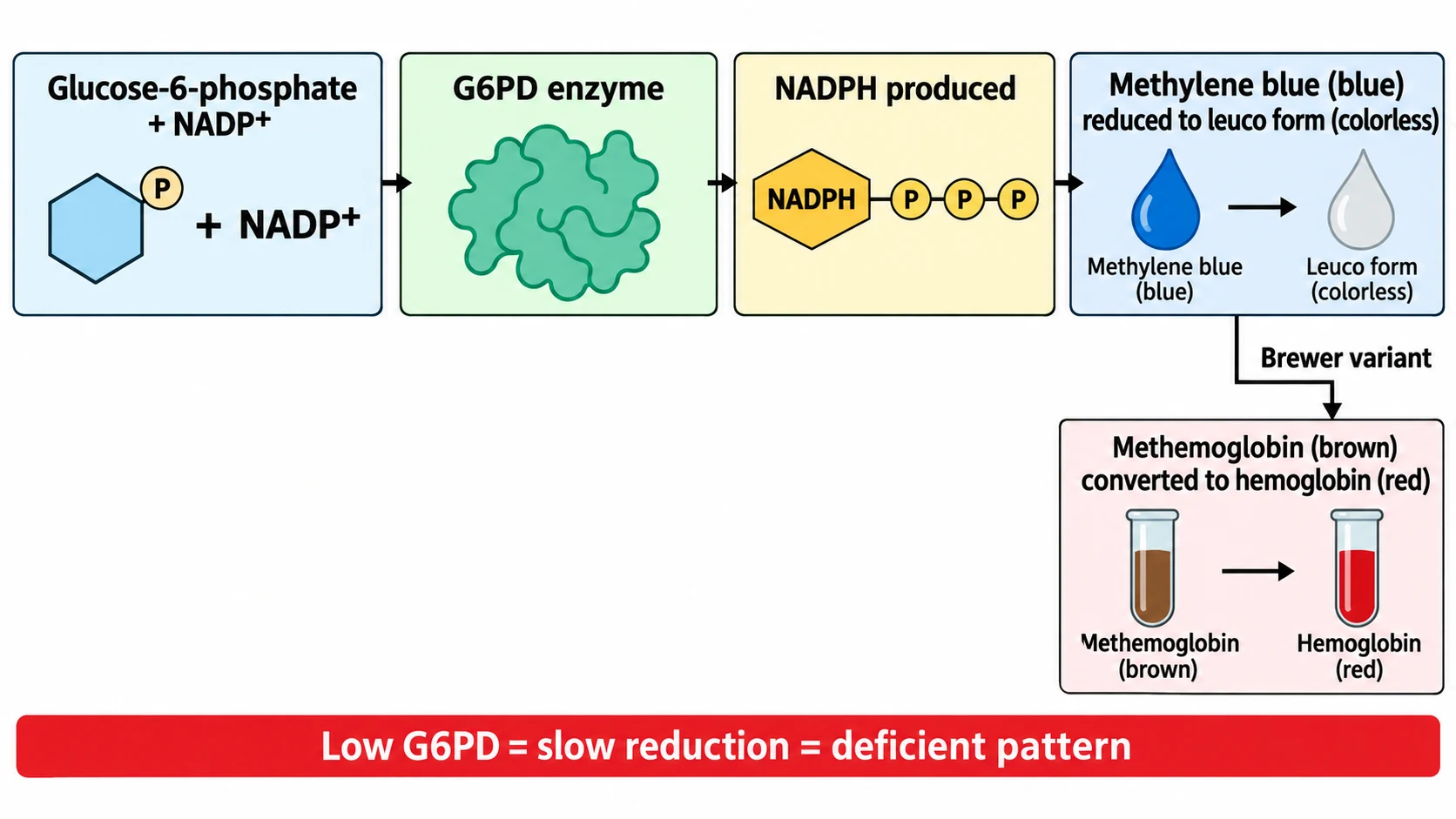
A student watches two tubes after incubation. One has turned the expected color. The other has not. The student concludes the second patient is deficient in glucose-6-phosphate dehydrogenase. That conclusion may be right, but the reasoning skips the concept that makes the test work. Nothing in the tube measures the enzyme directly. The tube measures what the enzyme produces, using methylene blue as a go-between.
Once that indirection is clear, the procedure, the controls, and the long list of false results all stop looking arbitrary.
The short answer
Methylene blue in G6PD testing is a redox indicator. The enzyme G6PD generates NADPH, and NADPH-dependent systems reduce blue methylene blue to its colorless leuco form (or use it to shuttle electrons). Normal enzyme activity gives efficient reduction. Deficient activity gives slow or incomplete reduction. That is the whole signal.
Two historical methods share the dye but differ in endpoint. Keep them apart and most confusion disappears.
Two tests, one dye, different endpoints
The first is the direct methylene blue reduction test, described by Sass and colleagues and later evaluated by Gibbs. Here the endpoint is the dye itself: oxidized blue methylene blue is reduced toward colorless leucomethylene blue, and the rate of that change reflects NADPH-generating capacity. The primary screening paper is indexed on PubMed under Sass et al. (1966).
The second is the Brewer methemoglobin reduction test. Sodium nitrite first oxidizes hemoglobin to brown methemoglobin. Methylene blue then acts as an electron-transfer mediator: reduced methylene blue passes reducing equivalents to methemoglobin, converting it back to red hemoglobin. The visible endpoint is therefore brown versus red, not blue versus colorless. The original method appeared in the Bulletin of the World Health Organization (Brewer et al., 1960).
Calling the Brewer assay a dye decolorization test is chemically imprecise. The dye cycles; hemoglobin changes color. The diagram below traces both pathways.

The underlying reaction
G6PD catalyzes the first oxidative step of the pentose phosphate pathway:
Glucose-6-phosphate + NADP+ becomes 6-phosphogluconolactone + NADPH + H+
Mature red cells depend heavily on this pathway for NADPH, as reviewed in Blood by Luzzatto and colleagues (2020). NADPH keeps glutathione reduced and protects the cell against oxidative damage. Methylene blue inserts itself into this economy as an electron acceptor. Adequate G6PD gives adequate NADPH, which gives efficient reduction. Low G6PD breaks the chain at its first link.
Think of the dye as a fuel gauge, not the engine. It reports on NADPH supply. Anything else that alters that supply, or the readout, can move the needle without any change in the gene.
How the Brewer-type procedure runs
Reagent recipes have varied across decades, so a teaching description must not be mistaken for a clinical protocol. Every laboratory follows its own validated standard operating procedure. With that caution, the classical tube setup works as follows, in a form documented with three tubes and 3-hour incubation in this published implementation:
- Patient tube: whole blood plus glucose, sodium nitrite, and methylene blue. Nitrite creates methemoglobin, glucose feeds red-cell metabolism, and the dye provides the NADPH-dependent shuttle.
- Oxidized control: blood plus nitrite and glucose but no methylene blue. It shows the brown methemoglobin endpoint, the deficient pattern.
- Hemoglobin control: blood plus water. It shows the red hemoglobin endpoint, the normal pattern.
- Incubate all tubes near 37 C, typically for 3 hours, mixing gently.
- Dilute a small aliquot of each tube into distilled water to lyse the cells and make the color comparison readable.
- Compare the patient tube against the two controls. Return toward red indicates substantial reducing capacity. Persistence toward brown suggests marked deficiency.
One historical formulation used about 1 mL of blood with reagents near 180 mM sodium nitrite, 280 mM dextrose, and 0.4 mM methylene blue. Treat those numbers as an example from the literature, not a universal specification.
Reading the result
| Pattern | Brewer appearance | Meaning |
|---|---|---|
| Normal | Patient tube approaches the red hemoglobin control | Reducing capacity above the screen threshold |
| Deficient | Patient tube stays brown, near the methemoglobin control | Screen suggests marked G6PD deficiency |
| Intermediate | Color falls between the controls | Do not assign a phenotype; measure activity quantitatively |
A qualitative screen discriminates best around 30 to 40 percent of normal activity, as discussed in this Malaria Journal review of G6PD detection gaps. A normal screening result therefore means the assay did not detect severe deficiency. It does not certify fully normal activity.
Why screening misleads
Reticulocytosis after hemolysis is the classic false normal. Young red cells and reticulocytes carry more G6PD than old cells. During an acute hemolytic episode the oldest, most deficient cells may already be destroyed, leaving a young population whose activity tests normal. The same biology fools quantitative assays, not only dye screens.
Recent transfusion masks deficiency because donor red cells contribute their own enzyme to the specimen. Mayo Clinic Laboratories lists transfusion among the causes of unreliable enzyme results, alongside reticulocytosis and leukocyte interference.
Heterozygous females are the hardest case. Random X-chromosome inactivation produces a mixture of normal and deficient red cells, and overall activity can land anywhere from severely deficient to apparently normal. Qualitative screens routinely miss intermediate phenotypes. The WHO documentation on G6PD testing discusses this limitation in detail.
Specimen age, storage temperature, visual endpoint subjectivity, hematocrit, and reagent quality add analytical noise on top of these biological effects.
Screening is not diagnosis
A deficient, intermediate, or clinically discordant screening result calls for a quantitative G6PD enzyme assay. WHO regards temperature-controlled quantitative spectrophotometry as the reference approach. In that assay, a red-cell hemolysate receives glucose-6-phosphate and NADP+, and the NADPH formed is tracked by rising absorbance at 340 nm. The rate of change is proportional to enzyme activity, usually normalized to hemoglobin as units per gram. Current clinical-laboratory practice is summarized in the Mayo test catalog entry for G6PD activity.
Interpretation uses the laboratory validated reference range, often framed relative to population normal activity, with deficiency commonly discussed near the 30 percent threshold and intermediate activity above it. The WHO target product profiles for G6PD tests (2022) set out that diagnostic framework. If recent hemolysis or transfusion makes phenotyping unreliable, repeat testing after the red-cell population re-equilibrates, and molecular testing can add information without replacing the enzyme measurement.
A boundary worth keeping
Methylene blue in these assays is an ex vivo laboratory reagent. That fact belongs to a different discussion than the safety of giving methylene blue to a patient, which is covered in guidance on methylene blue contraindications. The biochemistry connects them, since the drug also depends on NADPH-dependent reduction, but a dye in a test tube is not a dose in a bloodstream. For context on how the dye behaves as a stain in a different setting, see methylene blue staining principles, and for the chemistry family it belongs to, redox dyes in the laboratory.
Screening asks whether reducing capacity looks intact. Confirmation asks how much enzyme is actually there. The dye serves the first question well, provided nobody asks it to answer the second.