
Article
Methylene blue for DNA and RNA staining: gels and membrane applications
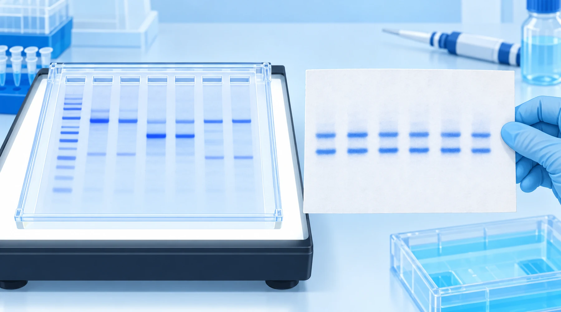
A student runs an agarose gel, stains it with methylene blue, and sees nothing where a bright band was expected. In the next bay, a postdoc stains a Northern membrane with the same blue bottle and gets sharp ribosomal bands in three minutes. The student concludes the gel failed. The gel did not fail. The two tasks were different, and the same dye answers them with different sensitivity, different background, and different rules for what happens next.
Methylene blue DNA stain and methylene blue RNA staining share chemistry but not purpose. In a gel, the dye reports how much nucleic acid sits inside a porous matrix. On a membrane, it reports whether transfer worked and whether lanes were loaded evenly. Once that split is clear, the protocols, detection limits, and compatibility limits fall into place.
The short answer
Methylene blue is a planar cationic dye that binds nucleic acids noncovalently and reversibly, through a mix of electrostatic attraction to phosphate, intercalation between base pairs, and minor groove association. That reversibility is the point. Bands can be stained with visible light, documented, then destained or cleaned up for cloning, hybridization, or reprobing.
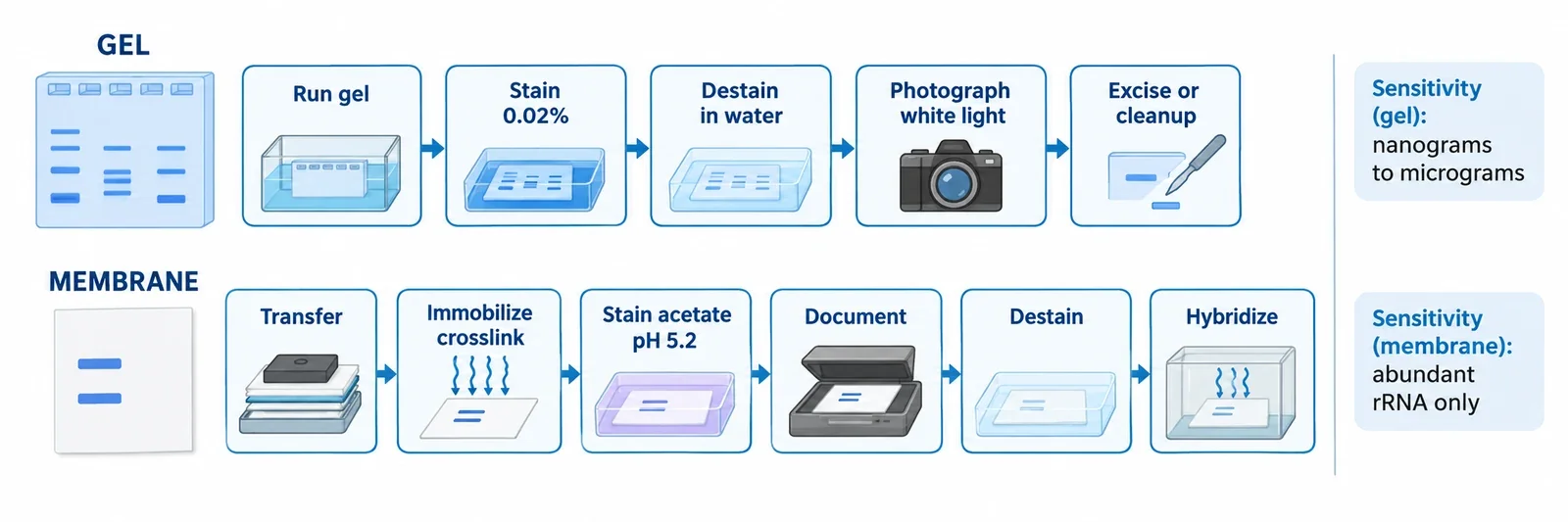
For methylene blue agarose gel staining of double stranded DNA, the standard rapid recipe is 0.02 percent w/v (200 micrograms per mL) in water, about 15 minutes of staining plus about 15 minutes of water destaining, viewed on a white light box. For membranes, the classic Northern blot recipe is 0.02 to 0.04 percent dye in sodium acetate near pH 5.2, several minutes of staining after transfer and immobilization, then a water rinse and full destaining before hybridization. Sensitivity is modest: tens to hundreds of nanograms per gel band under good conditions, and conservative guides list 0.5 to 1 microgram. That is orders of magnitude above ethidium bromide and SYBR stains. On membranes there is no useful single transcript detection limit because the stain mainly shows abundant ribosomal RNA and total loaded material. Do not substitute a retail supplement solution into either recipe. Validated methods assume analytical grade dye, defined buffers, and RNase free handling where RNA is involved.
How binding works, and where it misleads
The useful model has three parts. First, the positively charged ring system is attracted to the negatively charged phosphate backbone. Spectroscopic work finds this electrostatic term dominant at low salt, while modeling and molecular dynamics keep intercalation and minor groove binding as stable modes for double stranded DNA, as summarized in a spectroscopic study of methylene blue DNA binding. Second, some GC preference exists, with modeling favoring CpG and GpC steps and experiments reporting preferential association with GC rich duplexes in work on methylene blue intercalation preferences, but AT duplexes also bind. Routine staining is not sequence specific. Third, single stranded DNA and RNA bind mainly through electrostatics and stacking with accessible bases and duplex regions, which is why staining strength varies with length, sequence, and secondary structure.
Two consequences follow. Because binding is noncovalent, dye washes out. Polyacrylamide bands fade quickly unless deliberately fixed, and a dedicated permanent fixation variant exists precisely because normal staining is temporary, as described by Soto and Draper for PAGE gels. Because the dye is a photosensitizer, long bright illumination of stained DNA in the presence of oxygen can oxidize guanine and eventually nick strands, a mechanism detailed in work on methylene blue sensitized guanine oxidation. Brief white light viewing avoids the well known UV damage of classic ethidium bromide workflows, and that advantage is real for preparative recovery. Visible light is gentler, not harmless. Keep illumination short when the DNA will be cloned. For the wider photochemistry behind that caution, see the two pathways in methylene blue photochemistry. For the redox pair and charge states that explain why the oxidized form binds and the reduced form does not, see the chemistry of methylene blue structure and properties.
Task one: staining inside gels
Gel staining answers one question: is there enough nucleic acid in this band to see by eye. Double stranded DNA in agarose is the best characterized case. The original methylene blue gel method of Flores and colleagues validated the 0.02 percent rapid protocol on 1 percent agarose and showed successful recovery and cloning afterward. The same supplier protocol appears on the IBI Scientific methylene blue product page, which lists the 0.04 percent membrane recipe and the rapid gel variant side by side.
Practical workflow:
- Run the gel normally. Stain after electrophoresis, not during it, unless transfer or resolution has been validated for a modified method.
- Immerse in 0.02 percent w/v methylene blue in distilled water, with gentle agitation, about 15 minutes.
- Destain in several changes of water, about 15 minutes total, until the matrix background drops and bands stand out.
- Photograph promptly on a white light illuminator.
Agarose and polyacrylamide behave differently. Agarose retains dye in the matrix, so destaining is the slow step. Thin polyacrylamide gels stain fast but also lose dye fast, which helps preparative excision and hurts archival records. Synthetic oligonucleotides in polyacrylamide can be located with the same 0.02 percent recipe in 20 to 30 minutes with repeated water washes, following established Current Protocols style PAGE methods. Very dilute variants near 0.0002 percent in dilute TBE have been reported for preparative oligos over several hours, but the evidence base is weaker.
DNA versus RNA matters. Double stranded DNA gives the strongest structured binding. RNA and single stranded DNA stain, but more weakly and less predictably. For formaldehyde agarose RNA gels, most laboratories skip in gel blue staining and move the RNA to a membrane first, then stain there. That move trades a weak gel signal for a sharper membrane signal plus a loading record.
Task two: staining on membranes
Membrane staining answers a different question: did transfer work, and are lanes comparable. The classic application is the Herrin and Schmidt reversible Northern blot stain, originally 0.04 percent dye in 50 mM sodium acetate at pH 5.0 for 2 to 3 minutes with a water rinse, removable with dilute SDS in SSPE before hybridization.
The widely used modern variant runs as follows:
- Transfer RNA or denatured DNA to nylon or nitrocellulose, then immobilize by UV crosslinking or baking so the sample is not lost during staining.
- Optional but common: wet the membrane in 5 percent acetic acid for about 15 minutes.
- Stain 5 to 10 minutes in 0.02 to 0.04 percent methylene blue in 0.3 to 0.5 M sodium acetate near pH 5.2.
- Rinse and destain in water for 5 to 15 minutes, document, then remove residual dye before hybridization.
Positively charged nylon dominates Northern work. Nitrocellulose also works with the same acetic acid plus acetate workflow. The image is used to confirm transfer, to compare 28S and 18S bands (or 23S and 16S for bacterial RNA), to mark ladders and standards, and to keep a rough total RNA normalization record. Published Northern methods quantify stained ribosomal bands for loading checks, including dairy science work that used methylene blue ribosomal bands for normalization. Southern membranes can be visualized the same way, but the dye is far more established as a Northern loading and transfer check, and Southern DNA is denatured before transfer, so gel sensitivity figures do not carry over.
Detection limits and background: where blue loses
The comparison below is approximate by design. Stain performance depends on gel percentage, band width, optics, and destaining discipline. Treat each number as an order of magnitude, not a specification.
| Stain and format | Approximate detection per band | Reading |
|---|---|---|
| Methylene blue, dsDNA in agarose | 40 to 250 ng optimized; 0.5 to 1 microgram conservative | Good enough for preparative loads and teaching gels. Weak for trace bands. |
| Methylene blue, short ssDNA or RNA or oligos in PAGE | Roughly 0.1 to 0.5 microgram | Length and structure dependent. No universal limit. |
| Ethidium bromide, dsDNA agarose | About 1 to 10 ng | Roughly fourfold more sensitive than optimized blue in the Flores comparison. Needs UV. |
| SYBR Green I, dsDNA | About 60 pg | Fluorescence imaging required. |
| SYBR Gold, DNA and RNA | About 25 pg | More than tenfold more sensitive than ethidium bromide in several formats. |
| Silver stain, PAGE nucleic acids | About 0.3 to 0.9 ng; 100 pg RNA reported in an optimized blue to silver sequence | Very sensitive but chemically involved and poor for recovery. |
| Toluidine blue, PAGE RNA and oligos | Hundreds of ng working amounts, about 250 ng documented | Visible light like methylene blue, but no standardized limit comparable to SYBR specs. |
Background control decides whether the modest signal is usable. Free dye is intensely colored, so contrast comes from removing unbound dye without stripping bound dye. More concentrated stain rarely adds sensitivity. It mainly adds destaining time. Run a dilution series on the exact gel, membrane, and camera, and use only the range where integrated density stays proportional to input. Methylene blue supports semiquantitative loading normalization. It is not a high dynamic range quantitative assay because membrane capacity, transfer efficiency, saturation, and destaining vary lane to lane.
Compatibility limits: what survives staining
Cloning after blue staining is demonstrated. Flores and colleagues excised stained agarose bands, recovered the DNA, and ligated and transformed it, reporting better cloning than after ethidium bromide with UV. The likely reason is less photodamage, not a benefit from the dye itself. Keep white light exposure brief and pass recovered DNA through a normal gel extraction cleanup.
PCR and sequencing have thinner evidence. Residual dye binds nucleic acids, so destain and clean up before enzymatic steps. Properly purified DNA from a blue gel is expected to work, but direct carryover tolerance across polymerases and sequencing chemistries is not well quantified. Do not assume it.
Blot hybridization is compatible when the order is strict: transfer, immobilize, stain, document, destain, hybridize. That sequence is the Herrin and Schmidt design. Staining before transfer, or skipping destaining, is a different method and needs its own validation. After dye removal, standard probe stripping and reprobing proceed normally. For cell level staining with the same dye family and why fixed film logic does not transfer to live cell methods, see the microscopy staining atlas.
Band excision works for preparative agarose and for PAGE oligos located with blue, provided the permanent fixation variant is avoided. Electroelution after destaining is chemically plausible for a reversible binder, but controlled yield data specific to methylene blue are sparse. Desalt or precipitate after elution.
Failures that repeat
Faint gel bands usually mean too little mass for a colorimetric stain, especially for short single stranded or RNA targets. Load more, use fresh stain, extend staining before raising concentration, and stop destaining earlier. A solid blue gel with no contrast usually means overstaining or too few water changes. Fading PAGE bands are expected chemistry, not a failed stain. Photograph fast. Uneven membrane background points to folds, bubbles, fingerprints, uneven agitation, or patchy transfer. Do not read that as unequal loading without a hybridization control. A smeared weak ribosomal pattern can mean degradation, but also incomplete transfer, too little total RNA (typical lanes carry several to tens of micrograms), or excessive destaining. The dye reports abundant ribosomal RNA. It does not report a specific transcript. That job belongs to the probe.
Safety and handling without overclaiming
Methylene blue is simpler to handle than ethidium bromide, but it is not innocuous. A current MilliporeSigma safety data sheet classifies the dye as acute oral toxicity Category 4, harmful if swallowed, with disposal through an approved waste route per the Sigma-Aldrich methylene blue SDS. Ethidium bromide carries stronger genotoxicity warnings, including suspected germ cell effects in its Sigma-Aldrich ethidium bromide SDS, and its gels and solutions are commonly segregated as hazardous chemical waste as noted in the Thermo Fisher nucleic acid stain guide. Follow the SDS and institutional rules for both. Keep dry dye tightly closed and dry, reuse working stain only as the protocol allows, store stained membranes dry between filter sheets, and photograph gels before the bands diffuse away.
Remember the bench sentence. Gels test visibility at high load under white light. Membranes test transfer and loading before the probe does the sensitive work. One dye serves both, with modest sensitivity, reversible binding, and strict order of operations.