
Article
Methylene blue and ALS: why results differ across experimental models
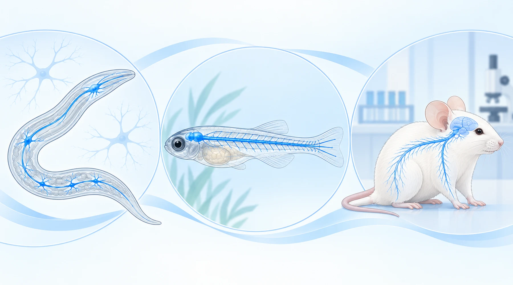
Search for methylene blue ALS and you find two claims that seem to contradict each other. One says the blue dye protected neurons in ALS models. The other says it did nothing in an ALS mouse. Both claims cite real papers, and both papers are worth taking seriously. The contradiction dissolves once you state the concept behind each experiment: the model is the claim.
A result in a worm is a claim about a worm. A result in a larval fish is a claim about a larval fish. Neither is a claim about a patient until a mammalian disease course says so. This article compares the two most cited papers side by side so you can see why their answers differ.
The two experiments in one table
Keep this table open while you read. Every important difference between the studies fits in it.
| TDP-43 and FUS in worm and fish | SOD1 in mouse | |
|---|---|---|
| Paper | Vaccaro and colleagues, 2012, in PLoS ONE | Lougheed and Turnbull, 2011, in PLoS ONE |
| Organism | C. elegans roundworm and zebrafish larvae | Congenic high copy human SOD1 G93A mouse |
| ALS genetics modeled | Mutant human TDP-43 (A315T in worms, G348C in fish) and mutant FUS (S57Δ in worms, R521H in fish) | Mutant human SOD1 G93A, a cause of familial ALS |
| Exposure | Bath exposure: 6 or 60 μM on plates from hatching, 600 μM in acute liquid screen, 30 to 60 μM overnight for fish embryos | About 25 mg/kg per day by mouth in flavored gelatin, daily from age 45 days until end stage |
| Endpoints | Paralysis, motor neuron process degeneration, synaptic assay, fish swimming and axon shape, oxidative stress readouts | Time to neurological onset, ordinal disease stage durations, survival to a defined end stage, body weight |
| Headline result | Broad rescue of movement and neuron structure | No significant delay in overall onset and no significant survival extension |
The genetics differ, the species differ, the dosing route differs, the timing differs, and the endpoints differ. Genotype is only one of five differences, so the pair cannot prove that methylene blue works selectively on one ALS pathway. It proves something narrower and still useful: strong rescue under early direct exposure in simple models did not carry into a chronic oral mammalian disease course.
The positive result: what the worm and fish study showed
The 2012 worm and fish study expressed mutant human TDP-43 or FUS in worm motor neurons and injected mutant human mRNA into fish embryos. These are proteotoxicity models. They reproduce toxic effects of the mutant protein on movement and neuron shape over days, not the full course of adult human ALS over years.
Treatment started very early. Worms grew on agar containing methylene blue from hatching. Fish embryos sat in drug solution overnight starting at 24 hours after fertilization, with behavior tested around two days later. No blood brain barrier stood between drug and neuron in the way it does in a mammal, and no plasma or brain drug level was measured.
Under those conditions the effects were large and consistent. Both 6 and 60 μM reduced age dependent paralysis in TDP-43 and FUS worms. At adult day 12 roughly 80 percent of untreated TDP-43 worms were paralyzed against about 10 percent of worms treated from hatching. A synaptic assay pointed the same way: TDP-43 worms were hypersensitive to aldicarb, a drug that probes neuromuscular transmission, and methylene blue shifted the response back toward normal. Counts of degenerating motor neuron processes fell, with the clearest effect at 60 μM. In fish, touch evoked swimming improved substantially and shortened motor axons grew back toward normal length at 30 or 60 μM, though rescue was partial rather than complete.
The oxidative stress data fit an antioxidant interpretation. Mutant worms and fish showed elevated reactive oxygen readouts that fell with treatment, and normal worms on methylene blue survived a pro-oxidant challenge far longer than untreated worms. But two limits matter. Total mutant TDP-43 and FUS protein levels did not fall, and the study did not directly quantify protein aggregates, so it shows functional rescue without proving aggregate clearance. Anyone following how redox and mitochondrial hypotheses are usually framed should treat the mechanism as supported but not settled.
Timing was the most instructive detail. When the authors waited until adult day 5 to start treatment, the day 12 paralysis rate was about 55 percent instead of about 10 percent with treatment from hatching. Same drug, same worm, same mutation, much smaller effect. Early prophylactic exposure carried much of the apparent benefit.
The null result: what the SOD1 mouse study showed
The 2011 mouse study asked a harder question in a stricter system. Congenic SOD1 G93A mice progress from hindlimb tremor through gait impairment and loss of righting reflex to a predefined terminal endpoint. Treatment began before symptoms at day 45 and continued daily, with observers scoring neurology on a 0 to 6 scale. This is a whole disease course experiment, which makes it closer to what a patient would care about than any paralysis curve in a worm.
The main answers were null. Overall onset arrived at 121 days in controls against 124 days with methylene blue, a difference that was not significant. Survival to end stage was 167 days against 170 days, also not significant. A few secondary slices favored treatment: females showed about a 7 day delay in onset, and treated animals spent somewhat more time in milder stages and less in severe ones. With small sex subgroups and many stage comparisons, those slices read as hypothesis generating rather than as proof. No neuron counts, no SOD1 aggregation assay, and no oxidative or inflammatory markers were measured, so the study also cannot say whether the drug reached its targets and failed or simply never engaged them at this dose. Only one dose was tested.
The authors themselves flagged the deepest limit, which applies in both directions. High copy SOD1 G93A models toxic SOD1 disease well, but SOD1 disease is a small slice of familial ALS and a different biology from most sporadic disease. They suggested retesting candidates in TDP-43 based mammalian models. That suggestion has since become more complicated, because the worm and fish authors note that a separate TDP-43 mouse study of methylene blue also failed to improve motor outcomes. A compound can therefore look strong in one TDP-43 system and flat in another TDP-43 system. The system matters as much as the gene.
Why improvement in a worm or fish cannot establish benefit in patients
Think of each model as answering a different question.
First, biology differs. TDP-43 and FUS are nucleic acid binding proteins with shared genetic interactions in these simple systems, while SOD1 toxicity runs through partly distinct proximal mechanisms. A drug that eases one proteotoxic stress need not ease the other, and success against embryonic overexpression need not predict success against slow adult neurodegeneration with inflammation, glial involvement, and systemic decline.
Second, exposure differs. Continuous bath exposure at micromolar concentrations is not oral dosing. Gut absorption, first pass metabolism, plasma protein binding, brain penetration, and clearance all sit between a swallowed dose and a spinal motor neuron. Neither study measured drug levels in the nervous system, so neither can connect its concentration to the other.
Third, timing differs. The worm and fish benefits depended heavily on treatment before pathology accumulated. The mouse treatment also started presymptomatically, which is a strength, yet human treatment would almost always start after symptom onset, which is later still. A prophylactic signal in a two day embryo assay is a starting point for research, not a model of clinical use.
Fourth, endpoints differ. Paralysis at 12 days, aldicarb sensitivity, axon length in micrometers, and swimming distance are proximal phenotypes. Onset and survival in a mammal integrate every compensating and complicating process in the body. Drugs routinely move proximal readouts without moving survival, which is why mammalian time to event endpoints exist.
Readers who want a checklist for the next headline can borrow one from how to read preclinical studies: ask the species, the transgene, the dose and route, whether drug levels were measured, when treatment started relative to pathology, whether the endpoint was a short term phenotype or a disease course, and whether the effect replicated in a second laboratory. The broader human evidence base for methylene blue remains the place to check whether any of these early signals matured into clinical data.
What this means for patients and families
No established methylene blue protocol for ALS follows from these papers. The positive study supports further research on oxidative and synaptic mechanisms in TDP-43 and FUS biology. The null study weighs against expecting disease modification from chronic oral dosing at this exposure in SOD1 type disease. Together they describe cross model inconsistency, not a treatment recommendation.
Safety deserves the same sober framing as efficacy. ALS studies in mice say nothing about safe human dosing, and methylene blue carries real risks through hemolysis in G6PD deficiency and through serotonin toxicity when combined with serotonergic drugs. Suspected G6PD deficiency, current antidepressant use, and any planned change in medication belong in a clinician conversation, as does review of general methylene blue safety data before anyone acts on laboratory headlines.
The bottom line fits in one sentence. Methylene blue produced strong early exposure rescue in TDP-43 and FUS worm and larval fish phenotypes and essentially no onset or survival benefit in a chronic oral SOD1 mouse study, so the honest interpretation is model dependent evidence that does not support use in patients.