
Article
Methylene blue and Achilles tendinopathy: what the 2026 study tested
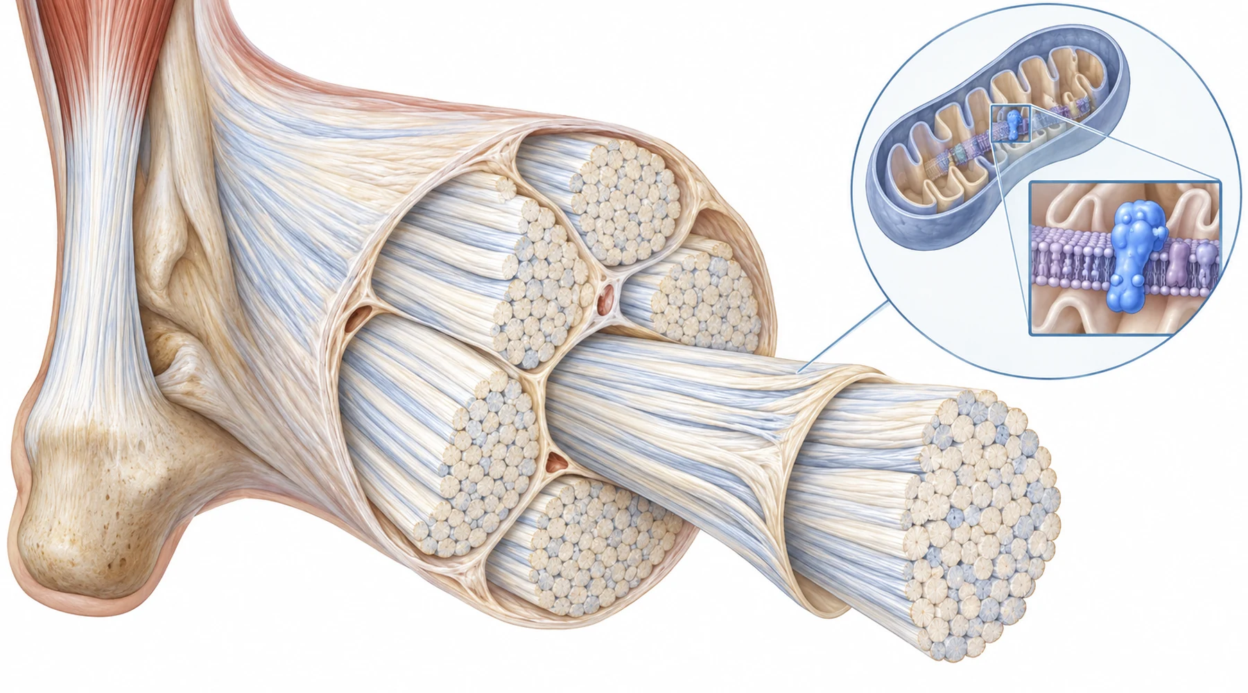
Search results for methylene blue tendonitis mix two different claims. One is that methylene blue helps tendon pain in people. The other is that methylene blue changed tendon chemistry in a laboratory preparation. Only the second claim was tested in the 2026 study of methylene blue in Achilles tendinopathy. No patients took methylene blue for tendon pain. That distinction organizes everything below.
The useful question is narrower. Diseased Achilles tendon showed a particular metabolic fault. Methylene blue corrected part of that fault in rats, in isolated human tendon pieces, and in cultured tendon cells. In one manipulation it corrected the chemistry without rescuing the tissue. That split result is the point of the paper.
Three branches, three different claims
The study has three branches. Each branch answers a different question, so results should not be moved across branches without notice.

The human branch is observational. Tendon came from amputations (used as comparison tissue) and from ruptured Achilles tendons (used as late stage disease tissue). The investigators measured chemistry and structure there. They did not dose those patients with methylene blue.
A second human preparation is interventional but still outside a body. Small pieces of human Achilles tendon were kept alive in a dish, injured with an oxidant, then exposed to methylene blue. This tests whether human tissue can respond in that preparation. It does not test distribution, clearance, whole body regulation, or pain. The general problem with this tier is explained in how to read methylene blue research.
The rat branch is interventional inside a living animal. Collagenase injected into the Achilles tendon produced matrix disruption, then methylene blue was given and gait, pain responses, chemistry, and histology were measured. Collagenase injury is abrupt chemical damage. Human Achilles disease usually develops gradually under load, age, circulation, and prior injury. The rat is therefore a model of disruption plus repair, not a small human with overuse injury.
The cell branch is mechanistic. Primary rat tenocytes were injured with TBHP, a peroxide that imposes oxidative stress, then treated with methylene blue at about 1 micromolar. Two further perturbations isolated parts of the pathway: rotenone to block mitochondrial complex I, and siRNA to lower AIF production. Those two perturbations are where the paper gets interesting.
What diseased tendon looked like
Healthy tendon is mostly type I collagen in long parallel bundles built for tensile load. Diseased tendon in this study showed the familiar shift. Type III collagen rose relative to type I, fibers lost parallel organization, mucin staining increased, vessels and rounded cells appeared, and Bonar scores reached grades 3 to 4. Apoptosis markers rose. This is matrix degeneration in the structural sense: not dissolution, but disordered remodeling toward a weaker fiber mix.
The chemistry moved in parallel. In diseased tendon, NAD+ level, NAD+ to NADH ratio, and total NAD pool fell while NADH accumulated. ATP fell. PARP and NOX4, two systems tied to NAD+ consumption and oxidant production, rose. Single cell RNA data pointed the same way: degradation, inflammation, and oxidative stress programs enriched in disease, while electron transfer, ATP metabolism, and oxidative phosphorylation programs enriched in healthy tendon.
Think of NAD+ and NADH as one shuttle in two states. NAD+ accepts electrons in metabolic reactions and becomes NADH. Mitochondria then oxidize NADH back to NAD+ at complex I and use the released energy to build the proton gradient that powers ATP synthesis. The ratio matters because traffic must flow in both directions. A low ratio plus a shrunken total pool means the shuttle fleet is both depleted and stuck in the loaded state.
Complex I sits at the center of that account. Two markers of its biogenesis and assembly, AIF and NDUFB8, were lower in diseased human tendon, and direct complex I activity (measured as NADH ferricyanide reductase) was reduced. The same pattern appeared in the rat model. This makes complex I weakness a plausible link between redox imbalance, excess reactive oxygen species (ROS), low ATP, and abnormal matrix. The link between electron transport and energy supply is part of the wider story of mitochondrial electron transport, ATP and energy metabolism, and oxidative stress and ROS control.
AIF needs a gloss because the name misleads. Apoptosis inducing factor sounds like a death protein only. Inside healthy mitochondria it supports complex I assembly and oxidative phosphorylation. Under severe injury it can leave mitochondria and take part in cell death. Location decides which role dominates.
What methylene blue changed
In the rat collagenase model, methylene blue restored NAD+ to NADH balance and total NAD chemistry toward control values, raised ATP, and lowered ROS. Mitochondrial membrane potential recovered by JC-1 staining. Histology improved: less fiber disarray and less stainable mucin, more type I collagen signal and less type III signal, lower PARP and NOX4 signals, and fewer apoptotic cells. Footprint gait measures (stride and step length) improved toward normal.
Human explants behaved similarly in the dish. After oxidant injury, methylene blue raised NAD+ chemistry and ATP, improved histology scores, shifted collagen staining back toward type I, lowered PARP and NOX4, and raised AIF and NDUFB8. Cultured rat tenocytes showed the same pattern: better viability after oxidant injury, restored NAD+ and ATP chemistry, lower ROS, better membrane potential, less apoptosis, and a lower type III to type I ratio.
None of this is a clinical outcome for methylene blue tendinopathy in people. It is a consistent tissue and cell response across three preparations: better redox and energy chemistry plus better matrix markers. Consistency across preparations strengthens a mechanism. It does not establish a dose, a route, or pain relief in patients.
Where the rescue stopped
The rotenone and AIF knockdown experiments test whether that mechanism depends on the proposed parts. They give opposite answers, and the contrast is the most informative part of the paper.
Rotenone is a selective complex I inhibitor. With complex I pharmacologically blocked, methylene blue still protected NAD+ chemistry and ATP related readouts, yet it failed to relieve ROS accumulation, failed to restore collagen and complex I markers, and failed to preserve cell viability. Mitochondrial depolarization persisted. In other words, the biochemical number moved while the tissue endpoints did not. A reader who equates restored NAD+ ratio with rescued tendon would misread this condition.
AIF knockdown gave the reverse pattern. Lowering AIF production itself disturbed complex I function, NAD+ chemistry, and ATP, and raised PARP, NOX4, ROS, depolarized mitochondria, and matrix degeneration. Here methylene blue rescued across the board: NAD+ balance, oxidative stress, membrane potential, NDUFB8 expression, and collagen I versus III markers. It even raised residual AIF signal under knockdown conditions, consistent with support for AIF dependent mitochondrial repair and complex I biogenesis.
Put side by side, the two results separate two ideas that are easy to fuse. NAD+ chemistry can be partly supported even when complex I is blocked, but that support alone does not fix oxidant load or matrix structure in this preparation. When the lesion is upstream at AIF, with complex I machinery still present but underbuilt, methylene blue support coincides with broader recovery. The pathway is therefore complex I dependent for tissue level rescue, even though one chemical readout is not.
This is also why the paper reports total NAD pool carefully. In some cell experiments methylene blue restored the NAD+ to NADH ratio and ATP without changing the total pool much. Ratio and pool are different quantities. A better ratio with the same pool means redistribution between the two states, not manufacture of new shuttle molecules.
What remains untested
Four limits should stay attached to any summary.
First, no human took methylene blue for tendon symptoms. Human data here are chemistry and histology from surgical tissue plus oxidant injured explants. Pain, rupture risk, return to activity, and adverse effects were not tested in people.
Second, the animal and cell injuries are controlled insults. Collagenase injection and TBHP exposure isolate matrix disruption and oxidative stress on purpose. They remove gradual loading history, circulation, immune context, nerve supply, and the mixed causes of real Achilles disease.
Third, concentrations do not translate by inspection. The cell work centered on about 1 micromolar methylene blue against 50 micromolar TBHP, with 5 micromolar rotenone where used. Dish concentrations are not oral doses, and the paper does not supply a human tendon dosing claim.
Fourth, methylene blue is redox active with its own dose response elsewhere in the literature. This study supports a mitochondrial and NAD+ mechanism in tendon preparations. It does not supply a home protocol, and tendon pain has evaluated options (load management, rehabilitation, clinician guided care) that this paper does not compare against.
The fair summary is therefore conditional. Where Achilles tendinopathy involves NAD+ imbalance and complex I weakness, methylene blue restored redox and energy chemistry and improved matrix markers in rat tendon, human tendon explants, and cultured tenocytes. When complex I was directly blocked, chemistry improved without tissue rescue. When AIF was lowered, broader rescue returned. That pattern supports targeting NAD+ metabolism, AIF, and complex I as a research strategy. It does not show that methylene blue treats sore Achilles tendons in people.