
Article
New methylene blue and reticulocyte staining: a separate dye and a living-cell method
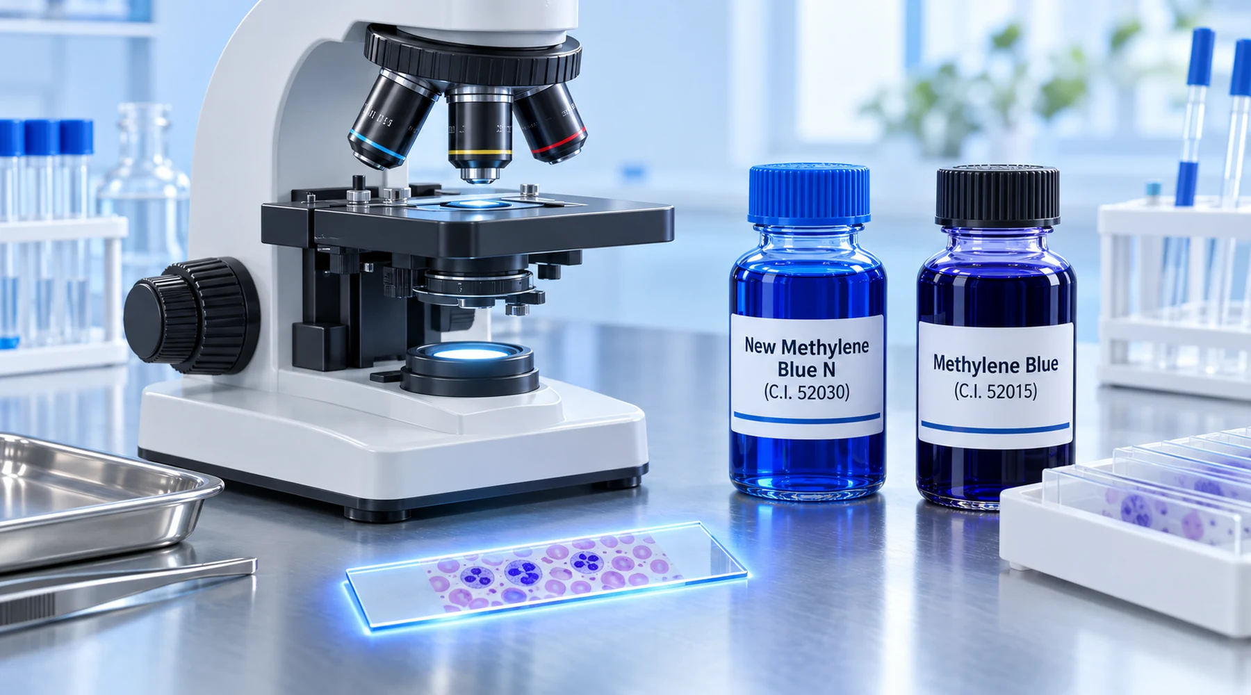
A student prepares a reticulocyte count, orders methylene blue, mixes it with blood, and sees almost nothing. A classmate uses a bottle labeled new methylene blue and gets crisp blue filaments inside a fraction of the red cells. The first student assumes the second bottle is fresher or more concentrated. It is neither. It contains a different molecule.
This is the concept error behind most confusion in this area. The name suggests that new methylene blue is an improved batch of methylene blue. The chemistry says otherwise. New methylene blue N is a separate phenothiazine dye with its own Colour Index number, its own substitution pattern, and its own standardized role in hematology. Once that distinction is clear, the rest of the method (why the cells must be alive when stained, what counts as a reticulocyte, where Heinz bodies fit, and when brilliant cresyl blue enters the picture) becomes straightforward.
The short answer
New methylene blue N, the hematology reagent, is generally identified as Basic Blue 24 in the PubChem record for new methylene blue, C.I. 52030, CAS 1934-16-3, formula C18H22ClN3S. Ordinary methylene blue is methylthioninium chloride, Basic Blue 9, in its PubChem record, C.I. 52015, CAS 61-73-4, formula C16H18ClN3S. Structurally, new methylene blue carries two ring methyl groups and ethylamino side chains where methylene blue carries dimethylamino groups. That difference changes staining behavior. Hematology texts describe ordinary methylene blue as a relatively poor reticulocyte stain and new methylene blue as deeper and more uniform, a comparison summarized in the ScienceDirect topic on new methylene blue.
Yes, new methylene blue is a supravital stain. Supravital staining means staining living cells after removal from the body and before fixation, as defined in the NCBI review of supravital staining principles. Both new methylene blue and brilliant cresyl blue belong to this category, and the CLSI reticulocyte standard covers new methylene blue explicitly as a supravital dye. Students sometimes file this with Romanowsky staining of fixed films. That mental model fails here. Fix first and the method stops working.
For readers tracking nearby dye names, methyl blue versus methylene blue covers a different and larger confusion between an anionic triarylmethane dye and the cationic phenothiazine family. The laboratory staining overview places supravital work among fixed-film methods, and the blood cell morphology guide shows where reticulocyte assessment fits in the wider differential.
| Reagent | Identity | C.I. / CAS | Role in reticulocyte work |
|---|---|---|---|
| New methylene blue N | Ethylamino, ring-dimethylated phenothiazine | C.I. 52030 (Basic Blue 24); CAS 1934-16-3 for the chloride | Standardized manual supravital stain, preferred comparator |
| Ordinary methylene blue | Bis(dimethylamino) phenothiazine (methylthioninium chloride) | C.I. 52015 (Basic Blue 9); CAS 61-73-4 | Poor reticulocyte stain, not a substitute |
| Brilliant cresyl blue | Oxazine dye, supplier formulation varies | C.I. 51010; CAS 81029-05-2 or 10127-36-3 depending on salt form | Acceptable supravital stain, less uniform, not the harmonized comparator |
One catalog caution: a separate zinc chloride double-salt form of new methylene blue exists under CAS 6586-05-6. A methods section should therefore name the salt form and supplier, not only the common name.
What supravital staining actually does
A reticulocyte is a young red cell that has lost its nucleus but retains residual ribosomal RNA. In an unstained wet preparation that RNA is invisible. Supravital dye enters the living cell and precipitates or aggregates that RNA into blue granules and filaments, the reticulofilamentous material seen under oil immersion. The network is therefore a staining product, not a preformed skeleton waiting for color. This is why protocol variables (dye concentration, buffer, blood to dye ratio, temperature, time) matter as much as the dye name.
The practical consequence is a strict order of operations: mix anticoagulated blood with dye first, incubate, then make films and examine them unfixed and without counterstain. Fixation before staining, or a counterstain that obscures faint filaments, destroys the information the count depends on.
A working reticulocyte protocol with new methylene blue
There is no single universal formulation, so report the one used. Three documented variants show the range.
The classic Brecher type procedure uses approximately 0.5 percent new methylene blue with potassium oxalate, mixed in equal volumes with well mixed EDTA blood, incubated about 10 to 15 minutes at room temperature. After gentle remixing, thin wedge films are prepared, air dried, and examined unfixed under the 100x oil objective. The NCBI procedure for reticulocyte counting describes this workflow and the counting frame used with it.
The Dacie and Lewis variant uses 1.0 percent new methylene blue in citrate saline, mixed with 2 to 4 volumes of blood and held at 37 C for 15 to 20 minutes before filming. A recent harmonization implementation for the CLSI H44 procedure used a leaner 0.1 percent solution in phosphate buffered saline at pH 7.4, with incubation at 20 C for 10 minutes and no fixation, as reported in the 2026 NMB harmonization study.
Counting rules are shared across variants. Score a cell as a reticulocyte when it contains at least two discrete blue staining granules or filament segments, which excludes isolated stain deposit. Count at least 1,000 red cells in routine work and report reticulocytes as a percentage. At low percentages the statistics demand more cells. A review of the CLSI H44 methodology notes that near 1 percent reticulocytes about 4,400 cells are needed to bring counting variation to 5 percent, in the CLSI H44 review at De Gruyter. A Miller ocular disc makes large counts practical. Filter or freshly prepare the dye, because precipitate mimics granular inclusions.

Heinz bodies in the same preparation
The same supravital film can also demonstrate Heinz bodies, which are aggregates of oxidized and denatured hemoglobin rather than residual RNA. In new methylene blue preparations they appear as round bodies, often single and near the membrane, staining a lighter blue than reticulofilamentous material. They survive poorly through fixation and counterstaining, so examine the unfixed film. A recent protocol using 20 microliters of EDTA blood with 20 microliters of 1 percent new methylene blue and 15 minutes incubation quantified Heinz body positive cells among 250 erythrocytes, in the PMC study reporting that procedure.
Two distinctions prevent misreads. First, methyl violet and crystal violet stain Heinz bodies more intensely than new methylene blue, as noted in the ScienceDirect Heinz body topic. New methylene blue is the convenient choice when reticulocytes and Heinz bodies are assessed together, not the strongest dedicated Heinz body dye. Second, HbH inclusions in alpha thalassemia trait produce numerous fine dots across the cell after supravital staining, while classic Heinz bodies are fewer, larger, and often membrane associated. Stain deposit is the third mimic: it lies outside the focal plane of the cells and varies between fields.
Brilliant cresyl blue versus new methylene blue
Both dyes are supravital stains for reticulocytes, but standardization favors one of them. The CLSI H44-A2 reticulocyte method, developed jointly with the International Council for Standardization in Haematology and listed on the CLSI H44 standard page, designates new methylene blue staining of EDTA blood as the manual comparative method. Classical hematology teaching agrees on appearance: new methylene blue gives deeper and more uniform filament staining, while brilliant cresyl blue varies more between batches and samples.
That status needs its qualifier. The ICSH analyzer evaluation guideline accepts manual new methylene blue microscopy as a reference comparison approach but calls it subjective and relatively imprecise, with flow cytometric reference procedures preferred where available. A 2022 comparison of 100 specimens found lower bias against its analyzer with brilliant cresyl blue than with new methylene blue, reported in the PMC comparison of staining methods. The defensible summary for students: use new methylene blue as the standardized manual comparator, verify any local brilliant cresyl blue method against it or against automation, and expect wider scatter from manual microscopy either way. Older standardization literature also advocated purified Azure B as a chemically cleaner alternative because commercial dye lots varied, in the Marshall study on Azure B, which explains why reagent grade still appears in methods discussions.
Avoiding the standard errors
Four mistakes account for most failed preparations. Ordering ordinary methylene blue instead of new methylene blue N produces weak staining by chemistry, not by technique. Fixing or counterstaining before reading removes the supravital signal. Counting stain precipitate as granules inflates the result, which the two granule rule and filtered dye prevent. Counting too few cells at low reticulocyte percentages produces a precise looking number with wide confidence limits. Each error has the same signature: the microscopist blames the microscope for what the reagent step decided.