
Article
How methylene blue differs from conventional antioxidants
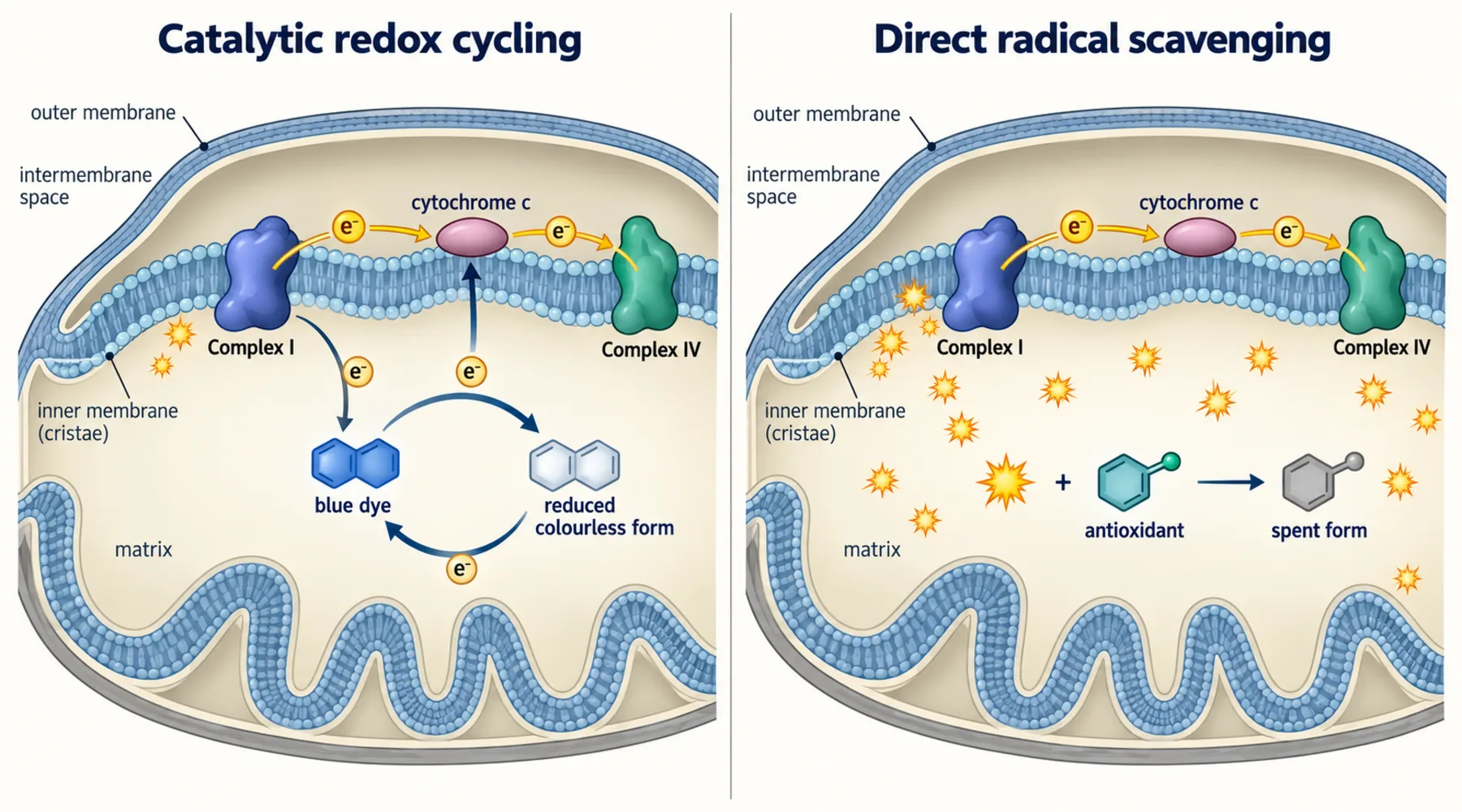
A conventional antioxidant meets a free radical exactly once. It donates an electron, neutralises its target, and is spent. The arithmetic is one to one, and protection stops when the supply runs out.
Methylene blue does not work that way. It accepts electrons, hands them on, and returns to its original form ready to cycle again. One molecule can act thousands of times. That difference explains its potency at low doses and its most instructive failures. What follows compares the two mechanisms through three published studies, keeping the null results in view.
A relay, not a sponge
The chemistry is a reversible couple. Oxidised methylene blue is blue, charged, and water soluble. Reduced methylene blue, called leuco-methylene blue, is colourless, uncharged, and fat soluble enough to cross membranes freely. Inside the mitochondrion, the oxidised dye takes electrons from NADH-linked carriers upstream and delivers them to cytochrome c and complex IV downstream, bypassing complexes I through III, which are the sites where electrons most often leak to oxygen and form superoxide. The dye emerges oxidised and repeats the loop. The mechanics of the couple itself are covered in the explainer on how methylene blue works.
A direct scavenger works downstream of the leak. It waits for superoxide or peroxide to form, then neutralises it, one molecule per radical. A redox cycler works upstream, diverting electron flow so less superoxide forms, without being consumed. The dose arithmetic follows: in the studies below, methylene blue acts near 100 nanomolar while the classic scavenger needs 100 micromolar.
A relay does nothing on its own, though. What it does depends on the donors and acceptors at each end. Change the system and the same molecule can be protective, useless, or irrelevant.
Neural cells: protection that requires working mitochondria
The cleanest dissection comes from a structure-activity study in HT-22 hippocampal neuronal cells, testing methylene blue and six relatives against four insults (Poteet et al., 2012). Against glutamate, rotenone (a complex I blocker), and iodoacetate, the dye protected at nanomolar concentrations. Against glucose oxidase, which generates hydrogen peroxide directly in the medium, it did nothing from 10 nanomolar to 10 micromolar, while the simple scavenger pyruvate protected. Where the insult is a broken electron chain, the relay helps. Where peroxide is already formed outside the cell, there is nothing for it to carry.
The same study showed why. In a mitochondrial lysate assay, two relatives lacking the side chains at positions 3 and 7, phenothiazine and 2-chlorophenothiazine, acted as direct antioxidants on their own. Methylene blue did not. It only quenched oxidation when both NADH and intact mitochondrial material were present. It was, in other words, not scavenging at all. It was shuttling, and the shuttle needs both a loading dock and a destination.
Two details sharpen the picture. Adding a side chain to the ring nitrogen at position 10 cut protective potency roughly a thousandfold, identifying the free nitrogen as load-bearing for the cycling chemistry. And chronic exposure raised complex IV expression and activity, an adaptation the directly scavenging relatives did not produce. Related detail on mitochondrial electron transfer is background for judging antioxidant claims.
Skin cells: a head-to-head comparison methylene blue wins
A separate study set methylene blue against three established antioxidants in primary human skin fibroblasts from healthy donors, progeria patients, and donors over 80 (Xiong et al., 2017). Over four weeks, methylene blue was dosed at 100 nanomolar, N-acetylcysteine at 100 micromolar, and MitoQ and mito-TEMPO at 100 nanomolar each.
The result was not close. Methylene blue reduced mitochondrial superoxide and supported proliferation across the cell lines. N-acetylcysteine did not lower mitochondrial ROS and appeared to slow proliferation. MitoQ, at the literature-standard dose, drastically increased mitochondrial ROS and inhibited proliferation. Mito-TEMPO showed moderate benefit in some lines. Of the four, the catalytic cycler was the most effective and the targeted stoichiometric antioxidant was actively worse than nothing, at least under month-long exposure in this system.
The authors flagged duration as a likely explanation, since shorter MitoQ exposures elsewhere looked better. That caveat generalises: stoichiometric antioxidants deplete, and their breakdown products can misbehave over long incubations. A catalyst does not deplete.
The same programme extended into reconstructed 3D human skin, where low-dose methylene blue thickened the dermis, improved hydration, raised elastin and collagen 2A1, suppressed MMP9, and sped wound closure. Encouraging, but it belongs to skin, and skin is not bone.
Bone: cell-dish promise that vanished in the animal
The skeletal-aging study is the other end of the spectrum (Poudel et al., 2024). In culture, both methylene blue and MitoQ inhibited osteoclast formation dose-dependently above 0.25 micromolar without harming stem cell viability or osteoblast formation. Reading only those panels, a researcher would predict stronger bones in treated animals.
The animals disagreed. Aged female C57BL/6J mice received 250 micromolar methylene blue in drinking water for 6 or 12 months. Diverse UM-HET3 mice received 28 ppm methylene blue in food from 7 to 22 months, or 100 ppm MitoQ from 4 to 22 months. Micro-CT of femoral cortical and trabecular bone plus lumbar vertebrae showed age-related loss proceeding identically in treated and control groups. Two strains, two routes, two antioxidants, months of exposure, no skeletal effect.
The authors' conclusion is blunt: cutting oxidative stress with an antioxidant alone appears insufficient to slow skeletal aging. Bone loss integrates hormones, loading, inflammation, and senescent-cell clearance. Fixing electron leak addresses one input among many, and here the others dominated. Context on how antioxidant strategies have fared against aging phenotypes frames this null.
Reading the three studies together
Three patterns survive contact with all three papers.
First, mechanism predicts the boundary. Methylene blue helps where electrons are misrouted inside mitochondria and a shuttle can reroute them. It fails where oxidants are already formed and remote from the chain. The glucose oxidase null and the bone null are not embarrassments to be explained away. They are the theory working as specified.
Second, catalytic arithmetic beats stoichiometric arithmetic under chronic exposure. A molecule that is not consumed keeps working at nanomolar levels for weeks, while one-shot scavengers need hundred-micromolar doses and can invert into harm over long incubations.
Notes on dose, exposure route, and tissue distribution explain part of why a nanomolar cell-culture winner can become an in vivo null.
For clinicians and researchers, the upshot is narrow but firm. Methylene blue is not a stronger antioxidant but a different category: a regenerable shuttle that needs intact mitochondria, available donors, and a downstream acceptor. Judge any proposed use by asking whether that circuit exists in the target tissue, and by demanding the control the Poteet study ran, a direct peroxide challenge a true scavenger passes and a shuttle should fail. That is also why investigator-led comparisons of redox-active compounds keep returning to head-to-head designs.
Sources
The three comparisons above come from a structure-activity study of methylene blue and six derivatives in neuronal cells, a four-antioxidant comparison in human skin fibroblasts and 3D skin models, and a long-term methylene blue and MitoQ skeletal-aging study in two mouse strains. This article reports their findings and does not make treatment recommendations.