
Article
Methylene blue for sentinel lymph-node mapping: dyes, tracers, and accuracy
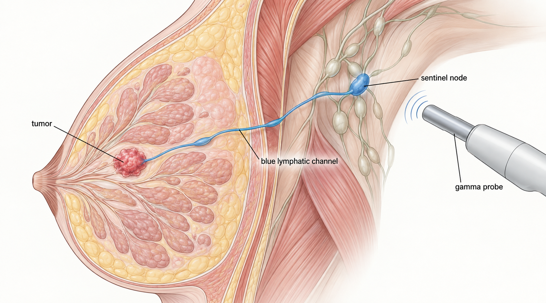
A surgeon treating early breast cancer faces a narrow question: has the tumor reached the lymph nodes under the arm? Removing every node answers it, but at the cost of lymphedema and lasting arm problems for many patients who turn out to be node negative. The sentinel-node concept offers a narrower operation. The first node (or small group of nodes) receiving drainage from the tumor area is identified during surgery, removed, and examined. If it is free of tumor, the rest of the basin is likely free too, and the full dissection can be skipped.
Everything therefore depends on finding that node reliably. That is what tracers are for, and where methylene blue enters the picture.
Two tracers, two physical signals
A common confusion pairs the blue dye with the radioactive tracer as if they were versions of one thing. They are separate concepts that produce separate signals.
The blue dye (methylene blue, isosulfan blue, or patent blue) is injected near the tumor or areola, travels through lymphatic channels, and stains the channel and node a visible blue. The surgeon follows the color with the naked eye.
The radiotracer (usually technetium-99m sulfur colloid) is injected separately, travels the same drainage path, and emits gamma photons detected with a handheld probe. It finds nodes that are deep, faintly stained, or invisible.
Combining them is called the dual-tracer technique, and it remains the conventional reference approach in modern practice. Each method covers the failure mode of the other: the probe hears what the eye cannot see, and the dye shows what the probe localizes only approximately. The rest of this article compares methylene blue against the alternatives within that frame.
Methylene blue alone: high identification, uneasy false negatives
The strongest pooled estimate for methylene blue used on its own comes from a systematic review and meta-analysis of 18 studies in 1,559 breast cancer patients with clinically negative axillae. Studies using other blue dyes or combined radioisotope techniques were excluded, so this is a clean estimate of methylene-blue-alone performance.
The pooled identification rate was 91% (95% CI 88 to 94%). In plain terms: in about 9 of 10 attempted procedures, at least one sentinel node was found. Individual studies ranged from 75 to 100%, with substantial heterogeneity (I² about 68%), reflecting different concentrations (mostly 1%), volumes (0.1 to 10 mL, commonly 2 or 5 mL), injection sites (peritumoral or subareolar), pathology methods, and surgeon experience.
The pooled false-negative rate was 13% (95% CI 9 to 18%). The false-negative rate answers a different question from the identification rate: among patients who truly had nodal disease, in what fraction did the sentinel node incorrectly test negative? A technique can score well on the first measure and poorly on the second, and that is exactly what happened here. Sensitivity was 87%, negative predictive value 91%, and overall accuracy 94%.
The authors judged 91% identification acceptable but 13% false negatives too high against the surgical standard they cited, and urged caution about relying on methylene blue alone. Subgroup comparisons found no significant difference between 2 mL and 5 mL doses or between peritumoral and subareolar injection, but the confidence intervals were wide enough that real differences cannot be excluded. No optimal concentration, volume, or injection site emerged from the evidence. For context on how dosing language is read in clinical papers, see how to read a methylene blue clinical protocol.
Methylene blue alone versus methylene blue plus radiotracer
A single-center randomized trial in 60 early-stage breast cancer patients in Hyderabad assigned 30 patients to 5 mL methylene blue alone and 30 to the same dye plus 1 mCi filtered technetium-99m sulfur colloid, with every patient then receiving completion axillary dissection as the reference standard. Both arms identified at least one sentinel node in all 30 patients (100% identification each), with nearly identical mean node yields (3.16 versus 3.20 nodes).
That perfect identification rate looks persuasive until the sample size is considered. Only 7 sentinel-node-positive patients sat in each arm, so every diagnostic estimate rests on a handful of events and no confidence intervals were reported. The paper's stated "false-negative" figures (8.6% versus 4.3%) also use a nonstandard denominator: they divide false negatives by all negative test results rather than by all truly node-positive patients, which makes them closer to one minus the negative predictive value than to the conventional false-negative rate. The sensitivity and specificity calculations appear oriented toward predicting additional non-sentinel disease rather than detecting any axillary metastasis. These figures should therefore not be pooled with conventional estimates such as the 13% above.
The defensible reading of this trial is narrow: in one experienced center, both techniques found at least one node in every patient. Its claim of comparable diagnostic accuracy is not secured by the design, and absence of a significant difference in 60 patients does not establish equivalence. The trial's practical value lies in its setting. Where nuclear medicine is unavailable, methylene blue alone still mapped a node in every case here, which is why the authors proposed it as a workable fallback rather than a proven equal.
A note on the third reference
The brief for this article listed a 2025 systematic review comparing methylene blue across cancer sites. That paper has since been retracted in February 2026 over invalid references, with the corresponding author attributing errors to AI-assisted tools. Its pooled numbers are excluded from this review. Mentioning it matters for one reason: older copies of its figures still circulate online, and they should not be quoted.
Isosulfan blue versus methylene blue
For methylene blue dye for lymphatic mapping compared with isosulfan blue, the tradeoff the literature supports is consistent: systemic allergy favors methylene blue, local tissue injury favors neither and slightly disfavors superficial methylene blue injection.
A systematic review covering 61,951 sentinel-node procedures found 94 anaphylactic events (weighted overall rate about 0.061%), with isosulfan blue carrying the highest estimated rate at about 0.16%. Lower dye volumes and intradermal administration were associated with lower rates. Older clinical series point the same way, with one breast-center series reporting allergic reactions in 3 of 351 isosulfan blue patients. Methylene blue is therefore generally described as less anaphylactogenic, though its risk is not zero.
Methylene blue's characteristic problem is local: inflammation, ulceration, and skin or fat necrosis, particularly after superficial or intradermal injection. Within the 18-study methylene blue meta-analysis, two patients in one study developed injection-site skin necrosis, one patient in another developed skin and subcutaneous necrosis after subareolar injection, and three patients in a third study had skin allergic reactions. No life-threatening methylene blue allergy appeared in that dataset. The 60-patient randomized trial reported no methylene blue side effects at all, but with 30 exposed patients per arm that observation carries little weight.
How long does the blue staining last
Patients often ask this, and the honest answer separates the dyes because most long-term staining data concerns patent blue, not the dye the patient received.
Temporary tattooing after methylene blue is well recognized, and one large methylene blue series reported it as the only complication of note. For isosulfan blue, published reports describe skin staining lasting from hours to weeks. The prolonged staining figures that circulate (visible staining in 36.5% of patients at 12 months, 23.6% at 24 months, and 8.6% beyond 36 months) come from a prospective series using patent blue, a related but distinct dye, and should not be quoted as methylene blue figures. One cohort that compared the two directly found discoloration in 14.1% with methylene blue versus 23.7% with patent blue. Direct long-term evidence comparing methylene blue specifically against isosulfan blue is limited, so any article that assigns a month count to methylene blue staining without naming the dye and the study is guessing. Blue or green urine after systemic dye absorption is transient and documented with blue dyes generally, but none of the reviewed sentinel-node studies quantified it, so no incidence belongs here. Readers weighing the broader safety picture may also want the methylene blue adverse-effect overview and its drug-interaction profile.
Tracer-and-endpoint comparison
| Tracer strategy | Identification rate | False-negative rate | Adverse-effect pattern | Setting and certainty |
|---|---|---|---|---|
| Methylene blue alone | 91% pooled (95% CI 88 to 94%), 18 studies, n = 1,559 | 13% conventional FNR (95% CI 9 to 18%) | Local necrosis or inflammation in a small minority; temporary tattooing common; no life-threatening allergy in dataset | Breast cancer, clinically node-negative; high heterogeneity, all studies at high selection-bias risk |
| Methylene blue plus technetium-99m | 100% in both arms of one n = 60 trial; dual-tracer rates generally high in wider literature | Trial figures use a nonstandard denominator; do not pool | Same dye risks plus radiation exposure and nuclear-medicine logistics | Single experienced center; 7 positive cases per arm; accuracy comparison underpowered |
| Isosulfan blue (comparator) | Comparable detection in wider literature; not estimated here | Not estimated here | Anaphylaxis about 0.16% in large pooled data; staining hours to weeks | Large pooled safety data; direct long-term staining comparison against methylene blue limited |
Reading the evidence like a buyer of certainty
Three distinctions carry the whole topic. First, identification rate measures whether the technique works mechanically; false-negative rate measures whether a negative result can be trusted. Methylene blue alone passes the first test more comfortably than the second. Second, the blue dye and the radiotracer are independent detection mechanisms, so "dye versus tracer" is the wrong contest; the real choice is dye alone (necessary where nuclear medicine is absent) versus dye plus tracer (the reference where available). Third, safety comparisons must name the dye: isosulfan blue owns the systemic allergy disadvantage, methylene blue owns the local necrosis problem after superficial injection, and the longest staining figures belong to patent blue.
For a patient or caregiver, the questions to bring to the surgeon are concrete: which dye will be used, is a radiotracer combined with it, how many sentinel nodes are typically removed, and what was this team's identification and false-negative experience. For background on what the dye does systemically and how it is studied, see methylene blue pharmacology and distribution and how methylene blue clinical research is evaluated. And as the retraction episode shows, check the date and status of any review you are shown: in a fast-moving evidence base, even a one-year-old pooled figure can already be withdrawn.