
Article
Methylene blue in plasma pathogen reduction: how the process works
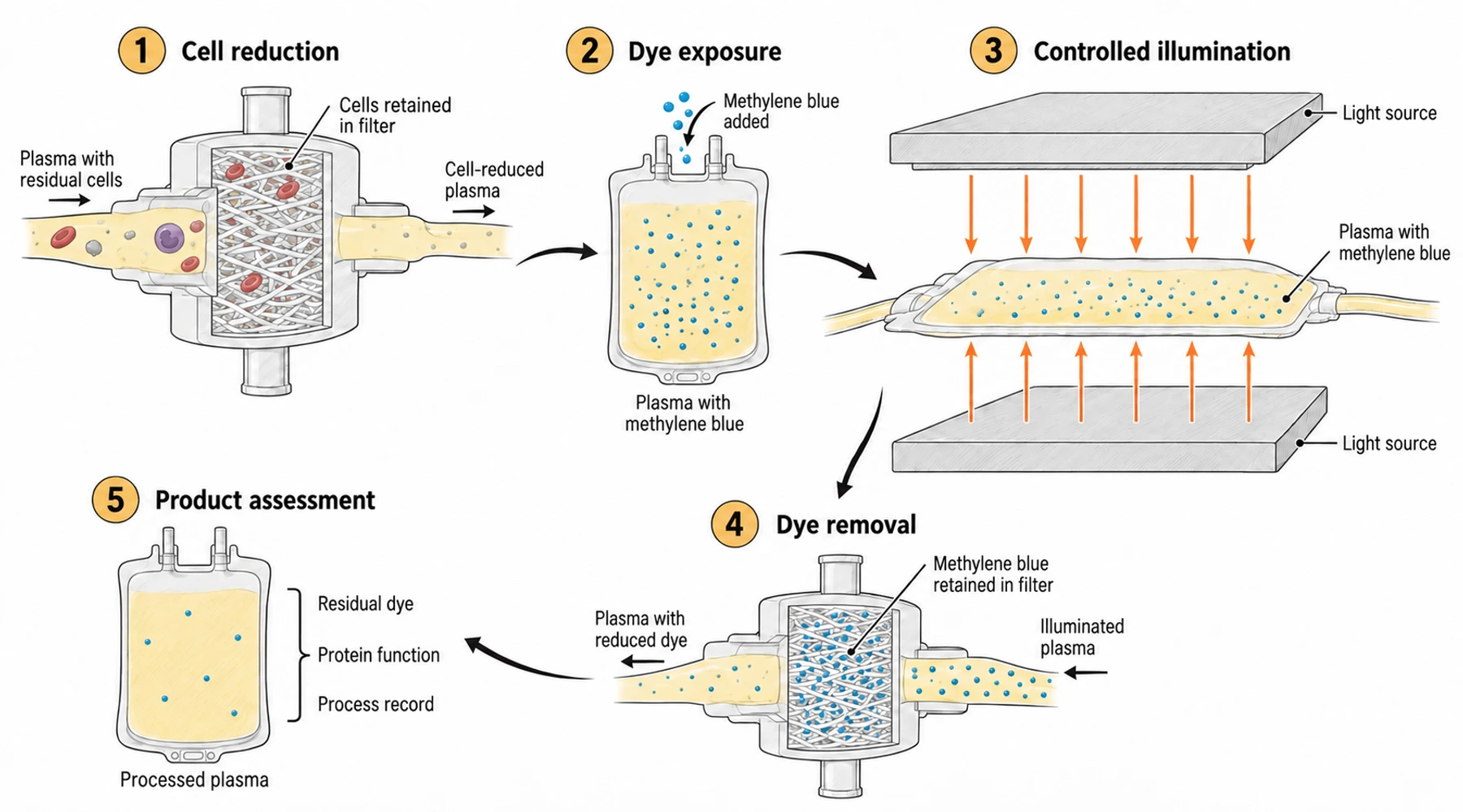
Methylene blue can help reduce pathogens in donated plasma when it forms part of a controlled processing system. The treatment occurs outside the body, before the plasma is released for transfusion. It combines specified equipment, dye exposure, illumination, filtration, and checks on the resulting blood component.
That distinction determines what the evidence means. A study showing that a processing system reduces infectious virus in a plasma bag establishes a result for that material and process. It does not establish that taking methylene blue treats an infection in a person.
The clinically relevant question is also two-sided: how much pathogen reduction does the process achieve, and how much of the plasma's useful function remains?
Start with plasma, not an unspecified blood product
Plasma is the liquid blood component that contains proteins, including coagulation factors. A plasma-processing system should not be assumed suitable for whole blood, red-cell concentrates, or platelet components. Those materials have different compositions and functions to preserve.
For a concrete example, Macopharma's THERAFLEX MB-Plasma system treats single-donor plasma units. Its documented sequence includes cell reduction, methylene blue addition, illumination, dye removal, and storage. The manufacturer describes different configurations for plasma derived from whole blood and for larger apheresis collections. The starting material and the disposable set therefore matter before illumination begins.
“Methylene blue plasma” is a description of a processed component, not a complete specification. A useful comparison names the system, configuration, plasma source, and version of the operating instructions.
Follow the material through processing and assessment
The illustration above separates the stages. This companion table explains what each stage contributes and what it cannot establish by itself.
| Processing stage | Main purpose | Separate question to check |
|---|---|---|
| Cell reduction before exposure | Remove residual blood cells and aggregates from the plasma | Was the starting material suitable for this configuration? |
| Dye exposure | Bring the photosensitizer into contact with the material to be treated | Was exposure achieved within the specified process conditions? |
| Controlled illumination | Drive photochemical reactions that reduce susceptible pathogens | Were the validated light and temperature conditions achieved? |
| Dye and photoproduct removal | Reduce residual processing chemicals before storage | Did the removal step meet the system's requirements? |
| Product assessment and release | Assess processing records and component quality under the blood service's procedures | Does the finished component satisfy its applicable specification? |
The table is a reading guide for a validated process, not a substitute operating procedure. In particular, the removal filter and the light source do different jobs. Macopharma describes Blueflex as reducing methylene blue and its derivatives, including azure compounds and thionine. A claim of chemical removal should be read as a measured reduction, not a promise that no molecule remains.
Why the light source is part of the intervention
Methylene blue is photoactive. Illumination can cause chemical damage to biological targets, which is why the combination has been investigated for virus inactivation. In Mohr and colleagues' study of phenothiazine dyes and light, changing the illumination source altered both virus inactivation and plasma-protein damage.
This prevents a common shortcut: treating the dye name as if it described the whole intervention. A different lamp, bag, optical path, or material can change the exposure. The broader guide to how methylene blue photodynamic treatment is studied explains why formulation and illumination belong in the same description.
Temperature provides an especially useful example. A 2018 study of THERAFLEX processing at different temperatures compared plasma near 5°C, 22°C, and 30°C. At the lowest temperature, investigators observed poorer tablet dissolution and lower inactivation of the three viruses studied. Higher temperatures increased changes in clotting time and losses of coagulation-factor activity.
Those findings do not supply a new operating range. They explain why a blood service must use the specified conditions: improving one outcome by changing a parameter may impair another.
Which part of the system reduced the organism?
Pathogen reduction measured after the complete process cannot automatically be attributed to light-activated dye alone.
In a 2015 challenge study using deliberately contaminated plasma, investigators sampled the material after individual processing stages. Cell filtration accounted for much of the bacterial reduction. For the small bacterium Brevundimonas diminuta, that first step achieved a 1.7-log reduction; subsequent light treatment and dye filtration brought the overall reduction to at least 3.7 logs, below the assay's detection limit.
This is a result for a combined system. It does not show that an ordinary methylene blue solution produces the same bacterial reduction. The distinction also applies when reading broader methylene blue antimicrobial research.
A log reduction describes the ratio between measured starting and remaining infectious material. A three-log reduction is a thousandfold reduction. “Below the detection limit” means the assay could not quantify what remained under its test conditions. It is not proof of absolute sterility or universal activity against untested organisms. Virus species, starting burden, sample matrix, and assay sensitivity remain necessary parts of the claim.
Preserve the function for which the plasma will be used
A process can reduce infectivity while changing proteins that matter clinically. Measuring pathogen reduction alone therefore leaves the assessment incomplete.
In a 2014 comparison of plasma sources and processing times, factor VIII recovery after methylene blue treatment ranged from 78% to 89% across the study series. Recovery depended on plasma source, and the investigators also assessed fibrinogen. These are laboratory measurements on the processed component. They are not percentages of clinical effectiveness in patients.
Consider two plasma units with different starting factor levels. Even if they retain the same fraction of activity during treatment, their final levels will differ. Conversely, a higher percentage recovery need not mean a higher final concentration. Read both the before-and-after measurements and the applicable release specification.
Patient outcomes require another kind of evidence. A component-quality study cannot, by itself, establish equivalent bleeding control, transfusion requirements, or adverse-event rates in a particular patient population.
Keep processing evidence separate from treatment claims
The plasma bag makes controlled exposure possible: it has a defined volume, geometry, processing sequence, and measurable output. A person has tissues, circulation, metabolism, and sites of infection that this experiment does not reproduce.
For that reason, HIV treatment claims versus plasma-inactivation evidence and methylene blue evidence for COVID-19 and long COVID require their own clinical assessment. Neither question is answered by demonstrating reduced infectivity in donated material.
For blood-service use, evaluate the exact component and locally permitted system, its organism-specific validation, retained protein function, residual chemicals, and release controls. Product availability differs by country, as the manufacturer states. Historical use or a successful experiment does not establish current authorization everywhere. The useful unit of evidence is the complete validated process applied to its specified plasma component.