
Article
Methylene blue in pilonidal sinus surgery: mapping and recurrence evidence
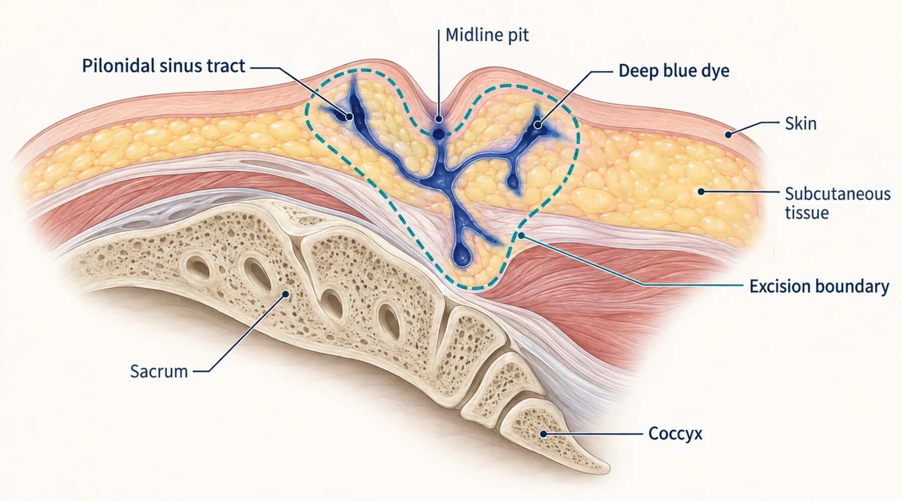
A pilonidal sinus is a tract under the skin near the tailbone, often with side branches that are hard to see during surgery. The operation fails when diseased tissue is left behind, so surgeons want a reliable way to find every branch before they cut. Injecting methylene blue into the sinus opening stains the tract system blue and gives the surgeon a visible map to follow.
That map is a visualization aid, not a treatment. This distinction matters because the literature on methylene blue pilonidal surgery mixes two different questions. The first is whether the dye accurately marks all diseased tissue. The second is whether patients whose operations used the dye have fewer recurrences years later. A dye can fail the first test in the microscope yet still be associated with better outcomes in the clinic, or the reverse. The two published studies most often cited on this topic each answer only one of these questions, with different designs and different follow-up.
What the surgeon is trying to see
Think of the dye as a label on a hidden structure. The surgeon injects blue liquid through the skin opening, waits for it to spread through connected tracts, then excises the stained tissue. Anything the dye did not reach, or any diseased tissue that was never connected to the injected tract, stays unstained and can be missed.
Three things can therefore go wrong independently. The dye may not fill every branch. The surgeon may not remove everything that is stained. And tissue outside the stained boundary may still contain microscopic disease. The studies below isolate different parts of this chain, which is why their results cannot be averaged together.
Readers comparing operative techniques may also want the broader overview of surgical uses of methylene blue and the separate explainers on how dye staining differs from tissue treatment and how recurrence is defined and measured.
Long-term recurrence: an observational cohort
The recurrence evidence comes from a retrospective cohort reported by Doll and colleagues in the International Journal of Colorectal Disease, in a paper whose title states the headline result directly and whose long-term follow-up of methylene blue in acute pilonidal disease runs far longer than most surgical studies.
The investigators identified 1,960 patients first operated for pilonidal disease at three German military hospitals between 1980 and 1996, then selected 247 for detailed telephone follow-up. Mean follow-up was about 15 years, with a range of roughly 9 to 25 years. The population was mostly young military men. Surgical technique varied: about two thirds had excision with open healing, one quarter had excision with primary midline closure, and the rest had marsupialization.
Exposure was simple: 197 patients had methylene blue injected at the start of surgery to delineate the tract system, and 50 did not. The dye was described as a single careful injection, with no reported concentration or volume. Use was not randomized. Surgeons chose dye far more often for chronic fistulating disease than for acute abscesses, and more often when there were more sinus openings, so the two groups started out different.
Recurrence was assessed by a prespecified telephone questionnaire. Patients counted as recurrent only after initial healing, with no later tailbone trauma, plus either one hard criterion (physician diagnosis, repeat incision or excision, new sinus, hair in an opening, purulent discharge) or two soft criteria (redness, swelling, pain, discharge).
Crude recurrence over the whole period was 32 of 197 with dye (16 percent) against 15 of 50 without dye (30 percent). The Kaplan-Meier estimates at 20 years, which account for staggered follow-up, were about 20 percent with dye and 38 percent without, with a log-rank p value of 0.018. In a multivariable Cox model the authors reported an odds-like ratio of 0.45 for dye use. Most recurrences appeared within five years, but some appeared as late as 17 years after surgery, which is the reason long follow-up matters for methylene blue pilonidal recurrence claims.
The subgroup pattern deserves care. In acute abscess-forming disease, 20-year estimates were roughly 19 percent with dye against 43 percent without, but with only 79 patients the difference did not reach significance (p = 0.078). In chronic disease, estimates were roughly 19 percent against 24 percent (p = 0.35), essentially no demonstrable difference. The overall association was therefore driven by a subgroup too small to stand on its own, in a study where treatment assignment was not random and the control group held only 50 patients.
What this study does not show is equally important. It did not measure mapping accuracy, it did not compare histology between dye and no-dye operations, and it reported no usable dye-versus-no-dye comparison for wound complications or healing time. It is evidence of an association between dye use and lower long-term recurrence in one historical cohort, not proof that the dye caused it. Confounding by indication, era effects, and technique mix all remain open.
What the dye actually stains: a paired-specimen study
The second study asks the mapping question directly, but with a design that is easy to misread. Idiz and colleagues published what they called a prospective randomized trial, and the paired histopathology study of methylene blue guided pilonidal excision is still sometimes cited as randomized evidence. The trial registry for the same study lists a single group with no allocation and no masking, so the honest classification is a prospective single-arm study with within-patient paired specimens, not a randomized comparison of dye against no dye.
Thirty-three patients with uncomplicated primary disease took part. Acute inflammation, abscess, and recurrent disease were excluded, so this population is almost the mirror image of the acute subgroup where the Doll study suggested the largest effect. Each patient received at least 4 mL of methylene blue through each sinus opening under enough pressure for dye to leak from the tract. The surgeon then excised all blue tissue plus the sinus opening as one specimen (Group 1), inspected the remaining cavity, and separately excised any macroscopically suspicious unstained granulation tissue as a second specimen (Group 2).
A blinded pathologist scored five microscopic features in both specimens: enlarged hair follicles, dispersed keratin lamellae, inflammatory infiltration, hair shafts, and fibrosis.
The dye concentrated the worst disease but did not bound it. Abnormalities remained common in the unstained second specimen: enlarged follicles in 31 of 33, keratin lamellae in 29 of 33, inflammation in 30 of 33. Hair shafts were the clearest separator, present in 29 of 33 stained specimens but only 5 of 33 unstained ones. Mean intensity scores were higher in stained tissue for every feature. In plain terms, the blue boundary marked the center of disease without enclosing all histologically abnormal tissue.
Two limits qualify even that conclusion. The second specimen was not a systematic sample of all unstained tissue. It was taken only when the surgeon saw suspicious granulation tissue and chose to re-excise, so it preferentially sampled the worst-looking unstained areas and cannot yield a true sensitivity or false-negative rate for dye mapping. And the study reported no clinical outcomes at all: no recurrence rate, no infection rate, no dehiscence rate, no healing time. Microscopic abnormality beyond the blue edge is a surrogate, not proof that those patients would have recurred.
Putting the two studies side by side
| Doll et al., 2008 | Idiz et al., 2014 | |
|---|---|---|
| Design | Retrospective observational cohort, nonrandomized dye vs no dye | Prospective single-arm, within-patient paired specimens |
| Patients | 247, mixed acute and chronic, military hospitals | 33, uncomplicated primary disease only |
| Dye protocol | Single injection, dose not reported | Minimum 4 mL per opening, concentration not reported |
| Main endpoint | Recurrence by questionnaire, mean 15 years | Residual microscopic disease beyond dye boundary, no clinical follow-up |
| Wound outcomes | No usable dye vs no-dye comparison | None reported |
| Headline result | 20-year recurrence about 20% with dye vs 38% without | Unstained tissue still abnormal, though less intensely |
These rows do not combine into one verdict. The cohort study measures a clinical outcome over decades without testing mapping accuracy. The paired study tests mapping completeness under the microscope without measuring any clinical outcome. Neither is a randomized comparison of dye-guided against non-guided surgery with both recurrence and wound endpoints, and no such trial is present in this evidence pair.
That leaves three honest statements for anyone discussing methylene blue pilonidal sinus options with a surgeon. First, one long but nonrandomized study associates dye use with lower recurrence, concentrated in acute presentations, with wide uncertainty. Second, one small mechanistic study shows dye staining underestimates the histologic extent of disease, without showing that the extra microscopic findings cause recurrence. Third, neither study supports claims about wound infection, healing speed, or complication rates for dye-guided surgery, because neither measured those comparisons.
The practical question to bring to consultation is therefore narrow: given the planned technique (open healing, closure, or flap), does the surgeon use dye to find tracts, and what do they do about suspicious unstained tissue at the cavity edge? The evidence gives the dye a plausible but unproven recurrence association and a documented limit as an anatomical marker. Everything beyond that is extrapolation.