
Article
Methylene blue, Nrf2, and mitochondrial biogenesis: what was measured and what is still proposed
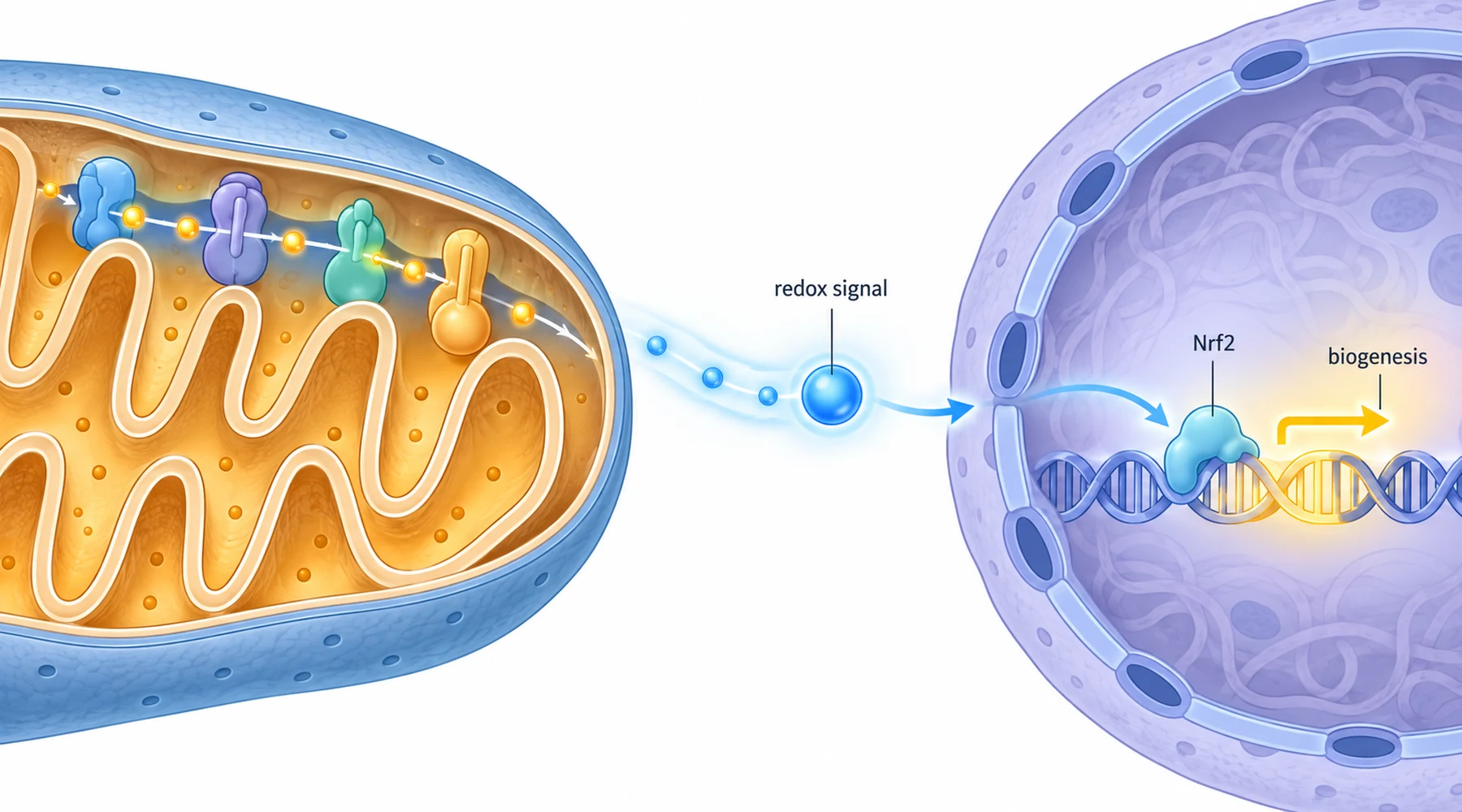
A single sentence shows up often in writing about methylene blue: it activates Nrf2 and builds new mitochondria. The sentence reads as one claim, but it contains two different concepts, studied in different systems, with different kinds of evidence. The first is a signaling pathway, a sensor that turns on defense genes. The second is a construction program, a cell making more mitochondrial machinery. A signal can trigger construction, but showing the signal fired is not the same as showing the building went up, and showing more building material is not the same as showing a better power supply. This article keeps those three things apart.
The Nrf2 concept: a sensor, not a shield
Nrf2 (nuclear factor erythroid 2-related factor 2) is a transcription factor, a protein whose job is to turn on genes. Under resting conditions it is held in the cytoplasm by a sensor protein called Keap1, which hands it to the cell's disposal machinery for continuous destruction. When oxidants or electrophiles modify reactive cysteines on Keap1, the disposal stops. Newly made Nrf2 accumulates, moves into the nucleus, pairs with small Maf proteins, and binds DNA sequences called antioxidant response elements. That binding switches on a battery of defense genes.
The canonical readouts of this pathway are specific. NQO1 (NAD(P)H quinone dehydrogenase 1) and TrxR1 (thioredoxin reductase 1) are among the most reliable Nrf2 target genes. HO-1 (heme oxygenase-1) is strongly inducible but answers to other regulators too. Enzymes such as SOD and catalase are antioxidant machinery, but their levels are controlled by many transcription factors, so a rise in SOD does not by itself prove Nrf2 was the cause. The evidence closest to the signaling event is nuclear Nrf2 accumulation, activation of an ARE reporter, or induction of canonical targets that disappears when Nrf2 is removed. Everything else, lower oxidant levels, better survival under stress, higher glutathione, is a downstream effect that many pathways can produce.
The earliest methylene blue result in this story fits the near end of that scale. Fibroblasts exposed to nanomolar methylene blue showed delayed senescence alongside higher complex IV content, higher oxygen consumption, and induction of the phase-2 enzymes TrxR1 and NQO1, as reported in the study that first tied methylene blue to mitochondrial enhancement and senescence delay. TrxR1 and NQO1 are exactly the canonical Nrf2 targets a careful reader wants to see. What the study did not show is the signaling event itself: no nuclear Nrf2 measurement, no ARE reporter, no Nrf2 knockout control. So the honest reading is that methylene blue induces known Nrf2 target genes in this system, which is consistent with Nrf2 activation but does not isolate Nrf2 as the cause. Induction of a target gene is one step downstream of the switch.
The biogenesis concept: a program with three checkpoints
Mitochondrial biogenesis is the program by which a cell makes more mitochondrial capacity. Its textbook hierarchy runs through three checkpoints. First, the coactivator PGC-1alpha switches on the program. Second, nuclear transcription factors (confusingly, one is named NRF1, nuclear respiratory factor 1, a different protein from Nrf2) drive expression of mitochondrial genes, including TFAM, which maintains and replicates mitochondrial DNA. Third, the cell ends up with more mitochondrial genomes and more respiratory machinery, measurable as mtDNA copy number, citrate synthase activity, or respiratory complex abundance and activity.
Each checkpoint is a weaker or stronger claim than the last. Higher PGC-1alpha or TFAM messenger RNA shows the program was requested, not that it completed. More mtDNA or more complex IV protein shows more material, but remodeled mitochondria can look the same way. Better respiration, higher oxygen consumption or complex activity, shows function changed, but function can improve without any new mitochondria at all. A strong biogenesis claim combines all three: regulator, content, and function.
The fibroblast follow-up study walked through most of that chain. In human IMR90 cells treated with 100 nanomolar methylene blue, the NAD to NADH ratio rose transiently within 15 minutes and returned to baseline within a day. That transient energy signal was followed by transient phosphorylation of the energy sensor AMPK, then induction of PGC-1alpha and of SURF1, a protein required to assemble working complex IV. Complex IV activity roughly doubled, cellular oxidants fell by about 28 percent, and telomeres eroded more slowly. The full sequence is laid out in the paper connecting methylene blue's redox activity to AMPK and mitochondrial biogenesis.
Two details in that paper deserve attention because they are the kind of detail that usually gets dropped. First, the pattern mattered more than the presence: methylene blue switched AMPK on transiently, while the reference drug AICAR switched it on chronically, and the transient activator delayed senescence about eight times more effectively. A claim of the form "methylene blue activates AMPK" is therefore incomplete; the operative claim is that it activates AMPK transiently, and the comparison suggests chronic activation does not do the same work. Second, methylene blue did not alter the cell cycle, which rules out the most boring alternative explanation, that the cells simply divided more slowly and looked younger for it.
The mouse branch: behavior, genomes, and a deliberately paradoxical trigger
The cell-culture work was extended into animals by a group studying mid-aged mice. After 60 days of oral methylene blue, the treated mice kept more of their physical endurance and exploratory behavior and showed less anxiety-like behavior than untreated aged mice. In brain tissue, genes that aging had suppressed, NRF1, TFAM, SOD2, and the complex IV subunit COX1, were restored toward adult levels, and mtDNA copy number rose. Those results, behavior plus regulator plus genome abundance, are reported in the mid-age mouse study on methylene blue and brain mitochondrial biogenesis.
The proposed trigger is the interesting part, because it runs against the simple story that methylene blue is an antioxidant. The treated mice showed higher reactive oxygen species generation in brain mitochondria, specifically hydrogen peroxide. The authors' proposal: a mild peroxide signal is what activates the Nrf2 pathway, which then drives the biogenesis program. In this model methylene blue is a pro-oxidant at the trigger step and an antioxidant at the outcome step, a pattern pharmacologists call hormesis, where a small stress provokes a defense larger than the stress itself. Whether that peroxide rise actually causes the Nrf2 activation was not directly tested; it is the proposed bridge, not a measured link.
A companion study from the same group stress-tested the safety side of that proposal. In isolated brain mitochondria, methylene blue raised hydrogen peroxide production at concentrations as low as 50 nanomolar, yet therapeutic concentrations caused no detectable damage to mitochondrial DNA. Only at 100 micromolar, far above therapeutic levels, did selected mtDNA fragments show damage, and the combination of 20 micromolar dye with red light produced singlet oxygen that damaged mtDNA broadly, which is the known photosensitizer behavior of the dye rather than its dark pharmacology. Pretreatment even protected ventral midbrain mitochondria against rotenone-induced mtDNA lesions. The dose response and the genotoxicity controls are detailed in the study on methylene blue's non-genotoxic peroxide production.
Measured versus proposed: reading the diagram
The pathway diagram below was drawn for this article to enforce the distinction the text has been making. Boxes with solid borders are steps with direct experimental support in the methylene blue literature: redox cycling that shifts NAD/NADH, transient AMPK phosphorylation, PGC-1alpha and SURF1 induction, higher complex IV activity, induction of NQO1 and TrxR1, restored biogenesis gene expression and mtDNA copy number in mouse brain, and peroxide elevation without mtDNA damage at therapeutic doses. Boxes with dashed borders are the proposed links: Keap1 sensing the peroxide signal, Nrf2 nuclear accumulation driving the defense genes, and the causal arrow from Nrf2 activation to the biogenesis program. Those dashed steps are plausible, consistent with the data, and standard textbook biology, but in these papers they were not measured with the controls (nuclear Nrf2, ARE reporters, Nrf2 knockout) that would promote them to solid boxes.
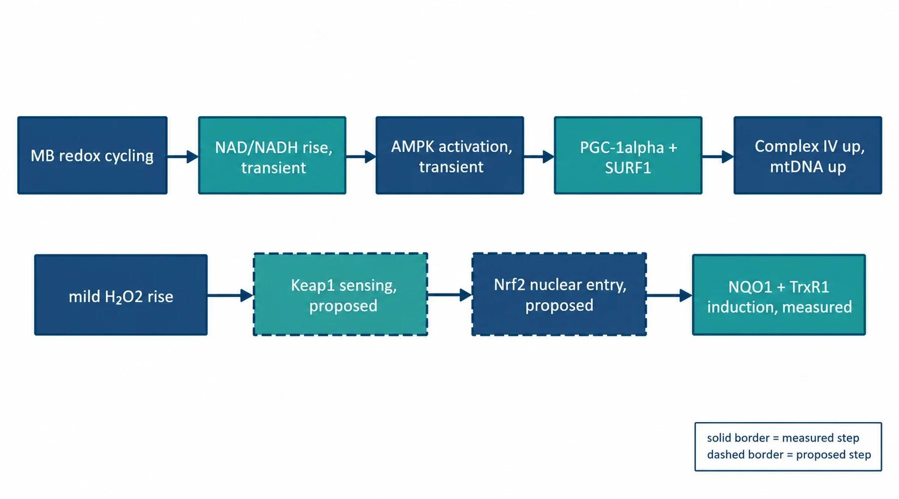
Three confusions become visible once the diagram is in front of you. First, NRF1 and Nrf2 are different proteins with near-identical names; the mouse study measured nuclear respiratory factor 1, a biogenesis regulator, not the antioxidant transcription factor, and conflating them collapses the whole diagram into a single node. Second, gene expression is the start of the story, not the end; restored TFAM or COX1 messages support a biogenesis request, while mtDNA copy number and complex IV activity are the evidence the request was fulfilled. Third, the antioxidant outcome does not prove the Nrf2 route; the 28 percent oxidant decline in fibroblasts and the rotenone protection in mice are real downstream effects, but several pathways converge on them.
What would promote a dashed box to solid
The missing experiments are standard ones. Nuclear Nrf2 protein after methylene blue exposure, with a time course matched to the transient AMPK signal, would test whether the two arms actually fire together, as the synchronized-activation hypothesis claims. An ARE-driven reporter would confirm transcriptional activity rather than inferring it from target genes. Repeating the biogenesis readouts with Nrf2 knocked down or knocked out would test the causal arrow directly: if complex IV induction and mtDNA gains survive without Nrf2, the bridge is wrong and the AMPK arm carries the effect alone. On the animal side, a chronic-dosing study with the same three checkpoints, regulator, content, function, would test whether the program stays on or adapts. Until then, the synchronized activation of energy and defense pathways remains what its authors called it: a possible key factor, not an established mechanism.
Readers coming from the broader pharmacology may want the surrounding context: how methylene blue shuttles electrons at the mitochondrial level, what else it does to mitochondria and cellular energy, where the senescence findings sit in aging research, how biogenesis claims relate to cognitive performance, and the safety record that bounds every dosing discussion. Each of those is a separate concept with its own evidence, and each deserves the same measured-versus-proposed treatment this article gave Nrf2.