
Article
Methylene blue and nitric oxide: NOS, cGMP, and vascular signaling
The question sounds simple: does methylene blue inhibit nitric oxide? In the laboratory the answer splits into four separate questions, and each has a different experimental test. Confusing them is the main reason writing about methylene blue and nitric oxide reads as confident but vague.
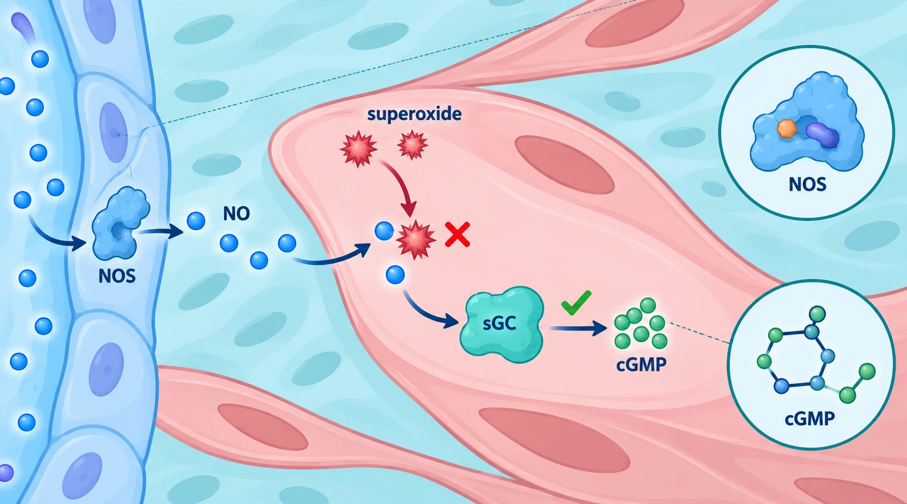
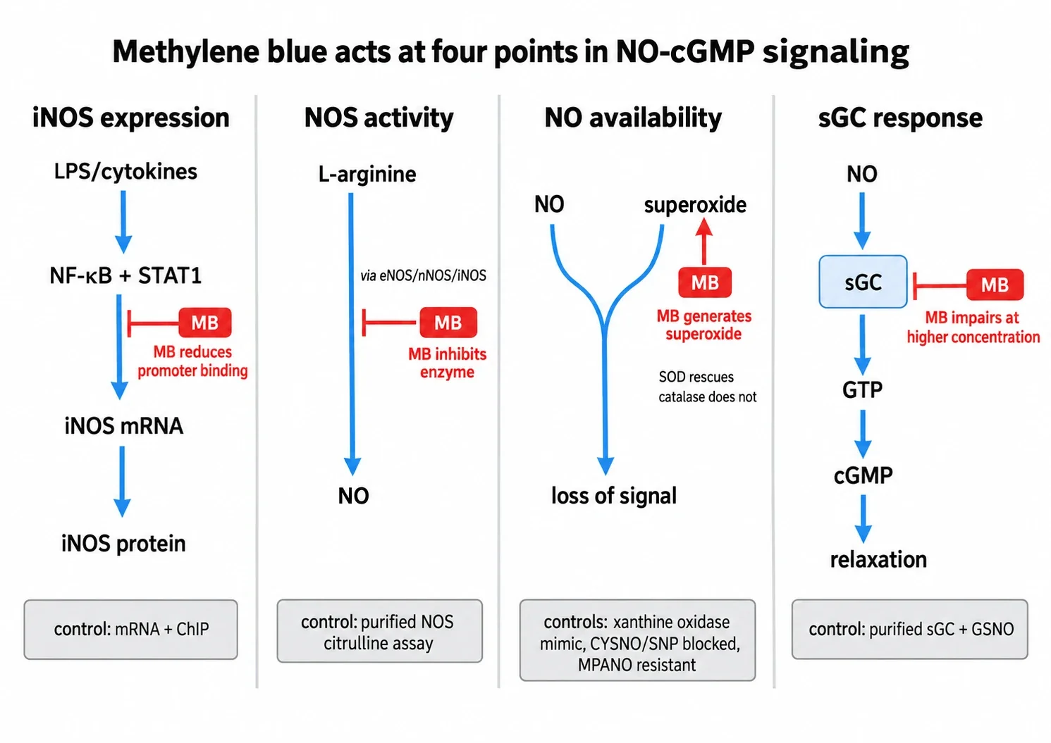
Think of the nitric oxide signal as a chain with four links. A gene is transcribed to make nitric oxide synthase. The enzyme converts arginine to nitric oxide. The gas diffuses to its target before it reacts with something else. Soluble guanylate cyclase detects it and makes cyclic GMP. Methylene blue has published effects at every link, but the evidence comes from different preparations, different concentrations, and different isoforms. A result at one link does not prove action at another.
The canonical chain, stated plainly
In a blood vessel wall the standard sequence runs like this. A stimulus acts on an endothelial cell. Endothelial nitric oxide synthase, called eNOS or NOS3, makes a small pulse of nitric oxide. The gas diffuses into the neighboring smooth muscle cell, binds the reduced iron heme of soluble guanylate cyclase, and accelerates conversion of GTP to cyclic GMP. Cyclic GMP activates protein kinase G, calcium handling shifts, and the muscle relaxes.
There is a second chain worth keeping separate. During infection or cytokine exposure, cells transcribe a different enzyme, inducible nitric oxide synthase or iNOS, also called NOS2. It produces larger and longer lasting nitric oxide fluxes than eNOS. A third isoform, neuronal NOS or nNOS, belongs to yet another context. When a paper says nitric oxide synthase without specifying the isoform, stop and check which one was measured. The overview of how methylene blue moves electrons makes the same point for redox targets generally: the relay does nothing on its own, and the partner at each end decides the outcome.
Measured cyclic GMP is also an integrated readout, not a direct enzyme assay. Its level reflects synthesis by cyclase minus breakdown by phosphodiesterases, especially PDE5 in vascular smooth muscle. A fall in cyclic GMP therefore means less signal reached the detector. It does not by itself name the link that failed.
Link 1: less iNOS enzyme is made
In mouse macrophages stimulated with bacterial lipopolysaccharide, interferon gamma, or both, methylene blue prevented the expected rise in iNOS protein and strongly suppressed iNOS messenger RNA in the macrophage transcription experiments. The informative part is what did not change. Degradation of the inhibitor I kappa B alpha, phosphorylation of NF kappa B and STAT1, and movement of those factors into the nucleus stayed largely intact. What fell was their binding to DNA, confirmed by chromatin immunoprecipitation showing reduced occupancy at the iNOS promoter.
That is a gene regulatory effect upstream of enzyme abundance, not acute blockade of an existing enzyme. It belongs to inflammatory signaling, where transcription factors must bind a promoter before protein appears. It says nothing direct about constitutive eNOS in a resting vessel, and it should be described in those terms.
Link 2: the existing NOS enzyme runs slower
A rat study tested a different level. Conscious male rats received lipopolysaccharide intravenously, then three hours later received either saline or methylene blue at 3 mg per kg intravenously, and nitric oxide synthase activity in the medial basal hypothalamus was measured by conversion of labeled arginine to citrulline in the hypothalamic NOS activity study. Stress plus saline alone induced some calcium independent activity, and lipopolysaccharide raised it to about ten times the control level. Methylene blue eliminated both induced components. Calcium dependent constitutive activity did not change in that tissue at that dose and time point.
Read that result exactly as written: suppressed measured iNOS type activity under those conditions. It is not a measurement of gene expression, and it is not proof of permanent selectivity for iNOS over eNOS or nNOS. Purified enzyme work sharpens the caution. In direct assays of purified nitric oxide synthase, methylene blue fully blocked citrulline formation at 30 micromolar, with a half inhibitory concentration near 5.3 micromolar without superoxide dismutase and 9.2 micromolar with it, as reported in the purified NOS and guanylate cyclase comparison. A separate purified nNOS study implicated oxidation of enzyme bound heme. The practical lesson is that direct NOS inhibition is real in isolated systems, but the hypothalamus result cannot be stretched into a general claim that methylene blue spares all constitutive NOS everywhere.
Link 3: nitric oxide is lost before it arrives
The cleanest demonstration that something other than enzyme blockade can explain a cyclic GMP fall comes from cultured rabbit pulmonary arterial smooth muscle cells. The design bypassed endogenous NOS entirely by adding nitric oxide donors directly, then asked whether methylene blue still suppressed the cyclic GMP response in the vascular smooth muscle superoxide study.
Three donors were tested from 1 to 100 micromolar: S nitroso L cysteine, sodium nitroprusside, and a stable deaminated analogue called MPANO. All raised cyclic GMP between roughly 1.5 fold and 12 fold. Methylene blue at 10 micromolar inhibited the first two by 51 to 100 percent and left the MPANO response unchanged. If methylene blue were simply blocking the cyclase catalytic site, that donor selectivity would be hard to explain, because all three converge on the same downstream enzyme.
Three controls then pinned the mechanism to superoxide. First, generating extracellular superoxide with xanthine plus xanthine oxidase reproduced the same pattern: CYSNO and nitroprusside blocked, MPANO spared. Second, methylene blue caused dose and time dependent superoxide production from the smooth muscle cells, measured by cytochrome c reduction. Third, superoxide dismutase fully prevented the inhibition while catalase did not, which points to superoxide rather than hydrogen peroxide as the proximate species in that preparation.
The co culture version of the experiment is worth stating because it locates the effect in space. Endothelial cells on coverslips were briefly co cultured with smooth muscle cells, so basally released endothelial factor raised smooth muscle cyclic GMP three to eight fold. Pretreating either compartment with methylene blue reduced that signal, and superoxide dismutase partly rescued the endothelial pretreatment effect. In other words, methylene blue need not act inside the smooth muscle detector cell at all. It can lower the signal by removing the messenger in transit. The guide to reading methylene blue research treats this as a general rule: name the compartment where the intervention happened before naming a molecular target.
Calling this NOS inhibition would be wrong. No synthase step was required for the donors to work, yet the signal still fell.
Link 4: the detector itself responds less
Direct effects on soluble guanylate cyclase exist, but the concentrations matter. In the purified enzyme comparison cited above, nitric oxide stimulated cyclase needed about 60 micromolar methylene blue for half inhibition, against NOS half inhibition near 5 to 9 micromolar, and even 1 millimolar produced only partial maximal blockade. That is roughly a six to eleven fold separation. A low micromolar cell experiment that lowers cyclic GMP therefore cannot be labeled selective cyclase inhibition on the strength of the cyclic GMP number alone.
A later redox study added a second caution: methylene blue deactivated nitric oxide activated cyclase by a mechanism distinct from simple air oxidation of the heme iron. So the detector can be impaired, but the chemistry is not settled, and the concentration gap means the superoxide and NOS effects deserve to be ruled out first at low micromolar exposures. This is also where phosphodiesterase control belongs. Unless breakdown was measured or blocked, reduced cyclic GMP accumulation is consistent with less synthesis, faster degradation, or both.
Why this does not reduce to one blood pressure claim
Each link was established in a different system. Macrophage transcription is not hypothalamic enzyme activity. Hypothalamic activity after lipopolysaccharide is not resting endothelial tone. Basally released endothelial factor in co culture is not agonist stimulated vasodilation. Cultured pulmonary arterial cells at 10 micromolar methylene blue are not a human circulation. Collapsing them into a statement like methylene blue raises blood pressure by blocking nitric oxide discards exactly the distinctions the controls were built to preserve.
Three checks keep the story honest. First, name the isoform: eNOS for resting vascular tone, iNOS for induced inflammatory flux, nNOS for neuronal and some purified enzyme contexts. Second, name the delivery of the nitric oxide signal: endogenous synthesis, basal endothelial release, or a specific donor such as CYSNO, nitroprusside, or MPANO, since the stable donor resisted methylene blue while the labile ones did not. Third, name the concentration and the rescue: superoxide dismutase rescue implicates superoxide, catalase failure argues against hydrogen peroxide in that preparation, xanthine oxidase mimicry shows sufficiency, and purified enzyme assays separate direct catalysis effects from transit losses.
The compact synthesis is this. Methylene blue is better treated as a multi site perturbant of nitric oxide to cyclic GMP signaling than as a selective cyclase blocker. Depending on preparation and concentration, it can reduce iNOS transcription factor binding, directly inhibit NOS enzymes, lower nitric oxide availability through superoxide generation, and impair nitric oxide activated cyclase. The role of cyclic GMP signaling in vascular studies and the comparison of nitric oxide donors used in cell assays each cover one of those branches in more detail, and the methods used to separate redox effects from enzyme effects explains why superoxide dismutase plus catalase plus a stable donor control appears so often in this literature.
A low cyclic GMP value after methylene blue identifies a broken chain. Only the controls say which link broke.