
Article
Methylene blue and light exposure: wavelength, irradiance, and treatment geometry
Two papers report exposing methylene blue to 10 J per square cm of light. One kills bacteria. The other does nothing measurable. A reader comparing only the dose concludes the field is inconsistent. The field is consistent. The dose was never the whole exposure.
A light exposure is a small system of parameters that act together: wavelength, irradiance, duration, the resulting fluence, oxygen availability, dye concentration and aggregation state, the timing between dye and light, and the optical path the light actually travels through the sample. Change one and the photochemistry changes. This article works through each parameter, then distinguishes near infrared photobiomodulation from direct dye photoactivation, because those two regimes are the most commonly confused.
Wavelength decides whether the dye participates
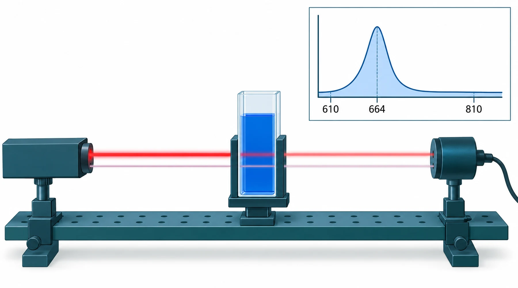
Methylene blue in water absorbs red light strongly as a monomer, with a peak near 664 to 665 nm and a shorter wavelength shoulder near 610 nm that grows as the dye aggregates into dimers and larger stacks. Molar extinction near the peak is on the order of 65,000 to 85,000 M-1 cm-1, so micromolar solutions are already intensely blue. The early photochemical review of methylene blue with DNA and biological substrates documents this absorption behavior and the excited state chemistry that follows: absorption populates a short lived singlet state, crossing to a longer lived triplet state with reported yields near 0.5, which in aerated solution is quenched by oxygen within about 2 microseconds.
That quenching step is where the two photochemical paths diverge. In Type II chemistry the triplet dye transfers energy to ground state oxygen and produces singlet oxygen, with commonly cited yields near 0.52. In Type I chemistry the triplet transfers an electron or hydrogen to a substrate instead, producing radicals including superoxide and hydroxyl species. Both can run at once, and conventional photodynamic effect stays strongly oxygen dependent even when Type I contributes.
The consequence for methylene blue wavelength selection is a simple rule. Red light from about 630 to 680 nm overlaps the dye band, with 660 to 665 nm near the monomer maximum, so it can drive photosensitizer chemistry wherever appreciable dye is present. Near infrared at 810 nm lies well outside the ground state absorption band, so it is a poor wavelength for direct excitation of the dye. Concentration complicates the picture in one honest way: as concentration rises, more dye sits in dimeric and aggregated forms that absorb toward 610 nm and photosensitize less efficiently per molecule, so doubling the dye does not double the effect. The companion review of methylene blue with red light therapy treats this absorption rule as the precondition for any photosensitized reaction.
Irradiance, duration, and fluence are three parameters, not one
Fluence, in J per square cm, is irradiance multiplied by time. Reporting fluence alone hides the rate at which the energy arrived, and the rate matters.
A high irradiance delivered briefly and a low irradiance delivered for a long time can give the same fluence with different chemistry. High irradiance consumes dissolved oxygen faster than diffusion replaces it, which throttles the oxygen dependent Type II path partway through the exposure. It also deposits heat, and tissue responds to heating with blood flow changes that alter both oxygenation and dye distribution. Low irradiance over a long exposure keeps oxygen supplied but gives repair and reoxygenation processes time to act during the exposure itself. Photochemistry textbooks call the assumption that only total dose matters reciprocity. It fails whenever oxygen, heating, or biology participates, which in tissue means nearly always.
Human antimicrobial photodynamic exposures with methylene blue commonly use 6 to 18 J per square cm, which overlaps photobiomodulation values. Fluence alone therefore cannot identify the regime. A 10 J per square cm exposure at 660 nm into dye loaded tissue is photosensitizer driven photochemistry. The same fluence at 810 nm into tissue with trace dye is something else entirely. The number matches. The concept differs.
Optical path and sample geometry decide what the light meets
Beer Lambert behavior means the front of a sample sees far more light than the back. In a cuvette with tens of micromolar dye, the first millimeter can absorb most of the incident red light, leaving deeper layers effectively unexposed. Shrink the same solution into a shallow well at low volume and the exposure becomes nearly uniform. Same dye, same lamp, same fluence at the surface, different result.
Tissue adds scattering on top of absorption. Red light near 660 nm penetrates on the order of millimeters, with the exact depth set by blood content, pigmentation, and tissue type. Wells add their own artifacts: meniscus lensing, reflection from walls, shading of edge wells, and warming of small volumes under the source. Two laboratories reporting identical lamp settings but different plate types, volumes, or source distances have run different experiments.
Distance deserves emphasis because it is the most common silent variable. Irradiance falls rapidly with distance from a non collimated source, so a lamp moved from 2 cm to 5 cm above the plate can cut irradiance several fold while the methods section still reads the same. Without a measured irradiance at the sample surface, published fluences computed from manufacturer power ratings are estimates, not measurements.
Annotated parameter guide: what makes an exposure reproducible
The original asset for this page is a checklist. Any study light exposure that omits these entries cannot be compared with another, however similar the headline dose looks.
- Wavelength and bandwidth. Peak and full width, not a color name. Red LED covers 630 to 680 nm of very different dye absorption.
- Irradiance at the sample surface, measured, in mW per square cm. Manufacturer wattage is not irradiance.
- Duration and pulsing. Continuous versus pulsed delivery, including pulse frequency and duty cycle where applicable.
- Fluence computed from the measured pair above, in J per square cm.
- Dye concentration, formulation, and aggregation state. Monomer near 664 nm photosensitizes differently than aggregates near 610 nm.
- Drug light interval. Minutes to hours between dye administration and irradiation, which sets where the dye is when light arrives.
- Oxygenation. Aerated solution versus hypoxic tissue changes Type I and Type II balance directly.
- Geometry. Sample depth, volume, vessel type, source distance and angle, and tissue target depth.
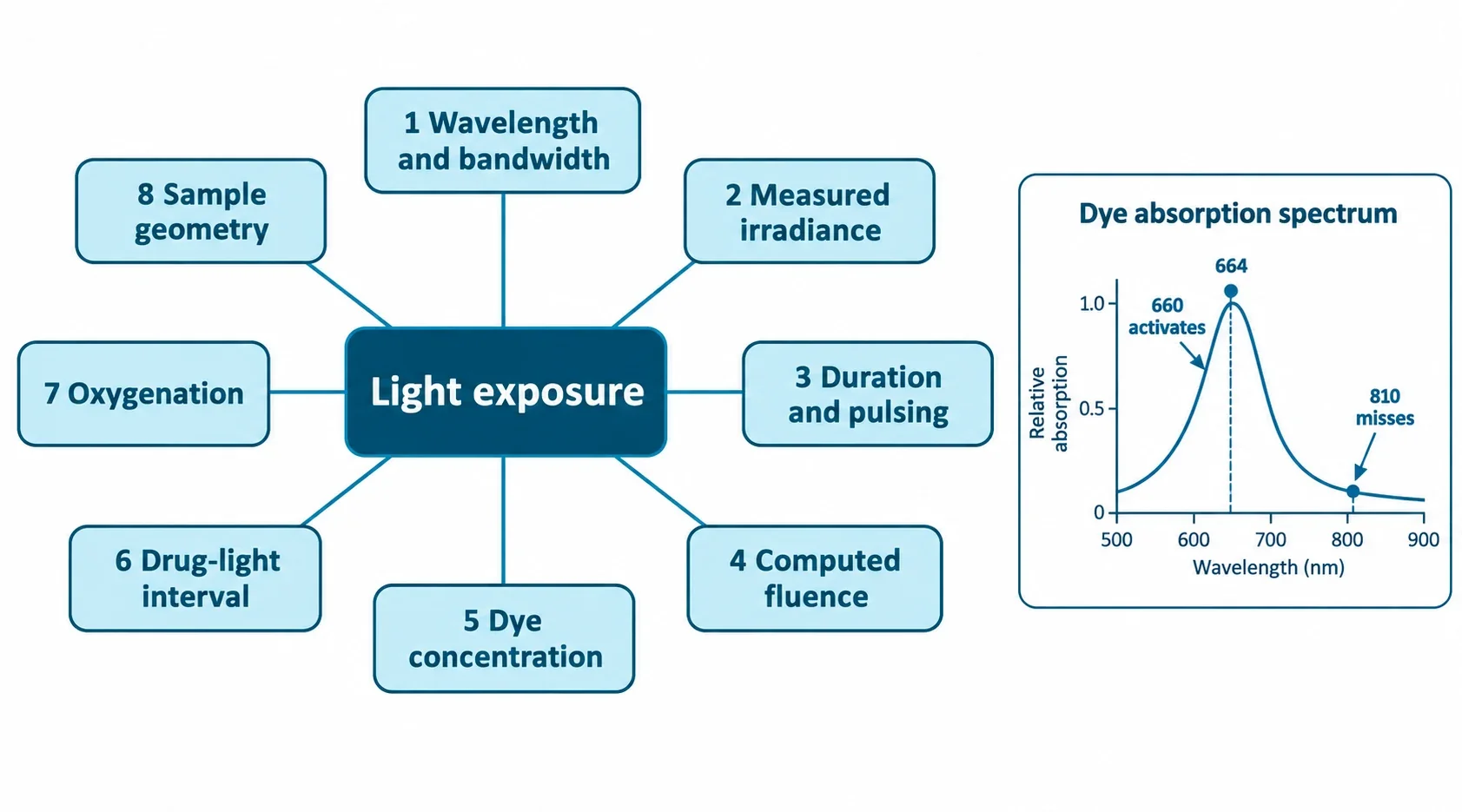
A useful habit when reading papers: check the drug light interval first. It reveals intent. Photodynamic designs irradiate dye loaded tissue deliberately. Combination designs may separate the two on purpose, as below.
Near infrared photobiomodulation is not dye photoactivation
Photobiomodulation, usually abbreviated PBM, uses red or near infrared light to modulate endogenous signaling without adding a photosensitizer. Photodynamic therapy, usually abbreviated PDT, uses three parts together: light the sensitizer absorbs, the sensitizer itself, and oxygen to create a burst of reactive species that damages the target. A review of mitochondria as a neuroprotection target pairs methylene blue with PBM because they converge on respiration from different directions: the dye acts as a catalytic redox cycler supporting electron flow, while PBM is hypothesized to promote dissociation of inhibitory nitric oxide from cytochrome c oxidase at Complex IV. That enzyme mechanism should stay labeled as a hypothesis.
The two most cited combination mouse studies both use 810 nm light, which is exactly why they do not demonstrate dye photoactivation. In a sleep deprivation pretreatment study, mice received daily methylene blue at 0.5 mg per kg, 810 nm pulsed laser every other day, or both before deprivation. Combined pretreatment protected memory behaviors and hippocampal synaptic proteins while single treatments largely failed. In a chronic stress study with concurrent treatment, mice received 810 nm PBM at 8 J per square cm alongside daily dye at 0.5 mg per kg, with the laser applied first and dye injected two hours later specifically to avoid irradiating dye loaded tissue. All three arms reversed learning deficits, with additive benefit mainly for serum cortisol and brain reactive oxygen species. These results test two mitochondrial interventions used together, a distinction the photodynamic study collection keeps visible by recording wavelength per indication.
Skin evidence sits on the other side. Human methylene blue plus light skin data clusters at 630 to 680 nm with topical or intralesional dye and destructive or remodeling intent, which is PDT by definition. Citing that clearance as proof that dye enhances PBM, or citing an 810 nm mouse rescue as proof that a 660 nm panel plus dye is a cognition protocol, crosses the regimes. The dosing and pharmacology reference and the safety and interaction guide carry the parts of this story about concentration and contraindications further.
Name the concept before comparing numbers. Wavelength determines whether the dye absorbs. Irradiance and duration determine what the chemistry does with that absorption. Geometry determines which part of the sample experiences any of it. Two exposures that match on one parameter and differ on the rest are different experiments, and treating them as interchangeable is the error this guide exists to prevent. No home protocol follows from any of this; wavelength compatible light plus systemic dye converts an otherwise modulatory exposure into photosensitizer driven photochemistry, which belongs in clinical hands.