
Article
Methylene blue as an indicator: what the color change actually reports
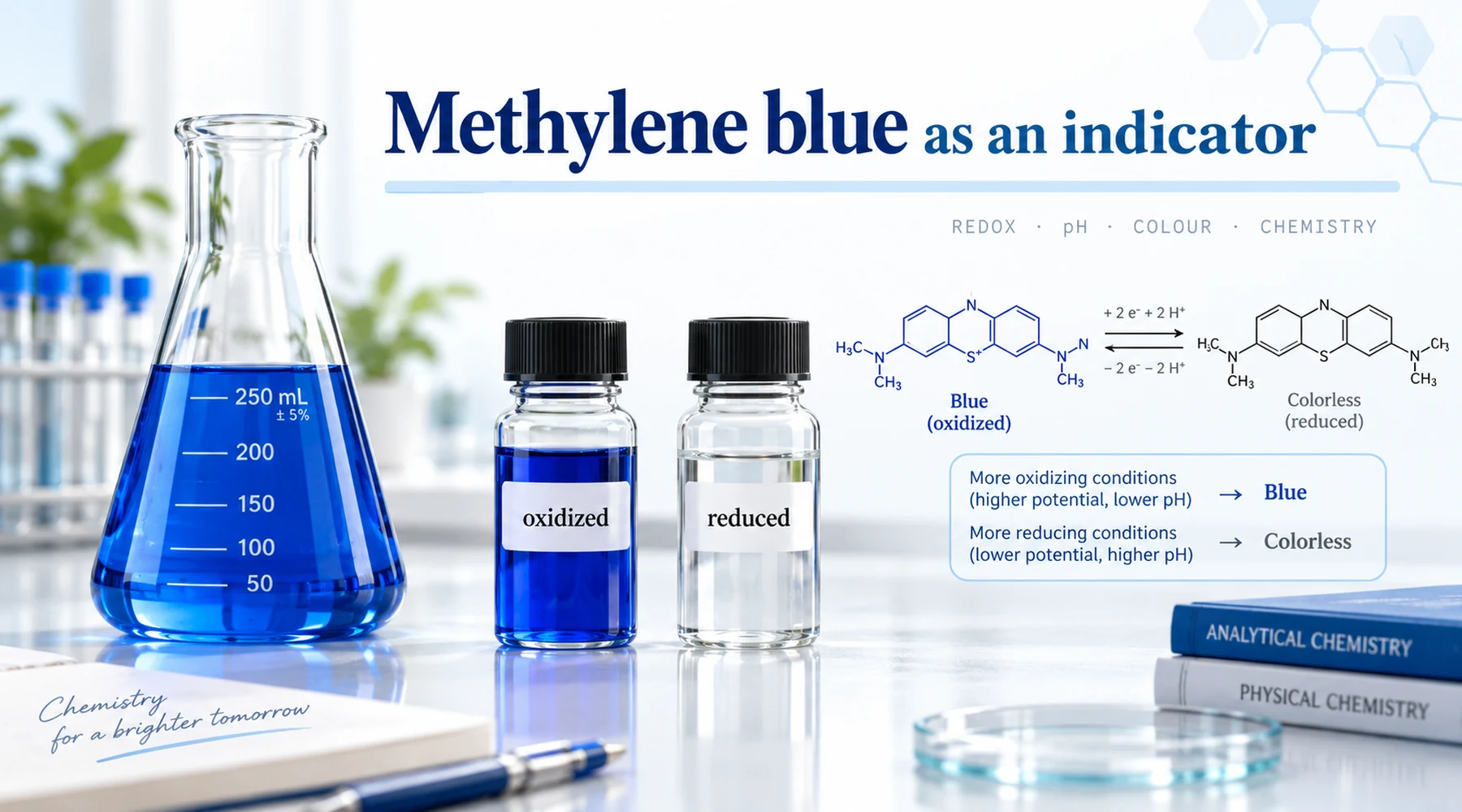
A technician adds methylene blue to a beaker, adjusts the pH, and waits for the color to report acidity. Nothing clean happens. She adds a drop of ascorbic acid solution instead, and the blue vanishes. She shakes the flask in air, and the blue returns. Same dye, two very different behaviors. The confusion is the whole subject of this article: methylene blue is a redox indicator that responds to electron transfer, not a pH indicator that responds to protons. Once that distinction is fixed, every other detail falls into place.
The practical rule fits in one sentence. Blue means the oxidized form is present. Colorless means the reduced form, leucomethylene blue, is present. Anything that moves electrons can flip the state. Acids and bases alone cannot, though pH changes the voltage at which the flip happens.
The couple behind the color
Oxidized methylene blue is the methylthioninium cation, a flat three-ring phenothiazinium system supplied as the chloride salt, which PubChem records as methylthioninium chloride. In dilute water the monomer absorbs near 664 nm, and that absorption is the blue the eye sees. The reduced partner, leucomethylene blue, has the same skeleton with the central ring nitrogen protonated and the chromophore broken, so it is nearly colorless with its strong band in the ultraviolet (PubChem entry for the leuco form).
The net reaction moves two electrons, but the proton count depends on pH because the reduced form protonates in acid. The reference treatment in visualizing redox chemistry gives the standard potential near +0.53 V at pH 0 and the formal potential near +0.011 V at pH 7 against the hydrogen electrode. Between those points the potential slides with pH. Modern voltammetry in the redox behavior and potentials of dyes study finds about 29.5 mV per pH unit from roughly pH 4 to 10, matching a two-electron one-proton regime, and about 88.5 mV per pH unit below pH 4, matching a two-electron three-proton regime. A reported potential without its pH is therefore meaningless. Always ask which slope and which reference electrode stand behind the number, since values quoted against silver references cannot be compared directly with values against the hydrogen scale. The underlying structure and properties set the chromophore, and a companion page on redox readout design extends the couple into formulation choices.
That middle position on the potential scale is what makes the dye useful. At pH 7 it sits among mild oxidants: strong enough to accept electrons from good donors such as ascorbate or NAD(P)H, weak enough to pass them on to dissolved oxygen or ferric centers. So it cycles instead of getting stuck. Reduction bleaches it. Air reoxidizes it. The path is the same in both directions.
Why pH questions keep coming up
Three sources of confusion recur in labs and classrooms, and each has a concrete correction.
First, pH shifts the potential, which looks like a pH response. Raise the pH and the couple becomes easier to reduce at a fixed electrode reading, so the same donor bleaches the dye faster. But no protonation step alone produces the clean blue to colorless transition over the normal range. A critical 2025 review of methylene blue properties reports the 665 nm maximum essentially unchanged from pH 2 to 12, with only modest absorbance changes. Protons tune the voltage. Electrons flip the color.
Second, the ontology label misleads. PubChem currently imports a ChEBI role calling the dye an acid-base indicator. That tag should not be read as evidence that methylene blue behaves like phenolphthalein or methyl red. In normal aqueous work it does not.
Third, the Tashiro mixture borrows the name. Tashiro indicator is a blend of methyl red and methylene blue in which methyl red supplies the acid-base response and methylene blue supplies a blue optical background, converting a red to yellow change into a sharper composite change. One commercial Tashiro formulation quotes pH 4.4 to 6.2, while a defined ITW Reagents formulation specifies red-violet at pH 4.4 to green at pH 5.8. Neither range is a transition range of methylene blue itself. The dye is the screening agent, not the sensor.
One edge case deserves a warning. Around pH 13, strongly alkaline conditions can generate red to pink methylene-violet type products by irreversible hydrolysis. That pink is degradation, not an indicator endpoint. Treat it as a signal to discard the solution, not to read it.
Where it works as a redox indicator
The classical use is the Lane-Eynon reducing-sugar titration. Hot alkaline Fehling solution is titrated with sugar, and methylene blue is added near the endpoint; the remaining blue disappears once the reducing capacity of the sugar exceeds that needed for copper(II). The AOAC Lane-Eynon procedure keeps the mixture boiling so steam evolution suppresses reoxidation by air, typically with a few drops of dye solution. The color change reports that no reducible copper remains, which is a redox statement about the flask, not a pH statement.
Older titrimetry used the same logic elsewhere. Dilute dye could substitute for starch in some iodimetric titrations where alcohol made starch unsuitable, at working strengths near 0.05 g per liter, though excess dye risks forming an iodine dye compound as Sinnatt showed in 1912. Handbook compilations also list the dye for titanometry and iodometry. The 1936 ascorbic acid method of Gal reduced the dye with vitamin C extracts and back-titrated residual dye with titanium trichloride. That method is now historical rather than routine, but it shows the pattern: donor reduces blue to colorless, and the disappearance of blue is the endpoint.
Dissolved oxygen methods run the same couple in reverse. One published approach first reduces the dye with glucose in alkaline aqueous ethanol, then lets dissolved oxygen oxidize the colorless leuco form back to blue almost instantly, reading near 600 nm up to about 13 micrograms per milliliter as described by Okumura and Hashitani. Do not confuse this with the classical Winkler dissolved-oxygen titration, whose endpoint uses starch; the NOAA Winkler SOP specifies about 1 percent starch. Methylene blue oxygen methods are a separate family with reversed color direction.
The classroom blue bottle is the same couple in miniature: glucose in alkaline solution reduces blue dye to the colorless form on standing, and shaking dissolves oxygen that reoxidizes it to blue. The full procedure and its controls belong to the blue bottle demonstration, and the band positions and aggregation shifts belong to the spectra and concentration effects page. This article keeps only the mechanism, because procedure and spectra each need their own conditions.
Color-state chart: states, conditions, limits
This table is the original reference for the article. Read across a row before trusting any endpoint.
| State | Appearance | Condition that produces it | What it reports | Limitation |
|---|---|---|---|---|
| Oxidized monomer, dilute, neutral | Deep blue, peak near 664 nm | Air-saturated solution, no donor present | Baseline ready state | At high concentration the peak shifts toward 610 nm by dimerization, so judge hue only after diluting |
| Reduced leuco form, neutral | Colorless | Donor present (glucose in base, ascorbate, dithionite, NAD(P)H with catalyst) | Reducing capacity exceeded indicator demand | Air reoxidizes it, so endpoints fade back to blue unless air is excluded or standardized |
| Leuco form, acidic | Colorless | Same donors in acid, with extra leuco protonation | Same as above at shifted potential | Potential slope steepens below pH 4, so a pH 7 calibration does not transfer |
| Tashiro mixture, acidic | Red-violet | pH below about 4.4 in methyl red plus methylene blue blend | Acidity, from methyl red | Reports methyl red, not methylene blue; do not quote as a methylene blue pH range |
| Tashiro mixture, near pH 5.8 to 6.2 | Green to gray-green | Same blend near its upper transition | Same as above | Formulation dependent; check the bottle, not the memory |
| Degraded, strongly alkaline | Pink to red | pH near 13, heat, prolonged base exposure | Irreversible hydrolysis to violet type products | Not reversible by shaking; discard and remake |
| Photobleached | Faded blue with altered spectrum | Strong visible light, especially with donors present | Light-driven side chemistry including demethylated products | Protect standards from illumination, as visible-light photoreduction work showed |

Failure modes that blur the endpoint
Air is the first suspect whenever an endpoint drifts. The leuco form reoxidizes readily, especially in alkaline systems, so a titration that went colorless can turn blue again on standing or vigorous swirling. Exclude air where persistence matters, standardize the shaking, and read at a fixed time.
Light is the second suspect. The dye is photochemically active, and irradiation with donors present can bleach it through pathways that include demethylated products. Store analytical dye solutions dark and do not run endpoints under intense bench lamps.
Concentration is the third. The oxidized cation stacks into dimers and higher aggregates with a shifted band near 610 to 615 nm, so a concentrated solution looks off peak without being impure. Dilute into the monomer regime before judging spectra or endpoints.
Two quieter biases deserve space because they punish low-concentration work specifically. The dye adsorbs measurably at glass water interfaces, which biases dilute standards and kinetics; polymer vessels or cuvets are the fix where accuracy matters. And commercial dye can carry related thiazine impurities including Azure B, which historical purification studies removed explicitly, so analytical work should specify grade rather than assuming every bottle labeled methylene blue is chemically identical. Spectral detail on the aggregation shift is kept on the spectra page by design.
A short bench checklist
Buffer the solution and write the pH next to every potential and every endpoint. Fix the oxygen regime: boiling with steam cover for Lane-Eynon style work, inert cover or fixed timing elsewhere. Work dilute enough to see the monomer, protect solutions from light, prefer polymer ware at low concentrations, and record the dye grade. If the goal is actually pH, reach for a true acid-base indicator or a defined Tashiro blend and quote the blend range as the blend range, never as a methylene blue range.
The concept survives this discipline intact. Methylene blue reports oxidation state with unusual clarity because its two states have wildly different visible absorption. Treat the color as a redox readout taken at a stated pH, under stated oxygen and light conditions, with a stated dye grade, and it earns its place in the titration. Treat it as litmus and it will mislead with perfect consistency.