
Article
Methylene blue against biofilms: what light-activated laboratory studies measure
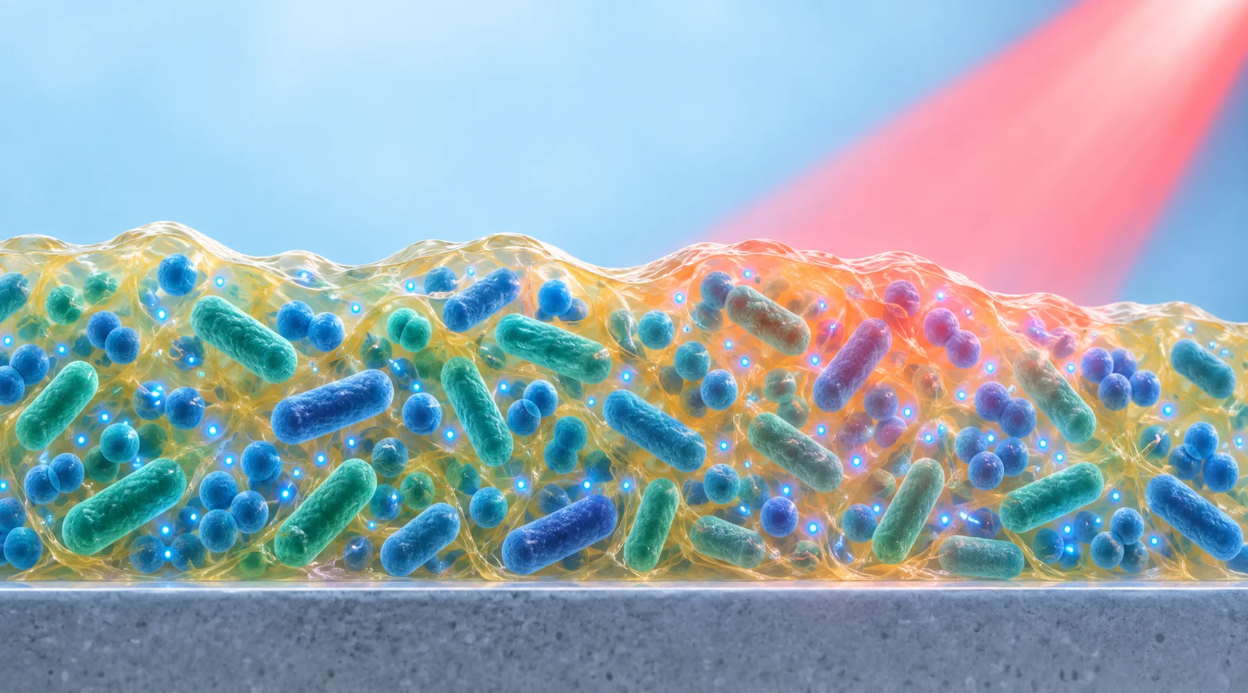
A biofilm is not a crowd of cells floating in liquid. It is a structure: cells attached to a surface, embedded in a self made matrix of polysaccharides, proteins, and extracellular DNA, with gradients of oxygen and nutrients from the outside to the inside. That structure is the reason a compound that kills suspended bacteria in minutes can fail against the same organisms grown as a film. Any claim about methylene blue and biofilms has to be read against that structure, because the matrix decides how much dye reaches the cells and how much light reaches the dye.
This review covers exactly three sources. One is background. Two are laboratory experiments. Keeping those roles separate is the whole method, and it prevents the most common error in this topic, which is quoting a dish result as though it were evidence for treating infection in a person.
The background source is chemistry, not killing
The oldest of the three is a narrative review of phenothiazinium photosensitizer development published in 2005. It traces methylene blue from a 19th century dye to a lead compound for photodynamic therapy of cancer and later for photodynamic antimicrobial chemotherapy. Its contribution is a structure activity story: methylation of the chromophore raises lipophilicity, singlet oxygen yield, and photobactericidal potency while slowing reduction to the less active leuco form. It also states the field's problem plainly. Most reports used standard commercial dyes, with relatively little derivative development.
Use that review for the mechanism section of your mental model: light excites the dye, the triplet state engages oxygen through Type II energy transfer to make singlet oxygen or through Type I electron transfer to make radicals, and that multifactorial attack is why conventional resistance mechanisms transfer poorly to photodynamic killing. Do not cite it for a log reduction. It measured none. The broader photochemistry behind that mechanism is laid out in the photodynamic therapy evidence collection, and the wavelength rules that decide whether a given lamp can drive it are covered in how methylene blue absorbs red light.
Two biofilm experiments, two different outcome constructs
The two experimental papers both test methylene blue photodynamic action on oral biofilms at 660 nm, and they agree on almost nothing else. Different communities, different ages, different dye formulations, different light doses, and above all different endpoints. That difference is the point of this article, and it is also the original comparison asset below.
The first is an in vitro study of a mature multispecies subgingival biofilm published in 2024. Thirty three species were grown together on polystyrene pins in a Calgary Biofilm Device, fed anaerobically, with medium replaced after 72 hours and growth continued to a nominal 7 days. Treatment was free methylene blue at 0.01 percent with a 660 nm red LED at about 330 mW per square cm for 5 minutes at 2 mm distance, which multiplies out to roughly 99 J per square cm. The design is a clean factorial with four arms: untreated control, dye without light, light without dye, and dye plus light.
The second is an in vitro study of early colonizing oral biofilm published in 2021. Four Streptococcus species were grown together for 24 hours in 96 well plates with 1 percent sucrose. The photosensitizer was methylene blue coupled to beta cyclodextrin nanoparticles at 4.65 micromolar, a delivery formulation meant to carry dye through biofilm barriers. Both light sources were red at 660 nm with 90 second exposures, but the doses diverged sharply: 320 J per square cm for the laser arm and 8.1 J per square cm for the LED arm. Controls included dye without light, laser only, LED only, saline negative control, and 0.2 percent chlorhexidine as an antimicrobial benchmark.
A 7 day, 33 species anaerobic community and a 24 hour, 4 species early colonizer film are different measurement systems. The numbers that follow cannot be averaged.
What each study actually measured
In the mature biofilm study, the endpoints were metabolic activity by TTC reduction read at 485 nm and bacterial abundance by checkerboard DNA DNA hybridization against probes for the 33 species, with a detection limit near 10,000 cells. There was no viable count plating, no crystal violet biomass, no live dead imaging, and no regrowth follow up. LED alone cut metabolic activity by about 50 percent relative to non LED groups, and dye plus LED cut it by about 55 percent. Dye alone grouped statistically with the untreated control. On DNA derived counts, dye alone, LED alone, and dye plus LED all sat significantly below control with no significant difference among the three. The authors state the consequence directly: the treatments did not destroy the established attached polymicrobial biofilm, and mature periodontal biofilm needs mechanical disorganization with photodynamics as an adjunct at most.
In the early colonizer study, the endpoint was viable microorganisms recovered on selective culture media, expressed on a logarithmic scale. The dye without light group did not differ from saline. Both photoactivated groups, laser plus encapsulated dye and LED plus encapsulated dye, reduced viable counts by up to about 4 log relative to the negative control, a 10,000 fold or 99.99 percent decrease on that measure, and neither differed statistically from chlorhexidine. Light only and dark only values were not reported as separate trustworthy log numbers, so the honest summary is the one the paper supports: photoactivated encapsulated dye produced the reduction, dark dye did not, and the size matched the antiseptic benchmark in this young film model.
Neither paper measured regrowth after treatment, residual biomass, or matrix structure. A CFU reduction says nothing about leftover matrix. A metabolic dip says nothing about survivors. A DNA count cannot say who is alive.
Assay and endpoint comparison
This table is the comparison asset for this article. Read across a row to see what one study proves, and read down a column to see which questions neither study answers.
| Variable | Mature subgingival model (2024) | Early colonizer model (2021) |
|---|---|---|
| Community and age | 33 species, about 7 days, anaerobic | 4 Streptococcus species, 24 hours |
| Substrate | Calgary device, polystyrene pins | 96 well plate surface |
| Photosensitizer | Free dye, 0.01 percent | Dye in beta cyclodextrin nanoparticles, 4.65 micromolar |
| Light | 660 nm LED, about 99 J per square cm in 5 min | 660 nm laser 320 J per square cm and LED 8.1 J per square cm, each 90 s |
| Dark dye control | Yes, grouped with untreated on metabolism | Yes, no difference from saline |
| Light only control | Yes, explained nearly all metabolic effect | Yes, separate laser only and LED only arms |
| Viable counts (CFU) | Not measured | Yes, up to about 4 log reduction with photoactivated dye |
| Metabolic activity | Yes, TTC, about 55 percent reduction for dye plus light | Not measured |
| DNA abundance | Yes, checkerboard hybridization, reduced with no separation among active arms | Not measured |
| Biomass, imaging, regrowth | None | None |
Three contrasts deserve emphasis. First, in the mature model the light alone arm accounts for nearly the whole metabolic effect, so the 55 percent figure is not a dye mediated kill. Second, in the young model the dark formulation is inert and the effect appears only with light, which is the signature of photoactivation. Third, an 80 percent fall in DNA derived counts is mathematically near 0.7 log only if the variable were viable counts, which it is not, so it must never be quoted as a log kill. Related dental biofilm context is collected in oral biofilm and dental research, and the general method for separating models, routes, and endpoints across studies is detailed in the laboratory methods guide.
Why biofilm studies need their own reading rules
Four variables decide whether a methylene blue biofilm result means anything beyond its own plate.
Biofilm age and matrix come first. Older films are thicker, more crosslinked, and harder for dye to penetrate, and the 2021 authors name penetration and poor light propagation through organized biofilm as the central constraints. Carrier strategies like cyclodextrin encapsulation are attempts to answer exactly that constraint, which is why formulation is an experimental variable rather than a detail.
Dose arithmetic comes second. Fluence values here span almost forty fold, from 8.1 to 320 J per square cm at the same wavelength, and irradiance spans from 90 mW to several W per square cm. Same wavelength does not mean same exposure, and neither study varies dose systematically enough to define a curve.
Controls come third. A study without a dark dye arm cannot exclude intrinsic toxicity, and a study without a light only arm cannot attribute anything to photoactivation. Both papers include both, which is to their credit, and the mature film paper shows why the light only arm matters: without it, the metabolic result would read as a dye effect.
Endpoints come fourth. Viability, metabolism, DNA abundance, biomass, structure, and regrowth are six different claims. No paper here measures more than two. Regrowth in particular is absent everywhere, so persistence of the effect is unmeasured rather than demonstrated.
What this does not establish about treating infection
None of these preparations contains circulation, immune cells, saliva flow, crevicular fluid turnover, or mechanical debridement. Dye concentration in a well is not a tissue concentration after any human route. A single 5 minute or 90 second exposure to a young or even mature laboratory film is not a course of therapy. The mature film authors say this themselves by framing photodynamics as a possible adjunct to mechanical removal rather than a standalone eradication method.
The correct summary is narrow and durable. Photoactivated methylene blue can suppress metabolism and DNA derived abundance in a mature laboratory oral community without destroying it, with light alone doing most of the metabolic work in that setup. Photoactivated cyclodextrin carried dye can reduce viable counts by orders of magnitude in a young laboratory streptococcal film to a degree comparable with chlorhexidine in the same model. Whether either observation transfers to infection in a person depends on delivery, light access, oxygen, anatomy, and clinical endpoints that these experiments were never designed to measure.